The Health Education Assets Library (HEAL) is a collection of over 22,000 freely available digital materials for health sciences education. The collection is now housed at the University of Utah J. Willard Marriott Digital Library.
Filters: Collection: ehsl_heal
| Title | Description | Subject | Collection | ||
|---|---|---|---|---|---|
| 1 |
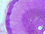 |
Adrenal | The classical organization of the adrenal gland with a thick capsule and the cortex composed of three layers: the outer zona glomerulosa, the middle zona fasciculata and the inner zona reticularis. Finally, in the center of the gland is the adrenal medulla. UCLA Histology Collection. | Adrenal | UCLA Histology |
| 2 |
 |
Adrenal | This is your best slide of the human adrenal cortex. The capsule is prominent as are the zones of the cortex, the zona glomerulosa, zona fasciculata, and zona reticularis. Deep to the zona reticularis should be the medulla but on this slide, instead of medulla there is a central zone of cortical cel... | Adrenal | UCLA Histology |
| 3 |
 |
Adrenal | Higher magnification of the outer layers of the adrenal cortex including the capsule. The boundary between zona glomerulosa and zona fasciculata is quite irregular. UCLA Histology Collection. | Adrenal | UCLA Histology |
| 4 |
|
Adrenal | The classical organization of the adrenal gland with a thick capsule and the cortex composed of three layers: the outer zona glomerulosa, the middle zona fasciculata and the inner zona reticularis. Finally, in the center of the gland is the adrenal medulla. UCLA Histology Collection. | Adrenal | UCLA Histology |
| 5 |
 |
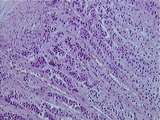
Adrenal - Adrenal Cortex | The layers of adrenal cortex beginning with that next to the capsule are the zona glomerulosa, zona fasciculata, and zona reticularis. The cells of the zona fasciculata look spongy because they contain abundant lipid (steroids) which are dissolved in processing the tissue for histological sectioning... | UCLA Histology | |
| 6 |
 |
Adrenal - Adrenal Cortex and Adrenal Medulla | This image shows the distinct boundary between the zona reticularis of the cortex and the basophilic chromaffin cells of the medulla. Note that in slide 178, it is difficult to differentiate the layers of the cortex and quite difficult to locate ganglion cells within the medulla. UCLA Histology Coll... | Adrenal | UCLA Histology |
| 7 |
 |
Adrenal - Adrenal Medulla | This high magnification view of the adrenal medulla shows its general lact of organization compared to that of the cortex. A ganglion cell complete with satellite cells is indicated. UCLA Histology Collection. | UCLA Histology | |
| 8 |
 |
Adrenal - Adrenal Medulla | In the center of this image is the atrophied medulla. Note the slight basophilia of the medulla compared ot the eosinophilia of the zona reticularis. UCLA Histology Collection. | adrenal | UCLA Histology |
| 9 |
 |
Adrenal - Adrenal Medulla | In the center of this image is the adrenal medulla. Note its basophilia in contrast to the eosinophilia of the zona reticularis. Compare this image to that of 177-10-1 to appreciate the abnormal atrophy of the medulla in that image. UCLA Histology Collection. | UCLA Histology | |
| 10 |
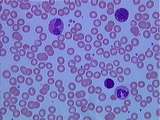 |
Blood | Due to their biconcave disc shape, erythrocytes appear to have a pale center. Small lymphocytes are roughly the same size as erythrocytes (7-8 microns). Platelets are cytoplasmic fragments of bone marrow megakaryocytes. Identify the three types of granulocytes: Neutrophil nuclei usually consist of 3... | Basophils; blood; lymphocytes; Neutrophil; Platelets | UCLA Histology |
| 11 |
 |
Blood | Erythrocytes or RBCs are devoid of organelles. Platelets often appear in clumps. Neutrophil nuclei assume many shapes; also note the sparse purple cytoplasmic granules. Monocytes are the largest leukocytes, and often display a large kidney-shaped nucleus. Because monocytes and neutrophils phagocytiz... | blood; Erythrocytes or RBCs; Monocyte; Neutrophil | UCLA Histology |
| 12 |
 |
Blood | Erythrocytes are roughly the same size as small lymphocytes. Lymphocytes have round, dark nuclei and a small basophilic cytoplasm. Lymphocytes account for roughly 30% of leukocytes, but this percentage can dramatically increase during viral infection. Neutrophils are the most abundant, comprising ab... | blood; Lymphocytes; Neutrophil | UCLA Histology |
| 13 |
 |
Blood | Be careful not to mistake the small purple granules of the neutrophil for the large red granules of the eosinophil. Identify the characteristic bilobed eosinophil nucleus, and the polymorphic neutrophil nucleus. UCLA Histology Collection. | blood; eosinophil; neutrophil | UCLA Histology |
| 14 |
 |
Blood | Note the clumped erythrocytes and platelets. Two neutrophils and one basophil. Basophils account for less than 1% of leukocytes. Granules of basophils are quite large and dark; they often make it impossible to see the bilobed nucleus. UCLA Histology Collection. | basophils; blood; Neutrophil; platelets | UCLA Histology |
| 15 |
 |
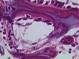
Bone | At a later stage of the developing skull, note the differences in cell density and organization between the immature or woven bone and the mature or lamellar bone. Often a cementing line separates the two types of bone. UCLA Histology Collection. | bone; lamellar bone; Skull | UCLA Histology |
| 16 |
 |
Bone | Identify the following: cartilage cell hypertrophy, calcified cartilage, bone spicules, and bone marrow space. UCLA Histology Collection. | bone; cartilage | UCLA Histology |
| 17 |
 |
Bone | Here we can identify dead bone, since there are no cells in the lacunae (osteocytes die once deprived of a blood supply). Woven bone and calcified cartilage are also present. UCLA Histology Collection. | bone | UCLA Histology |
| 18 |
 |
Bone | Intramembranous bone formation. In the fetal skull, plates of bone are laid down in the mesenchymal field. This image shows woven or immature bone, which tends to be more cellular than lamellar or adult bone. Osteoprogenitor cells differentiate into osteoblasts, which produce the osteoid or uncalcif... | bone; Intramembranous bone formation; Skull | UCLA Histology |
| 19 |
 |
Bone | A high power view of bone being laid down on calcified cartilage. Osteoblasts are laying down bone and osteoclasts are remodeling it. Pale osteoid is also visible. UCLA Histology Collection. | Bone; Osteoblasts; Osteoclasts | UCLA Histology |
| 20 |
 |
Bone - Canaliculi | Here the canaliculi of osteocytes can be seen. The canaliculi are little canals filled with the cytoplasmic extensions of the osteocytes which allow for exchange of nutrients with the central canal (CC). UCLA Histology Collection. | canaliculi | UCLA Histology |
| 21 |
|
Bone - Canaliculi | Here the canaliculi of osteocytes can be seen. The canaliculi are little canals filled with the cytoplasmic extensions of the osteocytes which allow for exchange of nutrients with the central canal (CC). UCLA Histology Collection. | canaliculi | UCLA Histology |
| 22 |
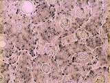 |
Bone - Haversian System | Haversian systems can be seen well in this image of dried compact bone. Haversian or central canals and concentrically arranged osteocytes are also visible. UCLA Histology Collection. | UCLA Histology | |
| 23 |
 |
Bone - Haversian System | This is an example of compact bone. A Haversian system consists of concentric layers (lamellae) of bone surrounding a central Haversian canal in which blood vessels and nerves are located. The osteocytes are found within small lacunae. Marrow cavity. UCLA Histology Collection. | compact bone | UCLA Histology |
| 24 |
|
Bone - Haversian System | Haversian systems can be seen well in this image of dried compact bone. Haversian or central canals and concentrically arranged osteocytes are also visible. UCLA Histology Collection. | UCLA Histology | |
| 25 |
 |
Bone - Haversian System | In this slide, the canaliculi of osteocytes are highly visible. Note that in each Haversian system, all osteocytes in that system are connected to the Haversian canal by the network of canaliculi. UCLA Histology Collection. | UCLA Histology |