The Health Education Assets Library (HEAL) is a collection of over 22,000 freely available digital materials for health sciences education. The collection is now housed at the University of Utah J. Willard Marriott Digital Library.
Filters: Collection: ehsl_heal
| Title | Description | Subject | Collection | ||
|---|---|---|---|---|---|
| 1 |
 |
Acellular cementum in the tooth (longitudinal segment of coronal half of root; high magnification; human, adult) | Stain: Hematoxylin and eosin. From left to right: periodontal ligament with collagen fibers; flattened cementoblasts followed by a thin seam of pink cementoid; darker stained acellular cementum, the so-called acellular extrinsic fiber cementum as collagen fibers protrude from the periodontal ligamen... | oral cavity; acellular cementum; Sharpey's fibers; incremental lines | Poja Histology Collection - Oral Cavity Subset |
| 2 |
 |
Acellular cementum in the tooth (longitudinal segment of coronal half of root; human, adult) | Stain: Hematoxylin and eosin. From left to right: periodontal ligament with blood vessels; flattened cementoblasts followed by a thin seam of pink cementoid; darker stained acellular cementum, the so-called acellular extrinsic fiber cementum as collagen fibres protrude from the periodontal ligament ... | oral cavity; acellular cementum; Sharpey's fibers; incremental lines | Poja Histology Collection - Oral Cavity Subset |
| 3 |
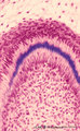 |
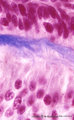
Advanced bell stage with ameloblasts and odontoblasts in tooth development - human embryo | Stain: Azan. From top to bottom: Stellate reticulum consisting of a non-vascularized network of ectoderm-derived cells; Cell layers of the stratum intermedium; Columnar (presecretory) ameloblasts with their upper side (nuclear area) in close contact with the stratum intermedium, and at the distal si... | oral cavity; predentin | Poja Histology Collection - Oral Cavity Subset |
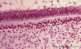
| 4 |
 |
Ameloblasts and odontoblasts in tooth development - advanced bell stage, human, embryo | Stain: Azan. From top to bottom: Stellate reticulum consisting of a loose network of ectoderm-derived cells; Cell layers of the stratum intermedium; Columnar presecretory ameloblasts with their nuclear area close to the stratum intermedium, and at the distal side (secretion area) oriented towards pr... | oral cavity; predentin | Poja Histology Collection - Oral Cavity Subset |
| 5 |
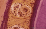
 |
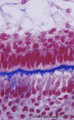
Ameloblasts and odontoblasts in tooth development - advanced bell stage, human, embryo | Stain: Hematoxylin and eosin. From top to bottom: Stellate reticulum consisting of a loose network of ectoderm-derived cells; Cell layers of the stratum intermedium; Palisade-arranged columnar presecretory ameloblasts at the distal side oriented towards the basal lamina enforced by deposited collage... | oral cavity | Poja Histology Collection - Oral Cavity Subset |
| 6 |
 |
Ameloblasts and odontoblasts in tooth development - advanced bell stage, human, embryo CR 80 mm | Scheme electronmicroscopy. From top to bottom: Stellate reticulum consisting of a non-vascularized network of ectoderm-derived cells continuous with the cell layers of the stratum intermedium; Columnar presecretory ameloblasts with their upper side (nuclear area) in close contact with the stratum in... | oral cavity; predentin | Poja Histology Collection - Oral Cavity Subset |
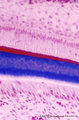
| 7 |
 |
Ameloblasts and odontoblasts in tooth development - advanced bell stage, human, embryo; high magnification | Stain: Azan. From top to bottom: A cell layer of the stratum intermedium; Columnar presecretory ameloblasts at the distal side (secretion area) oriented towards predentin (blue); Note a gradient from left to right in the amount of deposited collagen fibers; Tall columnar odontoblasts in an epithelio... | oral cavity | Poja Histology Collection - Oral Cavity Subset |
| 8 |
 |
Ameloblasts and odontoblasts in tooth development - bell stage, human, embryo | Stain: Azan. From top to bottom: Stellate reticulum consisting of a loose network of ectoderm-derived cells; Cell layers of the stratum intermedium; Palisade-arranged tall columnar secretory ameloblasts with their nuclear area close to the stratum intermedium. The distal side of the cell is oriented... | oral cavity; predentin | Poja Histology Collection - Oral Cavity Subset |
| 9 |
 |
Ameloblasts and odontoblasts in tooth development - early bell stage, human, embryo; high magnification | Stain: Azan. From top to bottom: Presecretory ameloblasts with the distal side (secretion area) oriented towards the basal lamina intertwined with the tangential cut blue stained Korff's fibers (collagen) in predentin. Columnar odontoblasts in a epithelioid arrangement with their secretion area clos... | oral cavity; predentin; Korff's fibers | Poja Histology Collection - Oral Cavity Subset |
| 10 |
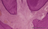 |
Apex of the tooth in osseus socket - alveolus; longitudinal section; human, adult | Stain: Hematoxylin and eosin. From top to bottom: apex region with two tips of dentin covered with cement (darker stained rim; left and right side); note at right tip a dark calcified spot (free cementicle); centrally pulp canal between the tips filled with connective tissue containing blood vessels... | oral cavity; alveolar bone; apical foramen; pulp canal | Poja Histology Collection - Oral Cavity Subset |
| 11 |
 |
Attached denticulus (pulp stone) in the tooth - longitudinal section of root; human, adult. | Stain: Hematoxylin and eosin. From left to right: dentin (purple stained) with dentin tubules; small lightly stained rim of predentin; layers of odontoblasts; irregular structured pulp stone attached to dentin; this false denticle consists of calcified layers with collagen fibers surrounding debris;... | oral cavity; denticulus; pulp stone | Poja Histology Collection - Oral Cavity Subset |
| 12 |
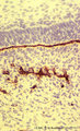 |
Basal lamina between ameloblasts and odontoblasts in tooth development - advanced bell stage, rat embryo | Stain: Anti-collagen IV immunoperoxidase and hematoxylin counterstained. From top to bottom: Stellate reticulum; Stratum intermedium; Ameloblasts at the distal side oriented towards the basal lamina intermingled with the Korffs' fibers (collagen, brown-black); Odontoblasts; Brown-black stained struc... | oral cavity; basal lamina; collagen IV | Poja Histology Collection - Oral Cavity Subset |
| 13 |
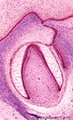 |
Bell stage of the tooth development - human, embryo; low magnification | Stain: Azan. From top to bottom: Stratified ectoderm with a distinct basal layer (red line) of cuboid cells; Dental lamina giving rise to the bell stage (left) and to the primordium of permanent tooth (right); Odontogenic organ (future deciduous tooth) surrounded by fibrous tooth follicle; Out... | oral cavity; dental lamina; predentin | Poja Histology Collection - Oral Cavity Subset |
| 14 |
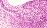 |
Bud stage outgrowth in tooth development - tooth germ, human, embryo | Stain: hematoxylin. At the top stratified ectoderm continuous with the former epithelial tooth bud that abuts toward the underlying neural crest-derived mesenchymal cells. A basal lamina delimits the palisade-arranged epithelial cells from the surrounding dense aggregate of mesenchymal cells. | oral cavity; tooth bud; tooth development | Poja Histology Collection - Oral Cavity Subset |
| 15 |
 |
Cellular cementum of the tooth - longitudinal section of root; human, adult. Thin ground section. | Stain: Hematoxylin and eosin. From left to right: remnants of fibers in periodontal ligament (dark band); cementum wih dark lacunae with projecting canaliculi, originally occupied by cementocytes and their processes (broad area); dentinocemental junction; and superficial dentin with S-shaped course ... | oral cavity; cementocytes | Poja Histology Collection - Oral Cavity Subset |
| 16 |
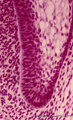 |
Cervical loop in tooth development - late cap stage, human, embryo | Stain: Azan. From left to right: Dental papilla; Thin basement membrane; Columnar inner dental epithelium (presecretory ameloblasts); Stellate reticulum; Cuboidal outer dental epithelium; Fibrous tooth follicle; Note the transition from inner to outer dental epithelium (cervical loop). | oral cavity; cervical loop | Poja Histology Collection - Oral Cavity Subset |
| 17 |
 |
Cross section of tooth in alveolar bone - cat | Stain: Picric acid and hematoxylin. From left to right: alveolar bone covered with a woven type bone (bundle bone) tissue (stained darkly purple); yellow stained periodontal ligament with heavy collagen fibers (horizontal fibers) and blood vessels; bone-related part with Sharpey's fibers followed by... | oral cavity; alveolar bone | Poja Histology Collection - Oral Cavity Subset |
| 18 |
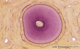 |
Cross section of tooth in alveolar bone - cat; low magnification | Stain: Picric acid and hematoxylin. From left to right: alveolar bone tissue with osteons; periodontal ligament with blood vessels; acellular cementum (dark purple rim); dentin with (purple) radiair arrangement of dentinal tubules; fine incremental (imbrication) lines of von Ebner run at right angl... | oral cavity; incremental lines; alveolar bone | Poja Histology Collection - Oral Cavity Subset |
| 19 |
 |
Cross-section of tooth in alveolar bone (cat, adult) | Stain: Picric acid and hematoxylin. From left to right: Periodontal ligament with blood vessels. Acellular cementum (dark purple rim). Dentin with radiair arrangement of dentinal tubules; fine incremental (imbrication) lines of von Ebner run at right angles to these tubules. These lines represent di... | oral cavity; incremental lines; von Ebner | Poja Histology Collection - Oral Cavity Subset |
| 20 |
 |
Dentin in cross section of tooth (human, adult) | Stain: Hematoxylin and eosin. Longitudinally sectioned dentinal tubules are parallely arranged, and numerous side branches are visible. | oral cavity; dentinal tubules | Poja Histology Collection - Oral Cavity Subset |
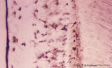
| 21 |
 |
Dentin in cross section of tooth - human, adult | Stain: Hematoxylin and eosin. Cross-sectioned dentinal tubules demonstrate: a light stained center with the odontoblastic process (Tomes' fiber); darker stained peritubular dentin (highly mineralized), also called Neuman's sheath. Intertubular dentin (less mineralized) is present between the tubule... | oral cavity; dentinal tubules; secondary dentin | Poja Histology Collection - Oral Cavity Subset |
| 22 |
 |
Dentinal tubules in longitudinal section of tooth - human, adult. Thin ground section, polarizing microscopy - optical axes of the polarizing plates are crossed at 60. | Slightly oblique section shows dentinal tubules depicted by brown-stained hollow fiber-like structures containing odontoblastic processes (Tomes' fibers) in a semi-three dimensional way. Numerous fine secondary branches of these processes are anastomosing with those of neighboring tubules. | oral cavity; Tomes' Fibers; dentinal tubules | Poja Histology Collection - Oral Cavity Subset |
| 23 |
 |
Dentinal tubules in longitudinal section of tooth - human, adult. Thin ground section, polarizing microscopy - optical axes of the polarizing plates are crossed at 60. | Slightly oblique section shows dentinal tubules depicted by brown-stained hollow fiber-like containing odontoblastic processes (Tomes' fibers) in a semi-three dimensional way ('stubble-field' aspect). Darker stained clusters represent anastomosing secondary branches. | oral cavity; Tomes' Fibers; dentinal tubules | Poja Histology Collection - Oral Cavity Subset |
| 24 |
 |
Dentinal tubules in longitudinal section of tooth - human, adult. Thin ground section, polarizing microscopy - optical axes of the polarizing plates are crossed at 60. | Slightly oblique section shows dentinal tubules depicted by brown-stained stubble structures containing odontoblastic processes (Tomes' fibers) in a semi-three dimensional way. Numerous fine branches of these processes are obvious in dentin. | oral cavity; Tomes' Fibers; dentinal tubules | Poja Histology Collection - Oral Cavity Subset |
| 25 |
 |
Dentinoenamel junction in longitudinal section of tooth (human, adult). Thin ground section. | From left to right: Enamel with fine striation (course of enamel rods or prisms). Few enamel tufts (left, dark) consisting of hypocalcified enamel rods and interprismatic substance arise from the junction. Scallop-like course of dentinoenamel junction. Dentin with dentinal tubules to the dentinoenam... | oral cavity; enamel tufts; dentinoenamel junction; dentinal tubules | Poja Histology Collection - Oral Cavity Subset |