The Health Education Assets Library (HEAL) is a collection of over 22,000 freely available digital materials for health sciences education. The collection is now housed at the University of Utah J. Willard Marriott Digital Library.
TO
Filters: Collection: ehsl_heal
| Title | Description | Subject | Collection | ||
|---|---|---|---|---|---|
| 201 |
 |
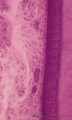
Papillae circumvallatae of the tongue (dorsal side, human) | Stain: Azan. A broad papilla with taste buds (lightly stained spots) facing the grooves in which the serous glands (von Ebner) drain. | oral cavity; von Ebner; circumvallate papillae | Poja Histology Collection - Oral Cavity Subset |
| 202 |
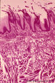 |
Papillae filliformes of the tongue (dorsal side, human) | Stain: Heidenhain light bordeaux. Threadlike keratinized extensions of the stratified epithelium. Primary connective tissue papillae with 2 to 3 secondary papillae. The skeletal muscle fibers are arranged in three directions. | oral cavity; filiform papillae | Poja Histology Collection - Oral Cavity Subset |
| 203 |
 |
Parotid gland (human) | Stain: Mallory trichrome. Survey: at the left bottom a large interlobular duct (in lumen remnants of secretion products) within a septum of dense connective tissue. At the top thinner (blue) septum, a thick (red-bluish) septum at the left. In the center three (intralobular) striated ducts between th... | oral cavity; serous gland | Poja Histology Collection - Oral Cavity Subset |
| 204 |
 |
Parotid gland (human) | Stain: Azan. The parotid gland: in most species the gland is composed entirely of serous acini. At the right a small (intralobular) striated duct; centrally one large interlobular duct with blood vessels. Scattered a few (white) fat cells. | oral cavity; serous gland | Poja Histology Collection - Oral Cavity Subset |
| 205 |
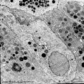 |
Parotid gland (rat) | Electronmicroscopy. Part of a serous acinus with characteristic secretion granules supranuclearly. Note different densities of the granules without any signs of fusion. | oral cavity; serous gland | Poja Histology Collection - Oral Cavity Subset |
| 206 |
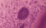 |
Periodontal ligament with epithelial rest of Malassez - longitudinal section of root of tooth, higher magnification; human, adult | Stain: Hematoxylin and eosin. Centrally within the connective tissue of the periodontal ligament a distinct darker stained epithelial islet with nuclei is present (epithelial rest of Malassez as a remnant of Hertwig's epithelial root sheath; in adults it might produce dental cyst). At the right side... | oral cavity; cementoblasts; epithelial rest of Malassez; cementoid | Poja Histology Collection - Oral Cavity Subset |
| 207 |
 |
Periodontal ligament with epithelial rests of Malassez - longitudinal section of root of tooth; human, adult | Stain: Hematoxylin and eosin. From left to right: connective tissue of periodontal ligament with epithelial rests of Malassez as the persistent remnants of the epithelium of Hertwig's epithelial root sheath. The three islets close to the cemental zone are slightly darker stained (note cluster of nuc... | oral cavity; cementoblasts; Sharpey's fibers; acellular cementum | Poja Histology Collection - Oral Cavity Subset |
| 208 |
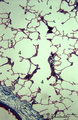 |
Peripheral alveolar area of the lung (human) | Stain: Azan. (1) Part of a pulmonary artery in a septum (2). (3) represents part of a bronchiolus respiratorius that continues into several alveolar ducts (4) and subsequently in alveolar sacs. Arrows (↓) indicate small foci of carbon deposits. | Respiratory bronchioli; Alveolar ducts; Alveolar sacs | Poja Histology Collection - Respiratory System Subset |
| 209 |
 |
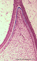
Permanent tooth - canine, human, adult; low magnification of labiolingual section | Stain: Hematoxylin and scarlet red. From top to bottom: Crown region with dentin but without enamel (decalcified specimens); Neck region at the attachment of the gingiva to dentin (left and right); Cementum is visible as a dark small rim from the neck region to the bottom of the tooth; Periodontal l... | oral cavity; alveolar process | Poja Histology Collection - Oral Cavity Subset |
| 210 |
 |
Predentin formation at the cuspal tip in tooth development - bell stage, human, embryo | Stain: Azan. From top to bottom: Stellate reticulum consisting of a network of ectoderm-derived cells; Cell layers of the stratum intermedium; Columnar (presecretory) ameloblasts with their upper side (nuclear area) in close contact with the stratum intermedium, and at the distal side (secretion are... | oral cavity; predentin | Poja Histology Collection - Oral Cavity Subset |
| 211 |
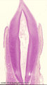 |
Premolar permanent tooth (human, adult; low magnification of labiolingual section) | Stain: Hematoxylin and eosin. From top to bottom: Crown region with enamel-dentin boundary (there is no enamel present in decalcified specimens); dentin (blue-pink) enwraps the whole pulp chamber (light) and here the lines of dentinal tubules appear S-shaped. Neck region at the attachment of the gin... | oral cavity; pulp canal | Poja Histology Collection - Oral Cavity Subset |
| 212 |
 |
Presecretory ameloblast in tooth development - mammalian embryo | Scheme electronmicroscopy. The ectodermal-derived cell appears as tall columnar with fingerlike extensions (dependent on the development stages) at their distal side (secreting area = 'functional base'). These extensions are formed as the cell withdraws during the production of initial enamel. The s... | oral cavity | Poja Histology Collection - Oral Cavity Subset |
| 213 |
 |
Presecretory ameloblasts in tooth development - bell stage, gerbil, postnatal | Electronmicroscopy. Well-arranged epithelial formation of presecretory ameloblasts (active nuclei) with their distal secretion sides towards the thin grey basal lamina. Predentin at the bottom close to the basal lamina and comprises collagen fibers, odontoblastic extensions and dispersed calcified m... | oral cavity; predentin; matrix vesicles | Poja Histology Collection - Oral Cavity Subset |
| 214 |
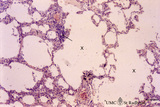 |
Progressing centrilobular lung emphysema (human, adult) | Stain: Hematoxylin and eosin. Emphysema is defined as enlargement of the air spaces (X) distal to the terminal bronchioles, with destruction of the alveolar walls. The remaining alveolar walls are thickened (1). There is an increased cellularity and locally signs of chronic inflammation (↓) are pr... | Poja Histology Collection - Respiratory System Subset | |
| 215 |
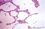 |
Progressing centrilobular lung emphysema (human, adult) | Stain: Hematoxylin and eosin. Emphysema is defined as enlargement of the airspaces (X) distal to the terminal bronchioles, with destruction of the alveolar walls. At (3) remnant of a respiratory bronchiolus. Alveolar walls are destroyed and other alveolar walls are thickened (1) and show an increas... | Alveolar tips | Poja Histology Collection - Respiratory System Subset |
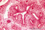
| 216 |
 |
Pseudoglandular - canalicular period of developing lung (human, fetus) | Stain: Azan. Part of a bronchus (1) with young cartilage (2). The hyaline cartilage presents solitary chondrocytes embedded in the blue intercellular substance surrounded by a perichondrium (3). Note that the bronchus as well as the bronchial tubes (4) show an apical position of the epithelial nucle... | Lung development; Pseudoglandular period; Canalicular period; Mesenchyme | Poja Histology Collection - Respiratory System Subset |
| 217 |
 |
Pseudoglandular - canalicular period of developing lung (human, fetus) | Stain: Azan. Terminal budding and elongation of the future bronchial tree: branching of a bronchial tubes (1) within an islet where the mesenchyme condenses (4). A large blood vessel (2) as well as a lymph vessel (3) are recognizable. Note in the mesenchyme formation of blood capillaries in the vici... | Lung development; Pseudoglandular period ; Canalicular period; Mesenchyme | Poja Histology Collection - Respiratory System Subset |
| 218 |
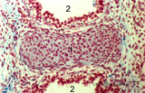 |
Pseudoglandular - canalicular period of developing lung (human, fetus) | Stain: Azan. Location of a cartilagineous ring (1) between two cross-sectioned bronchi (2). Note in the bronchial epithelial cells the apical position of the epithelial nuclei and the light-stained basal part (↑) containing glycogen. The young hyaline cartilage presents solitary chondrocytes embed... | Lung development; Pseudoglandular period; Canalicular period; Mesenchyme | Poja Histology Collection - Respiratory System Subset |
| 219 |
 |
Pseudoglandular - canalicular period of developing lung (human, fetus) | Stain: Azan. Longitudinal section through a large bronchus (1) with cartilagineous rings (2). At (3) developing glandular structures in islets of bronchial tubes surrounded by condensed mesenchyme. At (4) lymph vessels. | Lung development; Pseudoglandular period; Canalicular period; Mesenchyme; Bronchial tubes | Poja Histology Collection - Respiratory System Subset |
| 220 |
 |
Pseudoglandular - canalicular period of developing lung (human, fetus) | Stain: Azan. Part of a future lung lobe with cross-sectioned larger bronchi (1) in close association with islets of future smaller bronchial tubes (*). Note the branching of the bronchial tubes within an islet. The mesenchyme within the islets of future tubes becomes more condensed (↓). Blood vess... | Lung development; Visceral pleura; Pseudoglandular period; Canalicular period; Mesenchyme | Poja Histology Collection - Respiratory System Subset |
| 221 |
 |
Pseudoglandular period of developing lung (human, embryo) | Stain: Trichrome (Goldner). Cross-sectioned future bronchial tubes (1) of varying sizes, note the apical position of the nuclei with light-stained basal parts (glycogen). The surrounding mesenchyme condenses (↓) around the epithelium and in the neighbourhood small blood vessels (*) are present. At... | Lung development; Pseudoglandular period; Bronchial tubes; Mesenchyme | Poja Histology Collection - Respiratory System Subset |
| 222 |
 |
Pseudoglandular period of developing lung (human, embryo) | Stain: Trichrome (Goldner). Cross-sectioned future bronchial tubes (1) of varying sizes, note the apical position of the nuclei with light-stained basal parts (glycogen). The surrounding mesenchyme becomes condensed (↓) around the epithelium and in between small blood vessels (*) are present. | Lung development; Pseudoglandular period; Bronchial tubes; Mesenchyme | Poja Histology Collection - Respiratory System Subset |
| 223 |
 |
Pseudoglandular period of developing lung (human, embryo) | Stain: Hematoxylin and eosin. Two cross-sectioned future bronchial tubes. Note the basal position of the nuclei with light-stained apical cytoplasm (↓, glycogen). The surrounding mesenchyme condenses (1) around the epithelium and developing capillaries and small blood vessels (*) are present. Futu... | Lung development; Visceral pleura; Pseudoglandular period; Bronchial tubes; Mesenchyme | Poja Histology Collection - Respiratory System Subset |
| 224 |
 |
Pseudoglandular period of developing lung (human, embryo, low magnification) | Stain: Hematoxylin and eosin. The two future lung lobes contain many cross-sectioned future bronchial tubes (1). Note in these epithelial cells the apical position of the nuclei with light-stained basal parts (glycogen). The surrounding mesenchyme becomes more condensed (↓) around the epithelium a... | Lung development; Visceral pleura; Pseudoglandular period; Bronchial tubes; Mesenchyme | Poja Histology Collection - Respiratory System Subset |
| 225 |
 |
Pseudoglandular period of developing lung (human, embryo, low magnification) | Stain: Hematoxylin and eosin. Cross-sectioned future bronchial tubes (1), the surrounding mesenchyme becomes more condensed around the epithelium. The mesoderm of the future visceral pleura (2) as well as the future parietal pleura (3) and (4) indicates pleural cavity. The cartilagineous spinal colu... | Lung development; Visceral pleura; Parietal pleura; Pseudoglandular period; Bronchial tubes; Mesenchyme | Poja Histology Collection - Respiratory System Subset |