The Health Education Assets Library (HEAL) is a collection of over 22,000 freely available digital materials for health sciences education. The collection is now housed at the University of Utah J. Willard Marriott Digital Library.
Filters: Collection: ehsl_heal
| Title | Description | Subject | Collection | ||
|---|---|---|---|---|---|
| 151 |
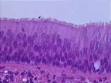 |
Epithelium - Trachea | The epithelium of the trachea is categorized as pseudostratified columnar with goblet cells. It is pseudostratified because, although all the cells contact the basal lamina, the basal cells do not reach the lumen. Cilia can be seen on most columnar cells. Two goblet cells are also evident. UCLA Hist... | pseudostratified columnar epithelium | UCLA Histology |
| 152 |
|
Epithelium - Trachea | The epithelium of the trachea is categorized as pseudostratified columnar with goblet cells. It is pseudostratified because, although all the cells contact the basal lamina, the basal cells do not reach the lumen. Cilia can be seen on most columnar cells. Two goblet cells are also evident. UCLA Hist... | pseudostratified columnar epithelium | UCLA Histology |
| 153 |
 |
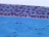
Epithelium - Urethra | The urethra is one of the few organs which is lined by stratified columnar epithelium. It is stratified columnar because there are many cell layers, and the cells bordering the lumen are columnar. UCLA Histology Collection. | stratified columnar epithelium | UCLA Histology |
| 154 |
|
Epithelium - Urethra | The urethra is one of the few organs which is lined by stratified columnar epithelium. It is stratified columnar because there are many cell layers, and the cells bordering the lumen are columnar. UCLA Histology Collection. | stratified columnar epithelium | UCLA Histology |
| 155 |
 |
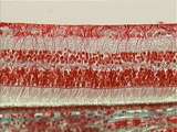
Epithelium - Urinary Bladder | When the urinary bladder is full of urine, the transitional epithelium lining this structure is said to be stretched. Note the squamous or flattened cells at the lumen. UCLA Histology Collection. | transitional epithelium; urinary bladder | UCLA Histology |
| 156 |
|
Epithelium - Urinary Bladder | When the urinary bladder is full of urine, the transitional epithelium lining this structure is said to be stretched. Note the squamous or flattened cells at the lumen. UCLA Histology Collection. | transitional epithelium; urinary bladder | UCLA Histology |
| 157 |
 |
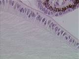
Epithelium - Vagina | The stratified squamous vaginal epithelium is characterized by the absence of glands and the fact that the glycogen is dissolved from the cells during histological processing. UCLA Histology Collection. | UCLA Histology | |
| 158 |
 |
Eye | The iris extends forward from its junction with the ciliary body. The double-layered epithelium of the ciliary body continues onto the posterior surface of the iris. Iris color is determined by the number of melanocytes in the stroma. Note the iris root and the iris epithelium. UCLA Histology Collec... | Eye; iris | UCLA Histology |
| 159 |
 |
Eye | The corneal epithelium is multi-layered and its continuously proliferating cells change their shape from columnar to squamous as they are displaced from a thick, specialized extracellular matrix called Bowman's membrane. The stromal cells are called keratocytes and they synthesize and organize the h... | cornea; Eye | UCLA Histology |
| 160 |
 |
Eye | Optic nerve showing the optic disc (blind spot with no retina) and the central artery. UCLA Histology Collection. | Eye; optic nerve; test | UCLA Histology |
| 161 |
 |
Eye | This slide provides a good view of the Mller glial cell processes. Their end feet rest on the inner limiting membrane of the retina and their microvilli extend external to the outer limiting membrane of the retina. UCLA Histology Collection. | Eye; retina | UCLA Histology |
| 162 |
 |
Eye | Further enlargement reveals the anterior lens capsule, area of proliferation (mitotic figures) and area of elongation. The developing ciliary epithelium (double layered) is seen in the top right corner. UCLA Histology Collection. | Eye; lens | UCLA Histology |
| 163 |
 |
Eye | The posterior layer of the iris epithelium is heavily pigmented and the anterior layer has reduced pigment when compared to the epithelium of the ciliary body. The anterior portion of each anterior layer cell is modified to collectively form the dilator muscle of the pupil (under sympathetic innerva... | Eye; iris | UCLA Histology |
| 164 |
 |
Eye | The limbus is the corneal-scleral junction. Present in this region is the conjunctival fornix, where the corneal epithelium is reflected onto the inner surface of the eyelid. Major anatomical features include the trabecular meshwork and Schlemm's canal, a major drainage site for aqueous humor from t... | Eye; limbus | UCLA Histology |
| 165 |
 |
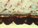
Eye - Choroid | The choroid is the vascular layer that feeds the retinal pigment epithelium and photoreceptors. It consists of a large vessel layer, capillary layer (choriocapillaris), and Brch's membrane. The latter is an important player in age-related macular degeneration. UCLA Histology Collection. | UCLA Histology | |
| 166 |
 |
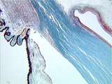
Eye - Ciliary Body | The ciliary body is a ring-shaped structure that extends around the eyeball, posterior to the corneal - scleral junction (limbus). It consists of a double-layered epithelium, smooth muscle and stroma. Canal of schlemm is a landmark for the limbal region. The epithelium covers a flat portion of the c... | canal of schlemm | UCLA Histology |
| 167 |
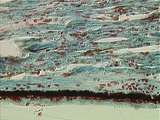 |
Eye - Ciliary Epithelium | Some features of the double-layered ciliary epithelium can be observed in the plars plana region. The innermost layer (closest to the posterior chamber) is non-pigmented and the outermost layer is pigmented. The ciliary epithelium forms aqueous humor and secretes it into the posterior chamber. Aqueo... | canal of Schlemm; ciliary epithelium | UCLA Histology |
| 168 |
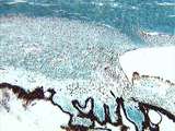 |
Eye - Ciliary Muscle | The ciliary muscle is used during accommodation. The muscle consists of longitudinal and circular fibers. The circular fibers perform a sphincter - like motion and the longitudinal fibers pull the choroid forward. In either case, tension on the zonular fibers of the lens is reduced and the lens bulg... | Ciliary Muscle | UCLA Histology |
| 169 |
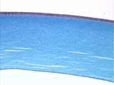 |
Eye - Cornea | The cornea is a multi-layered, transparent tissue that serves as the window to the eye and provides about two-thirds of its refractive power. Visible are the epithelium, stroma and endothelium. UCLA Histology Collection. | UCLA Histology | |
| 170 |
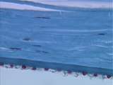 |
Eye - Corneal Endothelium | The corneal endothelium is a monolayer of non-proliferating cells. Between the endothelium and the stroma lies Descemet's membrane, a specialized basal lamina produced by the endothelial cells. UCLA Histology Collection. | UCLA Histology | |
| 171 |
 |
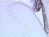
Eye - Developing Lens | The global architecture of the developing lens can be appreciated in the developing rat eye. This section was taken from an animal whose eyelids had not yet opened and the future site of separation can be seen. The developing cornea and lens epithelium can also be observed in this image. UCLA Histol... | UCLA Histology | |
| 172 |
 |
Eye - Iris | The sphincter of the iris is located near the pupil (a hole in the center of the iris) and is a ring-shaped, smooth muscle. The sphincter, which lies concentric to the iris, is innervated by parasympathetic fibers and acts during accommodation and in relatively bright light. UCLA Histology Collectio... | UCLA Histology | |
| 173 |
 |
Eye - Lens | The shape of the lens changes during the process of accommodation. When the eye focuses on an image while reading, the smooth muscle of the circular ciliary body contracts, reducing tension on the zonular fibers (collectively called the suspensory ligament), causing the lens to bulge, increasing its... | canal of Schlemm | UCLA Histology |
| 174 |
 |
Eye - Lens Epithelium | This enlargement of slide 236-2.5-2 reveals the lens epithelium in various phases: the anterior epithelium, area of proliferation, and area of elongation. The lens bow region consists of lens fiber nuclei in various stages of degeneration. UCLA Histology Collection. | UCLA Histology | |
| 175 |
 |
Eye - Lens Fibers | Lens fibers elongating as they move posterior. UCLA Histology Collection. | UCLA Histology |