The Health Education Assets Library (HEAL) is a collection of over 22,000 freely available digital materials for health sciences education. The collection is now housed at the University of Utah J. Willard Marriott Digital Library.
TO
Filters: Collection: ehsl_heal
| Title | Description | Subject | Collection | ||
|---|---|---|---|---|---|
| 26 |
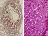 |
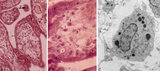
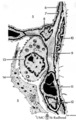
Survey of implanted blastocyst (human, ca. 3 weeks-old embryo) and detail of advanced stage of decidua cells (human, mid pregnancy) | Stain: Hematoxylin-eosin (A, left) and Azan (B, right). (A) Left: section through a slightly damaged implantation site shows at (1) the uterine lumen and (2) the decidua capsularis while at (3) several uterine glands are visible. At arrow (4) the amniotic cavity, (5) points to embryonic shield and... | placenta; uterus; syncytiotrophoblast; implantation; blastocyst | Poja Histology Collection - Placenta |
| 27 |
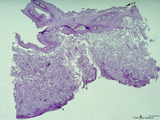 |
Survey of the chorionic plate and intervillous space (human placenta, full-term) | Stain: Perjodic acid-Schiff reaction (PAS). At the top the chorionic plate (1) with cross-sections of umbilical vessels (2) At (3) the folded amnion covering the chorionic plate. Ramifications of thicker stem villi demonstrate free-floating terminal villi (tertiary). At several places reddish-st... | placenta; tertiary villi; decidua; chorionic plate | Poja Histology Collection - Placenta |
| 28 |
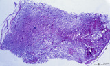 |
Survey of the intervillous space with villi (human placenta, full-term) | Stain: Perjodic Acid Schiff reaction (PAS). At the right and left side the chorionic plate (1) with few cross-sectioned umbilical vessels (2), at (3) a detached amnion. Cross-sections of thick stem villi (4) at the fetal side ramify into terminal villi (tertiary) that are found stuffed (due to fix... | placenta; stem villus; syncytiotrophoblast; amnion | Poja Histology Collection - Placenta |
| 29 |
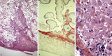 |
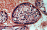
Surveys/detail of the decidua basalis and intervillous space of the placenta (human placenta, late midpregnancy) | Stain: Hematoxylin ?azophloxine (A, C), Trichrome (Goldner) (B). Left pictures (A, B): The maternal side at the bottom consists of a broad zone of the stripped basal decidua (1) intermingled with reddish matrix-type fibrinoid accumulations and streaks (Rohr) (2), close to the anchoring villi (3) an... | placenta; chorionic villi; fibrinoid; trophoblast shell | Poja Histology Collection - Placenta |
| 30 |
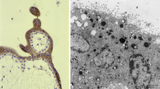 |
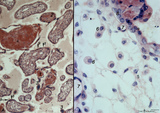
Syncytiotrophoblast cells of tertiary villi (human placenta, midpregnancy) | (A) Left: stain: Immunoperoxidase staining with diaminobenzidin (DAB) and human anti-chorionic gonadotrophin antibody (hCG-DAKO 231), counterstained with hematoxylin. (B) Right: electron microscopy. (A) At the left the exclusive reactivity (dark brown) in the cytoplasma of the multinucleated syn... | Syncytiotrophoblast; placenta ; chorionic villi; HCG; immunohistochemistry; electron microscopy | Poja Histology Collection - Placenta |
| 31 |
 |
Tertiary villi (human placenta, early pregnancy) | Stain: (A, left) Azan. (B, right) Immunoperoxidase staining with diaminobenzidin (DAB) and hematoxylin counterstaining for human anti-chorionic gonadotrophin (hCG-DAKO 231 antibody). (A) shows blue grey-stained postmitotic multinucleated syncytiotrophoblast cells (STC, 1b), partially cut tangential... | placenta; chorionic villi; placental barrier; HCG | Poja Histology Collection - Placenta |
| 32 |
 |
Tertiary villi (human placenta, full-term, cross-section) | Stain: Hematoxylin ? azophloxine. A few tertiary villi (1) within the intervillous space (2) contains capillary (3) and embryonic connective tissue (*). It remains covered by the postmitotic multinucleated syncytiotrophoblast cell (STC, 4). Distinct nuclear accumulation in the STC indicates the so-c... | placenta; tertiary villi; syncytiotrophoblast | Poja Histology Collection - Placenta |
| 33 |
 |
Tertiary villi (human placenta, full-term, cross-sectioned) | Stain: Hematoxylin-azophloxine. The central villus is lined by epithelial multinucleated syncytiotrophoblast cells (1) and sparsely light-stained cytotrophoblast cells (Langhans, (2)). The fetal stroma contains vessels and mostly fibroblasts, note capillaries (3) in close association with the epithe... | placenta; tertiary villus; syncytiotrophoblast | Poja Histology Collection - Placenta |
| 34 |
 |
Tertiary villi (human placenta, late midpregnancy) | : (A) Left: stain: Hematoxylin - azophloxine. (B) Right: electron microscopy (low magnification). At the left (A) multinucleated syncytiotrophoblast cells (STCs, 1) and vaguely cytotrophoblast cells (CTCs, 2) cover the loose fetal stroma (4). A large and a small arteriole (3) are embedded with stro... | placenta; tertiary villi; placental barrier; Hofbauer cell; vasculosyncytial membrane; syncytiotrophoblast | Poja Histology Collection - Placenta |
| 35 |
 |
Tertiary villi (human placenta, midpregnancy) | (A) Lower and (B) higher magnification. Stain: Hematoxylin - azophloxine. (C) electron microscopy of Hofbauer cell. (A, B) show tertiary villi and intervillous spaces. Squeezed between villi fibrinoid clots (1). The large arrow (2) points to either a detached, free circulating large STC (syncyt... | placenta; chorionic villi; Hofbauer cell; fibrinoid; syncytiotrophoblast | Poja Histology Collection - Placenta |
| 36 |
 |
Tertiary villi (human placenta, midpregnancy) | Stain: Hematoxylin - azophloxine. (A) Left a variety of tertiary villi and reddish-stained fibrinoid depositions (1). At (2): the embryonic connective tissue starts to fibrinize. Note syncytiotrophoblast knots (3) or sprouts of several villi .These knots might represent aggregations of degenerati... | placenta; fibrinoid; cytotrophoblast; syncytiotrophoblast | Poja Histology Collection - Placenta |
| 37 |
 |
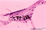
Trophoblast cell in lung alveolar interstitium (human, early midpregnancy) | Stain: Hematoxylin-eosin. It is well known that free circulating trophoblast cells can be found migrated into lung parenchyma during normal pregnancy. In this sample a large trophoblast cell (→) is localised within the normal alveolar interstitium. Alveolar phagocytes (*) are present in the alve... | placenta; trophoblast; lung | Poja Histology Collection - Placenta |
| 38 |
 |
Villi of complete hydatidiform mole (human) | (A) Macroscopy aggregation (1) of abnormal villi of hydatidiform mole intermingled with blood clots (2). (B) After dissection of a complete mole grape-like aggregations of dilated chorionic villi are presented as numerous liquid-filled vesicles (1) varying in diameters (from few mm up to 1 cm) surr... | placenta; trophoblast; hydatiform mole | Poja Histology Collection - Placenta |
| 39 |
 |
Acellular cementum in the tooth (longitudinal segment of coronal half of root; high magnification; human, adult) | Stain: Hematoxylin and eosin. From left to right: periodontal ligament with collagen fibers; flattened cementoblasts followed by a thin seam of pink cementoid; darker stained acellular cementum, the so-called acellular extrinsic fiber cementum as collagen fibers protrude from the periodontal ligamen... | oral cavity; acellular cementum; Sharpey's fibers; incremental lines | Poja Histology Collection - Oral Cavity Subset |
| 40 |
 |
Acellular cementum in the tooth (longitudinal segment of coronal half of root; human, adult) | Stain: Hematoxylin and eosin. From left to right: periodontal ligament with blood vessels; flattened cementoblasts followed by a thin seam of pink cementoid; darker stained acellular cementum, the so-called acellular extrinsic fiber cementum as collagen fibres protrude from the periodontal ligament ... | oral cavity; acellular cementum; Sharpey's fibers; incremental lines | Poja Histology Collection - Oral Cavity Subset |
| 41 |
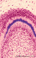 |
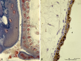
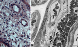
Advanced bell stage with ameloblasts and odontoblasts in tooth development - human embryo | Stain: Azan. From top to bottom: Stellate reticulum consisting of a non-vascularized network of ectoderm-derived cells; Cell layers of the stratum intermedium; Columnar (presecretory) ameloblasts with their upper side (nuclear area) in close contact with the stratum intermedium, and at the distal si... | oral cavity; predentin | Poja Histology Collection - Oral Cavity Subset |
| 42 |
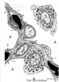 |
Air-blood barrier in the lung (mammals) | Scheme electron microscopy. (1, ↓) Represents type I pneumocytes lining alveolar spaces (A). Cell (2) represents a free alveolar macrophage. The type II pneumocyte (3) is adherent to type I pneumocyte extensions (note junctional connection), and contains multilamellar bodies (surfactant). A myofib... | Pneumocyte type I ; Pneumocyte type II | Poja Histology Collection - Respiratory System Subset |
| 43 |
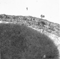 |
The air-blood barrier in the lung (rat) | Electron microscopy (high magnification). The alveolar space (1) and the capillary space (2) (with part of an erythrocyte, 3) are separated from each other respectively by cytoplasm of endothelial cell (5), the common basal lamina (6) with a lamina densa (6a) and the type I alveolar cell (4, pneumo... | Pneumocyte I; Alveolar cell type I | Poja Histology Collection - Respiratory System Subset |
| 44 |
 |
Alveolar cell type II (pneumocyte II) in an alveolus (dog) | Electron microscopy. At (X) the alveolar space, the bulging alveolar cell type II shows the characteristic multilamellar bodies (*, cytosomes) that contain precusor material of surfactant. The lamellar bodies are responsible for the vacuolated appearance of these cells, and they give rise to surfact... | Pneumocyte II; Alveolar cell type II | Poja Histology Collection - Respiratory System Subset |
| 45 |
 |
Alveolar cells in the lung (mammals) | Scheme electron microscopy. (5) alveolar space; (6) type I Pneumocyte; (7) basal lamina; (8) myofibroblast; (9) collagen and elastin fibers; (10) mesothelial cell of the visceral pleura; (11) capillary with erythrocyte; (12) endothelial cell lining the capillary; (13) type II pneum... | Pneumocyte type I ; Pneumocyte type I I | Poja Histology Collection - Respiratory System Subset |
| 46 |
 |
Alveolar duct in the lung (mouse) | Stain: PAS and hematoxylin. Part of an alveolar duct lumen (1) that shows bronchiolar characteristics such as cuboidal epithelium (2) covering a bundle of smooth muscle (3) and connective tissue containing macrophages (4) with black pigment deposits. Note PAS-positivity of these cuboidal cells (Clar... | Alveolar ducts; Clara cells | Poja Histology Collection - Respiratory System Subset |
| 47 |
 |
Alveolar macrophage in lung (rat) | Electron microscopy. A wandering alveolar macrophage (1) migrates through an alveolar pore from an alveolar space (A1) into another (A2). Note the dark lysosomal structures and abundant organelles in its cytoplasm. The thin lining type I alveolar cell is hardly discernable (thin arrows →). Interst... | Pneumocyte I; Alveolar macrophages | Poja Histology Collection - Respiratory System Subset |
| 48 |
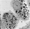 |
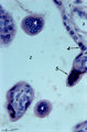
Alveolar macrophages in lung (human, adult) | Electron microscopy. Two macrophages in the alveolar space (N = nucleus). Note the heterogeneity of many lysosomal structures containing among others carbon particles and the abundancy of organelles. | Alveolar macrophages | Poja Histology Collection - Respiratory System Subset |
| 49 |
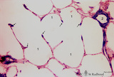 |
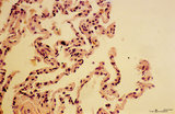
Alveolar sac in the lung (human) | Stain: Azan. Characteristic alveolar tips (arrows) of neighbouring thin-walled alveoli (1). The tips contain elastin masked by collagen (blue-stained). At (2) small pulmonary arteries. | Alveolar tips | Poja Histology Collection - Respiratory System Subset |
| 50 |
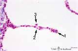 |
Alveolar septum in the lung (human, high magnification) | Stain: Azan. Two neighboring alveoli separated by a septum. (1) points to the tip containing bluish-pink elastin and collagen. The nucleus of a squamous alveolar cell (type I pneumocyte) is indicated by (2), and a free alveolar macrophage by (3). | Alveolar tip; Pneumocyte I | Poja Histology Collection - Respiratory System Subset |