The Health Education Assets Library (HEAL) is a collection of over 22,000 freely available digital materials for health sciences education. The collection is now housed at the University of Utah J. Willard Marriott Digital Library.
TO
Filters: Collection: ehsl_heal
| Title | Description | Subject | Collection | ||
|---|---|---|---|---|---|
| 351 |
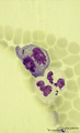 |
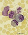
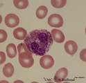
Dividing cells (mitosis) in bone marrow smear (human) | Stain: May-Grnwald-Giemsa (MGG). (1) Shows a dividing cell (mitotic figure) possibly an erythroblast cell type. (2) Shows two segmented neutrophils. | Poja Histology Collection - Blood & Bone Marrow Subset | |
| 352 |
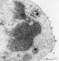 |
Endocytosis in lymphocyte (peripheral blood, rat) | Electron microscopy. Thorotrast is a suspension of thorium dioxide particles and was formerly used as a contrast medium in X-ray diagnostics. These particles were found to accumulate in spleen, lymph nodes and most likely in macrophages and phagocytizing reticular cells. Generally lymphocytes do not... | Poja Histology Collection - Blood & Bone Marrow Subset | |
| 353 |
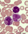 |
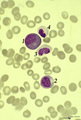
Eosinophil, monocyte and basophil in blood smear (human) | Stain: May-Grnwald-Giemsa (MGG). (1) eosinophilic granulocyte with two nuclear lobes and large eosinophilic granules in the cytoplasm. (2) monocyte with a large indented nucleus that is much more transparent than the nuclei of the two other cells. (3) basophilic granulocyte with aggregated dark purp... | Poja Histology Collection - Blood & Bone Marrow Subset | |
| 354 |
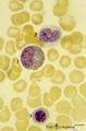 |
Eosinophilic (meta)myelocyte in bone marrow smear (human) | Stain: May-Grnwald-Giemsa (MGG). The granules of the eosinophilic (meta)myelocyte (1) are large and brown-blue stained in contrast to the hardly visible, dust-like granules in the neutrophilic band form (2). (3) orthochromatic erythroblast. | Poja Histology Collection - Blood & Bone Marrow Subset | |
| 355 |
 |
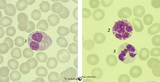
Eosinophilic (meta)myelocytes in bone marrow smear (human) | Stain: May-Grnwald-Giemsa (MGG). Three eosinophilic (meta)myelocytes (1) at slightly different maturation stages. Notice the brown blue large solitary granules. (2) neutrophilic metamyelocyte. (3) smudged cell. | Poja Histology Collection - Blood & Bone Marrow Subset | |
| 356 |
 |
Eosinophilic granulocyte | Scheme electron microscopy. A 11-15 m cell with a bilobed nucleus (1) moderate amount of organelles, mitochondria (2), Golgi area (3), many vesicles (5) and numerous specific eosinophilic granules (4). These granules contain a central electron-dense angular crystalloid core embedded in a finely gran... | Poja Histology Collection - Blood & Bone Marrow Subset | |
| 357 |
 |
Eosinophilic granulocyte (peripheral blood, human) | Electron microscopy. A 11-15 m cell with a partially cut bilobed nucleus (1) and moderate amount of organelles and vesicles. The specific eosinophilic granules (2) contain a central electron-dense angular crystalloid core (3) embedded in a finely granular matrix (4). The crystalloid consists of an a... | Poja Histology Collection - Blood & Bone Marrow Subset | |
| 358 |
 |
Eosinophilic granulocyte (peripheral blood, human) | Electron microscopy. (A) Overview of the cell. Note two nuclear lobes (1) due to the section. There are numerous specific eosinophilic granules (2) and a Golgi area (3). Few thin filopodia (↓, arrows) are present. (B) Detail: some vesicles and many specific eosinophilic granules of varying sizes. ... | Poja Histology Collection - Blood & Bone Marrow Subset | |
| 359 |
 |
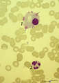
Eosinophilic granulocyte in peripheral blood smear (human) | Stain: May-Grnwald-Giemsa (MGG). The eosinophil (11-15 μm) contains a bilobed nucleus and numerous large solitary brown-orange granules. The eosinophils are the first line of defense against parasites but also take part in allergic reactions (bronchial asthma). Arrow (↓) points to two platelets. | Poja Histology Collection - Blood & Bone Marrow Subset | |
| 360 |
 |
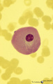
Eosinophilic granulocyte in peripheral blood smear (human) | Stain: May-Grnwald-Giemsa (MGG). The eosinophilic granulocyte usually has two to three nuclear lobes. The brown-orange granules are large, solitary and contain pharmacologically active mediators. The cell surface is occupied with IgE receptors. | Poja Histology Collection - Blood & Bone Marrow Subset | |
| 361 |
 |
Eosinophilic myelocyte and neutrophilic myelocyte in bone marrow smear (human) | Stain: May-Grnwald-Giemsa (MGG). The eosinophilic myelocyte (1) contains brown-like granules (not orange) in the bluish basophilic cytoplasm. Nucleoli are still visible. Maturation of eosinophils parallels that of neutrophils except for the production of the secondary, specific granules in myelocyte... | Poja Histology Collection - Blood & Bone Marrow Subset | |
| 362 |
 |
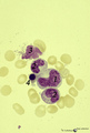
Eosinophilic, neutrophilic and basophilic granulocytes in peripheral blood smear (human) | Stain: May-Grnwald-Giemsa (MGG). The eosinophil (1) is slightly larger than the neutrophil with a diameter of 12-17 m. The nucleus is usually bilobed (occasionally trilobed). Eosinophil granules are considerably larger than those of neutrophils, and are stained reddish-orange. These cells are very f... | Poja Histology Collection - Blood & Bone Marrow Subset | |
| 363 |
 |
Erythroblast and neutrophilic myelocytes in bone marrow smear (human) | Stain: May-Grnwald-Giemsa (MGG). (1) an older basophilic erythroblast with slightly condensed nuclear chromatin. (2) two neutrophilic myelocytes with azurophilic primary granules. | Poja Histology Collection - Blood & Bone Marrow Subset | |
| 364 |
 |
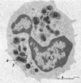
Erythroblastic island (bone marrow, rabbit) | Electron microscopy. In the bone marrow close associations of developing red blood cells with reticular cells are required during erythropoiesis. Different stages of erythroblasts (2, 3) are exposed in close vicinity of a reticular cell (1). (4) Reticulocyte. The centrally localized phagocytic retic... | Poja Histology Collection - Blood & Bone Marrow Subset | |
| 365 |
 |
Erythroblastic island in bone marrow | Scheme electron microscopy. Different stages of maturing erythroblasts (2) are exposed in this scheme. The centrally localized phagocytic reticular cell (1) has many long cytoplasmic extensions that form a network with similar cells within the bone marrow. Its nucleus is irregular. The cytoplasm has... | Poja Histology Collection - Blood & Bone Marrow Subset | |
| 366 |
 |
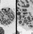
Erythroblasts (bone marrow, rabbit) | Electron microscopy. (1) shows basophilic erythroblasts or early normoblasts with clumped heterochromatin in the nucleus and numerous polysomes. (1m) points to a mitotic figure of a basophilic erythroblast. A late polychromatic erythroblast or intermediate normoblast (2) marks a maturing stage in wh... | Poja Histology Collection - Blood & Bone Marrow Subset | |
| 367 |
 |
Erythrocytes in peripheral blood smear (human) | Stain: May-Grnwald-Giemsa (MGG). Normal erythrocytes in a blood smear of peripheral blood. The mature human erythrocytes do not have nuclei. Their normal diameter is about 7.5 μm. Macrocytes have a diameter >9 μm, while microcytes are smaller than 6 μm. The thickness of an erythrocyte in the cent... | Poja Histology Collection - Blood & Bone Marrow Subset | |
| 368 |
 |
Erythron in bone marrow smear (human) | Stain: May-Grnwald-Giemsa (MGG). The erythron or erythroblastic island consists of a large reticulum cell (1) surrounded by erythroblastic cell types (2) at varoius stages of differentiation. The nucleus of the reticulum cell (histiocyte) usually contains a noticeable nucleolus. At distance, some my... | Poja Histology Collection - Blood & Bone Marrow Subset | |
| 369 |
 |
Erythron in bone marrow smear (human) | Stain: May-Grnwald-Giemsa (MGG). In the center of the slide an erythron that consists of a reticular cell (1) surrounded by erythroblasts (2) at various maturational stages. | Poja Histology Collection - Blood & Bone Marrow Subset | |
| 370 |
 |
Erythron or erythropoietic island in bone marrow smear (human) | Stain: May-Grnwald-Giemsa. The picture shows a reticular cell (1) surrounded by almost exclusively erythroblasts at different stages of development and maturation (2). The cells marked with (*) are myeloid cell types. The reticular cell has phagocytized nuclear debris of expulsed nuclei (→). The r... | Poja Histology Collection - Blood & Bone Marrow Subset | |
| 371 |
 |
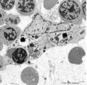
Fibrin phagocytosis by neutrophils (peripheral blood, mouse) | Electron microscopy. At site of tissue damage motile neutrophils are among the first to be involved actively in phagocytosis. In between a cluster of neutrophilic granulocytes fibrin depositions (1) are present. At (1→) fibrin accumulations are close attached to the surface of a neutrophil indicat... | Poja Histology Collection - Blood & Bone Marrow Subset | |
| 372 |
 |
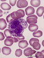
Flaming plasma cell in peripheral blood smear (human) | Stain: May-Grnwald-Giemsa (MGG). The so-called flaming plasma cell (1) is characterized by fiery fringes, which are formed by pseudopodic cytoplasmic projections (arrows) that stain with carmin red. These peripheral cytoplasmic spots contain numerous dilated endoplasmic reticulum cisterns, which are... | Poja Histology Collection - Blood & Bone Marrow Subset | |
| 373 |
 |
Giant neutrophilic metamyelocyte in bone marrow smear (human) | Stain: May-Grnwald-Giemsa (MGG). (1) Giant neutrophilic metamyelocyte. (2) Normal neutrophilic metamyelocytes. (3) Smudged eosinophilic granulocyte. (4) Orthochromatic erythroblast. | Poja Histology Collection - Blood & Bone Marrow Subset | |
| 374 |
 |
Giant plasma cell in peripheral blood smear (human) | Stain: May-Grnwald-Giemsa (MGG). A giant plasma cell with diffusely spread immunoglobulins in the cytoplasm. The Golgi area remains unstained (white area). | Poja Histology Collection - Blood & Bone Marrow Subset | |
| 375 |
 |
Granular megakaryocyte producing platelets in bone marrow smear (human) | Stain: May-Grnwald-Giemsa (MGG). The centrally located megakaryocyte (1) has a huge nucleus (polyploidy) and an extended cytoplasm from which platelets are released at the periphery (arrows). | Poja Histology Collection - Blood & Bone Marrow Subset |