The Health Education Assets Library (HEAL) is a collection of over 22,000 freely available digital materials for health sciences education. The collection is now housed at the University of Utah J. Willard Marriott Digital Library.
TO
Filters: Collection: ehsl_heal
| Title | Description | Subject | Collection | ||
|---|---|---|---|---|---|
| 126 |
 |
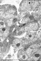
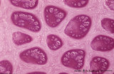
Exocrine gland (salivary gland) | Scheme electronmicroscopy. Part of an acinus of serous cells, basolaterally a well developed rough endoplasmic reticulum and apically secretion granules with different maturity towards the small lumen. On top stages of early formed secretion granule (left) and more matured ones (right). Note small j... | oral cavity; serous gland | Poja Histology Collection - Oral Cavity Subset |
| 127 |
 |
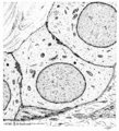
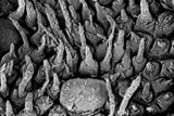
Exocrine gland - intercalated duct - submandibular gland, rat | Electronmicroscopy. The dark cells form an intercalated duct from the left lower corner to the right upper corner, and end in the acinus with lightly stained cells (serous). | oral cavity; intercalated duct; serous gland | Poja Histology Collection - Oral Cavity Subset |
| 128 |
 |
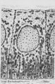
Exocrine gland - intercalated duct - salivary glands, pancreas | Scheme electronmicroscopy. Part of an intercalated duct, the low cuboidal epithelial cells contain sparsely organelles and some desmosomal structures. Between the lining cells and the basal lamina is squeezed part of a filament-rich myoepithelial cell. Lumen side top left quadrant. | oral cavity; intercalated duct | Poja Histology Collection - Oral Cavity Subset |
| 129 |
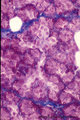 |
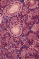
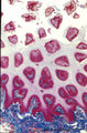
Exocrine gland - intercalated duct - submandibular gland, human | Stain: Azan. Branching intercalated duct draining several serous acini, the lining duct cells are lighter stained due to the content of organelles. Serous cells are darker stained (many organelles) with round nuclei. Note few fat cells, the septa are blue stained. | oral cavity; intercalated duct; serous gland | Poja Histology Collection - Oral Cavity Subset |
| 130 |
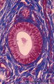 |
Exocrine gland - interlobular duct - submandibular gland, human | Stain: Azan. An interlobular duct with partly columnar as well as pseudostratified columnar epithelium. Note the dense connective tissue of the interlobular septum with small blood vessels. | oral cavity; seromucous glands | Poja Histology Collection - Oral Cavity Subset |
| 131 |
 |
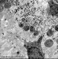
Exocrine gland - parotid gland, rat | Electronmicroscopy. Part of a striated duct with basally membrane infoldings and numerous mitochondria perpendicularly orientated to the basal membrane. Upper side is luminal side. Apically junctional complexes as well as mitochondria and many vesicles are present. | oral cavity; serous gland | Poja Histology Collection - Oral Cavity Subset |
| 132 |
 |
Exocrine gland - striated (intralobular) duct - salivary glands, pancreas | Scheme electronmicroscopy. Part of a striated duct with basally membrane infoldings and mitochondria accumulations in the tall columnar cells. Small junctional complexes as well as apically mitochondria and numerous vesicles are present. | oral cavity; striated duct | Poja Histology Collection - Oral Cavity Subset |
| 133 |
 |
Exocrine gland - striated (intralobular) ducts - parotid gland, human | Stain: Azan. Cross-sections of striated ducts in upper part between serous acini. Note at the right bottom part of (lighter stained) lining cells of an intercalated duct. Connective tissues between the structures are blue stained. | oral cavity; striated duct | Poja Histology Collection - Oral Cavity Subset |
| 134 |
 |
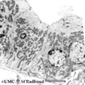
Exocrine gland - submandibular gland, gerbil | Electronmicroscopy. Part of a serous acinus of this mixed gland. Note characteristic electron-dense secretion granules, the mature ones are larger. At the lower bottom is shown part of a lining cell of the intercalated duct. | oral cavity; serous gland | Poja Histology Collection - Oral Cavity Subset |
| 135 |
 |
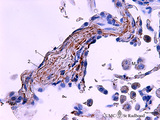
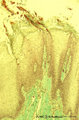
Fibrillin in the alveoli in lung tissue (human, adult) | Stain: imunoperoxidase staining with anti-fibrillin antibodies and diaminobenzidin reaction (frozen section). Fibrillin is one of the elastin-associated microfibrillar proteins that wraps the elastin protein core and is localized in normal lung structures such as alveolar septa and tips. The immuno-... | Alveolar tips; Elastin-associated proteins; Fibrillin | Poja Histology Collection - Respiratory System Subset |
| 136 |
 |
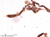
Fibrillin in the alveoli of lung emphysema (human, adult) | Stain: antifibrillin antibody immunoperoxidase staining with diaminobenzidin reaction. Fibrillin is one of the elastin-associated microfibrillar proteins, and marks therefore also the presence of elastin in lung tissue. In centrilobular emphysema (e.g., in lungs of smokers) the lesions are more com... | Alveolar septa; Elastin-associated proteins; Fibrillin | Poja Histology Collection - Respiratory System Subset |
| 137 |
 |
Fibrillin in the alveoli of lung emphysema (human, adult) | Stain: antifibrillin antibody immunoperoxidase staining with diaminobenzidin reaction. Fibrillin is one of the elastin-associated microfibrillar proteins, and marks therefore the presence of elastin in lung tissue. In centrilobular emphysema (e.g., in lungs of smokers) the lesions are more common an... | Alveolar tips; Elastin-associated proteins; Fibrillin | Poja Histology Collection - Respiratory System Subset |
| 138 |
 |
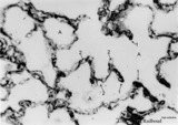
Fibrillin in the alveoli of the lung (human, adult) | Stain: antifibrillin antibody immunoperoxidase staining with diaminobenzidin reaction. Fibrillin is one of the elastin-associated microfibrillar proteins. Here fibrillin (and elastin) is localized as brown threads in the alveolar septa and alveolar tips (1). The elastin is visible as white threads (... | Alveolar tips; Elastin-associated proteins; Fibrillin | Poja Histology Collection - Respiratory System Subset |
| 139 |
 |
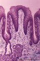
Filiform papillae of the tongue (dorsal side, human) | Stain: Heidenhain light bordeaux. Detail of the threadlike keratinized extensions of the stratified epithelium. Primary connective tissue papillae with 2 to 3 secondary papillae. Note the absence of taste buds in filiform papillae. | oral cavity; filiform papillae | Poja Histology Collection - Oral Cavity Subset |
| 140 |
 |
Filiform papillae of the tongue (dorsal side, human) | Stain: Azan. Oblique cross-section through the top of the filiform papillae. At the top of the picture keratinized extensions. Primary connective tissue papillae (blue) divide in several small secondary ones. | oral cavity; filiform papillae | Poja Histology Collection - Oral Cavity Subset |
| 141 |
 |
Filiform papillae of the tongue (dorsal side, human, neonate) | Scanning electronmicroscopy. Slender, thin threadlike extensions of the filiform papillae. In between a broad fungiform papilla. | oral cavity; filiform papillae; fungiform papilla | Poja Histology Collection - Oral Cavity Subset |
| 142 |
 |
Filiform papillae of the tongue - dorsal side, human | Stain: Goldner trichrome. Detail of the threadlike keratinized extensions (red) of the stratified epithelium. Primary connective tissue papillae (green) divide in several small secondary ones. Note the absence of taste buds in filiform papillae. | oral cavity; filiform papillae | Poja Histology Collection - Oral Cavity Subset |
| 143 |
 |
Filiform papillae of the tongue - dorsal side, human | Stain: Heidenhain light bordeaux. Transverse section through the middle of the filiform papillae showing secondary connective tissue papillae (lightly stained). | oral cavity; filiform papillae | Poja Histology Collection - Oral Cavity Subset |
| 144 |
 |
Foliate papillae of the tongue (dorsal side, rabbit) | Stain: Goldner trichrome. Foliate papil with primary and secondary connective tissue projections. Taste buds are localized in the lining, non-keratinized epithelium of the grooves. The serous gland of von Ebner (minor salivary gland) drains via an enlarged duct into the left groove of the papil. | oral cavity; foliate papillae; von Ebner | Poja Histology Collection - Oral Cavity Subset |
| 145 |
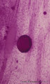 |
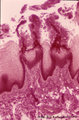
Free denticulus (pulp stone) in the tooth - longitudinal section of root pulp; human, adult | Stain: Hematoxylin and eosin. In the center of pulp connective tissue a free pulp stone (false denticulus). Dispersed through the pulp several small stones. Left of the central stone a longitudinal sectioned thin-walled blood vessel; right of the stone bundles of nerve fibers. | oral cavity; denticulus; pulp stone | Poja Histology Collection - Oral Cavity Subset |
| 146 |
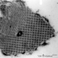 |
Free surfactant (tubular myelin) in alveolar space of the lung (rat) | Electron microscopy. After fixation the extracellular lining of surfactant (phosphatidylcholine, phosphoglycerol, cholesterol and proteins) will often be present as free packed lamellae in the alveolar space. This so-called tubular myelin (partly cross-sectioned), (1) is observed as stacks of lipid ... | Pneumocyte II; Tubular myelin | Poja Histology Collection - Respiratory System Subset |
| 147 |
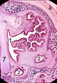 |
Frontal section of head (pig, fetus) | Stain: Azan. At the upper half the nasal septum (7) is a lightly stained plate (cartilago septi nasi). Developing conchae with supporting hyaline cartilage scaffolds (*) (light-stained) are present in the nasal chamber (1) and known as inferior (lowest), middle and superior turbinate bones. The whol... | Conchae nasales; Trabecular bone | Poja Histology Collection - Respiratory System Subset |
| 148 |
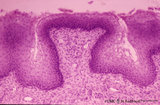 |
Fungiform papillae of the tongue (dorsal side, human) | Stain: Hematoxylin and eosin. A broad prominent papil of connective tissue (without secondary papils) covered by non-keratinized stratified epithelium. This specimen does not show any taste bud. Well vascularized lamina propria. | oral cavity; fungiform papillae | Poja Histology Collection - Oral Cavity Subset |
| 149 |
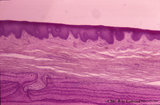 |
Gingiva ('attached' gingiva of decalcified alveolar bone, human, adult) | Stain: Hematoxylin and eosin. Thick layer of stratified squamous epithelium with parakeratosis (reddish) and shallow papillae of the lamina propria. At the bottom lamellar bone tissue (alveolar bone) with thickened periosteum. | oral cavity; alveolar bone | Poja Histology Collection - Oral Cavity Subset |
| 150 |
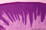 |
Gingiva ('free' gingiva of decalcified tooth, human, adult) | Stain: Hematoxylin and eosin. Thick layer of stratified squamous epithelium with parakeratosis (reddish) and many narrow papillae of the lamina propria. | oral cavity | Poja Histology Collection - Oral Cavity Subset |