The Health Education Assets Library (HEAL) is a collection of over 22,000 freely available digital materials for health sciences education. The collection is now housed at the University of Utah J. Willard Marriott Digital Library.
TO
Filters: Collection: ehsl_heal
| Title | Description | Subject | Collection | ||
|---|---|---|---|---|---|
| 626 |
 |
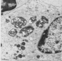
Thymus cortex (rat, young adult) | Electron microscopy. Type I epithelioreticular cells (4) separate connective tissue compartment (capsule, trabeculae, blood vessels) from the thymic parenchyma. At the left the capsule is bordered by a basal lamina (4a) of two projections (4) of type I epithelioreticular cells. Close to them, part o... | lymphoid tissue ; epithelioreticular cell type I; diapedesis | Poja Histology Collection - Lymphatic Tissues and Organs Subset |
| 627 |
 |
Thymus cortex (rat, young adult) | Electron microscopy. Two epithelioreticular cells type II or TEC2 (1) show the characteristic vacuoles (*) partly filled with granules (thymulin, lymphokines). At (--><--) small desmosomes. Apart from the mitochondria electron-dense lysosomal structures are present as well as tonofilaments (1, kera... | epithelioreticular cell II ; desmosome; MHC-II expression; lymphoid tissue | Poja Histology Collection - Lymphatic Tissues and Organs Subset |
| 628 |
 |
Thymus cortex (rat, young adult) | Electron microscopy. Type I epithelioreticular cells separate connective tissue compartment from the thymic parenchyma. With occludens junctions and desmosomes as barriers they form wide-mesh networks creating specific microenvironments for developing T cells. The extensions of type I cells (5) are... | thymus cortex; epithelioreticular cell type I; epithelioreticular cell type II; lymphoid tissue | Poja Histology Collection - Lymphatic Tissues and Organs Subset |
| 629 |
 |
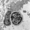
Thymus medulla (rat, neonate) | Electron microscopy. An interdigitating cell in the thymic corticomedullary region shows a large electron-light cytoplasm with a complex branching (7) at the periphery. The nucleus is sectioned twice (1). There is abundance of organelles as well as of quite uniform electron-dense lysosomal structure... | medullar epithelioreticular cell; interdigitating cell; corticomedullar region; lymphoid tissue | Poja Histology Collection - Lymphatic Tissues and Organs Subset |
| 630 |
 |
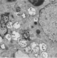
Thymus medulla (rat, young adult) | Electron microscopy. Epithelioreticular cells of the medulla (1) close to each other. The electron-light cytoplasm contains many small vesicles (1, Golgi area) as well as cross-sections of vacuoles (2) with small finger-like cytoplasmic extrusions in the lumen. Electron-dense lysosomal structures (3... | medullar epithelioreticular cell ; thymus medulla; lymphoid tissue | Poja Histology Collection - Lymphatic Tissues and Organs Subset |
| 631 |
 |
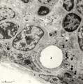
Thymus medulla (rat, young adult) | Electron microscopy. Surrounded by thymocytes (3) a medullary macrophage with an electron-light nucleus (1). The cytoplasm contains many electron-dense lysosomes of varying sizes and forms (2). | medullar macrophage; lymphoid tissue | Poja Histology Collection - Lymphatic Tissues and Organs Subset |
| 632 |
 |
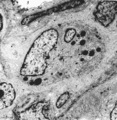
Thymus medulla (rat, young adult) | Electron microscopy. Epithelioreticular cell of the medulla with an electron-light cytoplasm contains cross-sections of vacuoles with small finger-like cytoplasmic extrusions in the lumen (1). Electron-dense lysosomal structures (2) are also present as well as bundles of intermediate filaments (kera... | medullar epithelioreticular cell ; keratin filaments; lymphoid tissue | Poja Histology Collection - Lymphatic Tissues and Organs Subset |
| 633 |
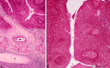 |
Tubal tonsil (human) | Stain: Azan. The tubal tonsil consists of a collection of lymphoid nodules near the auditory tube opening and forms part of the Waldeyers ring of defense in the nasopharyngeal cavity. This tonsil has fewer crypts (1), and the surface is covered by one to more layered ciliated epithelium (2). The la... | tubal tonsil; nasopharynx | Poja Histology Collection - Lymphatic Tissues and Organs Subset |
| 634 |
 |
Venous circulation pattern in perfused spleen (human) | Stain: Azan. The composed picture shows part of the splenic circulation system at several enlargements (inset, A, B). The open venous sinusoids (1) drain via short pulp veins into thin-walled trabecular veins (2), subsequently into thick-walled trabecular veins (4). The trabeculae originate from the... | splenic circulation; trabecular veins; sinusoid ; PALS | Poja Histology Collection - Lymphatic Tissues and Organs Subset |
| 635 |
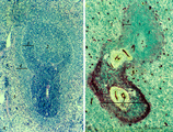 |
White pulp of spleen (mouse) | Stain: Hematoxylin & eosin in A and alkaline phosphatase in B (with substrate Naphtol Fast Blue RR). The general structure of the white pulp of the spleen and its specific microenvironment for T and B cells is well illustrated using alkaline phosphatase that strongly stains the capillaries around th... | alkaline phosphatase; PALS; T lymphocytes; white pulp | Poja Histology Collection - Lymphatic Tissues and Organs Subset |