The Health Education Assets Library (HEAL) is a collection of over 22,000 freely available digital materials for health sciences education. The collection is now housed at the University of Utah J. Willard Marriott Digital Library.
TO
Filters: Collection: ehsl_heal
| Title | Description | Subject | Collection | ||
|---|---|---|---|---|---|
| 426 |
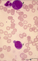 |
Myeloblast and promyelocyte in bone marrow smear (human) | Stain: May-Grnwald-Giemsa (MGG). The myeoloblast (1) measures 12-20 m and has a high nucleus-cytoplasm ratio and a round to oval nucleus. The nucleus has a fine disperse diffuse chromatin and one to five prominent nucleoli. The cytoplasm is pale blue (basophilic) with no or only scarce numbers of az... | Poja Histology Collection - Blood & Bone Marrow Subset | |
| 427 |
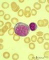 |
Myeloblast in bone marrow smear (human) | Stain: May-Grnwald-Giemsa (MGG). The myeloblast (1) shows a very transparent nucleus and several distinct nucleoli. The slightly basophilic cytoplasm is limited to a small rim in contrast to a promyelocyte. No granules are yet visible. (2) small lymphocyte. | Blood; Bone Marrow; Myeloblast; Lymphocyte | Poja Histology Collection - Blood & Bone Marrow Subset |
| 428 |
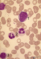 |
Myeloblast, neutrophilic granulocyte and lymphocyte in bone marrow smear (human) | Stain: May-Grnwald-Giemsa (MGG). The myeloblast (1) measures 12-20 m with 1-5 prominent nucleoli and fine diffuse chromatin. The slightly pale blue cytoplasm contains sometimes a few azurophilic granules (primary granules). (2) Indicates two segmented neutrophilic granulocytes with 3-5 nuclear lobes... | Poja Histology Collection - Blood & Bone Marrow Subset | |
| 429 |
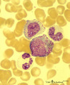 |
Myeloid cells in bone marrow smear (human) | Stain: May-Grnwald-Giemsa (MGG). (1) promyelocyte is the largest cell in the myeloid series. It has a transparent nucleus with nucleoli and ample cytoplasm with many azurophilic granules. (2) myelocyte. (3) metamyelocyte with an indented nucleus. (4) beginning of nucleus segmentation in the band neu... | Poja Histology Collection - Blood & Bone Marrow Subset | |
| 430 |
 |
Myelopoiesis in bone marrow smear (human) | Stain: May-Grnwald-Giemsa (MGG). The smear shows a group of myeloid cells in different stages of maturation, in which series the alterations in the nucleus can be noticed clearly going from (1) to (5), i.e. the nucleus gets more and more condensed, indented and subsequently segmented in lobes. (1) p... | Poja Histology Collection - Blood & Bone Marrow Subset | |
| 431 |
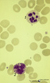 |
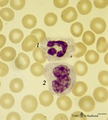
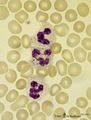
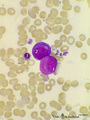
Neutrophilic and basophilic granulocytes in peripheral blood smear (human) | Stain: May-Grnwald-Giemsa (MGG). (1) A hypersegmented (>5 segments) neutrophilic granulocyte with clear fine granules. (2) Represents a mature basophilic granulocyte with clear distinguishable, coarse purple granules and a few vacuoles (because the granules dissolve in water during the staining proc... | Poja Histology Collection - Blood & Bone Marrow Subset | |
| 432 |
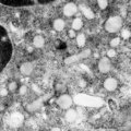 |
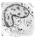
Neutrophilic granules in PMN (spleen, human) | Electron microscopy. A detail of the cytoplasm shows many granules of varying forms (round, elongated to dumb-bell), sizes and densities. The majority of these electron-grey to electron-lucent granules are the specific or secondary granules (diameter 0.2 up to length 0.8 μm). They contain substance... | Poja Histology Collection - Blood & Bone Marrow Subset | |
| 433 |
 |
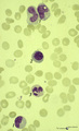
Neutrophilic granulocyte | Scheme electron microscopy. The neutrophil is a phagocytic cell (12-15 m) with a segmented lobular nucleus (3-5 lobes) and many cytoplasmic granules filled with degradative enzymes. These PMN cells (polymorph nuclear leukocytes) are the major cell types mediating acute inflammatory response to bacte... | Poja Histology Collection - Blood & Bone Marrow Subset | |
| 434 |
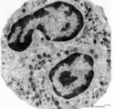 |
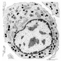
Neutrophilic granulocyte (peripheral blood, human) | Electron microscopy. Two nuclear lobes of the segmented nucleus are visible in a cytoplasm with a moderate amount of organelles but with abundant granules of varying sizes. The motile human neutrophil (9-14 μm) contains at least four types of granules. However, by routine electron microscopy primar... | Poja Histology Collection - Blood & Bone Marrow Subset | |
| 435 |
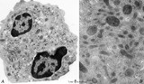 |
Neutrophilic granulocyte (peripheral blood, human) | Electron microscopy. Survey (A) and detail (B) of a neutrophilic granulocyte. Two nuclear lobes (1) of the segmented nucleus are visible in a cytoplasm with a moderate amount of organelles but with abundant granules (2, 3) of varying sizes. The detail shows in the cytoplasm large amounts of granules... | Poja Histology Collection - Blood & Bone Marrow Subset | |
| 436 |
 |
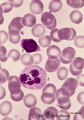
Neutrophilic granulocyte and monocyte in peripheral blood smear (human) | Stain: May-Grnwald-Giemsa (MGG). (1) the neutrophil (12-15 μm) shows a dark-stained partly lobulated nucleus with the smaller lobe connected by a fine strand of chromatin to the larger nuclear part. The cytoplasm reveals very faintly small azurophilic (primary) granules. (2) the monocyte (12-20 μm... | Poja Histology Collection - Blood & Bone Marrow Subset | |
| 437 |
 |
Neutrophilic granulocyte with drumstick in peripheral blood smear (human) | Stain: May-Grnwald-Giemsa (MGG). This neutrophil has a segmented lobulated nucleus with one drumstick (→) and one non-specific appendage (small club). The cytoplasm is filled with very fine granules. (2) small (smudged) lymphocyte with a dark condensed, indented nucleus and a small rim of cytopla... | Poja Histology Collection - Blood & Bone Marrow Subset | |
| 438 |
 |
Neutrophilic granulocytes in peripheral blood smear (human) | Stain: May-Grnwald-Giemsa (MGG). The nucleus of the neutrophilic granulocytes (also called polymorphonuclear leukocyte or PMN) is segmented into three to five connected lobules. The cytoplasm displays many fine (dust-like) azurophilic granules. The majority called specific granules are filled with e... | Poja Histology Collection - Blood & Bone Marrow Subset | |
| 439 |
 |
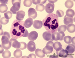
Neutrophilic granulocytes in peripheral blood smear (human) | Stain: May-Grnwald-Giemsa (MGG). The three neutrophilic granulocytes display segmented and lobulated nuclei. The lobes are connected with thin chromatin strands (→). The cytoplasm is ample filled with fine, dust-like granules and the majority are specific granules filled with enzymes such as lysoz... | Poja Histology Collection - Blood & Bone Marrow Subset | |
| 440 |
 |
Neutrophilic metamyelocyte | Scheme electron microscopy. From CFU-S (colony forming units-spleen) stem cells arise CFU-GM (colony forming unit-granulocyte/monocyte) stem cells. The latter divide by mitoses and differentiate via promyeloblasts and myeloblasts into neutrophilic myelocytes (the last proliferative stage). The next ... | Poja Histology Collection - Blood & Bone Marrow Subset | |
| 441 |
 |
Neutrophilic metamyelocyte in peripheral blood smear (human) | Stain: May-Grnwald-Giemsa (MGG). The neutrophilic metamyelocyte is differentiating nearly to a juvenile unsegmented (band form) neutrophil, but the nuclear chromatin is only partly condensed. Hardly visible dust-like granules can be observed in the still pale bluish cytoplasm. | Poja Histology Collection - Blood & Bone Marrow Subset | |
| 442 |
 |
Neutrophilic metamyelocytes in bone marrow smear (human) | Stain: May-Grnwald-Giemsa (MGG). (1) neutrophilic metamyelocytes. (2) polychromatic erythroblast. (3) segmented neutrophilic granulocyte. | Poja Histology Collection - Blood & Bone Marrow Subset | |
| 443 |
 |
Neutrophilic myelocyte | Scheme electron microscopy. From CFU-S (colony forming units-spleen) stem cells arise CFU-GM (colony forming unit-granulocyte/monocyte) stem cells. The latter divide by mitoses and differentiate via promyeloblasts and myeloblasts into myelocytes (the last proliferative stage). The neutrophilic myelo... | Blood; Bone Marrow; Electron microscopy; Neutrophilic myelocyte; Neutrophilic granulocyte; Primary granule; Secondary granule; Azurophilic granule; Lysosome | Poja Histology Collection - Blood & Bone Marrow Subset |
| 444 |
 |
Neutrophilic myelocyte and basophilic erythroblast in reactive bone marrow smear (human) | Stain: May-Grnwald-Giemsa (MGG). The neutrophilic myelocyte (1) has a nucleus (with nucleoli) located at one side of the cell. Cytoplasm contains many azurophilic granules. The early basophilic erythroblast (2) has a strong basophilic cytoplasm without granules, and a nucleus with condensed chromati... | Poja Histology Collection - Blood & Bone Marrow Subset | |
| 445 |
 |
Neutrophilic myelocyte in bone marrow smear (human) | Stain: May-Grnwald-Giemsa (MGG). In the neutrophilic myelocyte the nucleus is located eccentrically at one side of the cell. Nucleoli are visible as well as the primary azurophilic granules in the cytoplasm. | Poja Histology Collection - Blood & Bone Marrow Subset | |
| 446 |
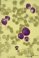 |
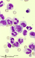
Neutrophilic myelocytes with strong toxic granulation in peripheral blood smear (human) | Stain: May-Grnwald-Giemsa (MGG). Neutrophilic myelocytes at various differentiation stages, but all cells show strong toxic granulation. | Poja Histology Collection - Blood & Bone Marrow Subset | |
| 447 |
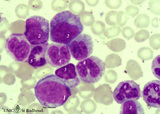 |
Neutrophilic myelopoiesis in bone marrow smear (human) | Stain: May-Grnwald-Giemsa (MGG). A group of neutrophilic myeloid cells are clustered together. (1) late myeloblast with nucleoli and hardly any azurophilic granules. (2) promyelocytes as the largest cells of the series, with ample cytoplasm filled with many azurophilic granules. (3) myelocytes with ... | Poja Histology Collection - Blood & Bone Marrow Subset | |
| 448 |
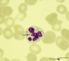 |
Normal mature neutrophilic granulocyte in peripheral blood smear (human) | Stain: May-Grnwald-Giemsa (MGG). Neutrophilic granulocyte with five nuclear lobes or segments, (not hypersegmented). The arrow indicates a drumstick. Note the fine dust-like granules. | Poja Histology Collection - Blood & Bone Marrow Subset | |
| 449 |
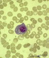 |
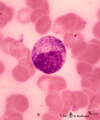
Normal plasma cell in peripheral blood smear (human) | Stain: May-Grnwald-Giemsa (MGG). The mature plasma cell has a large basophilic cytoplasma for IgG production, a clearly visible unstained (white) Golgi area, and a nucleus with well-condensed heterochromatin. Plasma cells can be found in the peripheral blood as a result of antigenic stimulation or c... | Poja Histology Collection - Blood & Bone Marrow Subset | |
| 450 |
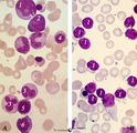 |
Normal pleiomorph and leukemic monomorph bone marrow smear (human) | Stain: May-Grnwald-Giemsa (MGG). In normal bone marrow smear (A) a pleimorph or heterogeneous collection of different cell types (A1-7) can be identified, while in the bone marrow smear of a leukemic patient a single type dominates (in this case lymphoid cells, B-1). | Poja Histology Collection - Blood & Bone Marrow Subset |