Videos, clinical notes and related presentations concerning neuro-ophthalmological and neurovisual disorders collected during Dr. Wray's work as the Director of Neuro-Visual Disorders at Massachusetts General Hospital.
Shirley H. Wray, M.D., Ph.D., FRCP, Professor of Neurology Harvard Medical School, Director, Unit for Neurovisual Disorders, Massachusetts General Hospital.
NOVEL: https://novel.utah.edu/
TO
Filters: Collection: "ehsl_novel_shw"
| Title | History | Type | ||
|---|---|---|---|---|
| 1 |
 |
Alcoholic Cerebellar Degeneration | The patient is a 72 year old woman who presented with a 4 year history of progressive difficulty with balance, frequent falls and unsteadiness walking. She required a cane to steady herself. Past History: Significant for alcohol abuse. In 1980, she came to Boston for a second opinion and was seen... | Text |
| 2 |
 |
Alexia Without Agraphia | The patient is a 69 year old left handed man with a history of hypertension, insulin dependent diabetes mellitus and atrial fibrillation. Treated with coumadin, adjusted to keep the INR between 2 and 3. On the morning of admission he awoke at 4 a.m., sat momentarily on the side of the bed and then... | Text |
| 3 |
 |
Alexia Without Agraphia | The patient is a 69 year old left handed man with a history of hypertension, insulin dependent diabetes mellitus and atrial fibrillation. Treated with coumadin, adjusted to keep the INR between 2 and 3. On the morning of admission he awoke at 4 a.m., sat momentarily on the side of the bed and then... | Image/MovingImage |
| 4 |
 |
Alzheimers Disease | The patient is a 78 year old left handed woman with a diagnosis of a left parietal infarct in 1995, bilateral carotid artery stenosis and hypertension. She was first seen in August 1997 for evaluation of involuntary movements of the lower face in the setting of rapidly progressive dementia and was... | Text |
| 5 |
 |
Amyotrophic Lateral Sclerosis (Guest Lecture) | Text | |
| 6 |
 |
Apraxia Eyelid Opening | This patient first presented to his PCP with increasing immobility. A diagnosis of Parkinson's disease was made. When his condition progressed, he was referred to the Neurology Clinic. Neuro-ophthalmological examination showed: Apraxia of eyelid opening Impaired initiation of horizontal saccades ... | Image/MovingImage |
| 7 |
 |
Apraxia of Eyelid Opening | In January 1997, This 73 year old patient was referred to the Neurovisual Clinic. At that time his speech was slurred and he stated that his eyes were his "biggest" complaint due to: 1. Impaired focusing "close up" 2. His eyes shut spontaneously much of the time 3. Bright sunlight provoked eye ... | Image/MovingImage |
| 8 |
 |
Ataxic Gait | The patient is a 72 year old woman who presented with a 4 year history of progressive difficulty with balance, frequent falls and unsteadiness walking. She required a cane to steady herself. Past History: Significant for alcohol abuse. In 1980, she came to Boston for a second opinion and was seen... | Image/MovingImage |
| 9 |
 |
Benign Essential Blepharospasm | The patient is a 60 year old estate manager with a history of retinal laser therapy, dry eyes and age related bilateral ptosis. He carries a diagnosis of hilar lymphadenopathy due to sarcoid and has had cancer of the kidney. He presented in 1995 with a 6 month history of frequent blinking and sp... | Image/MovingImage |
| 10 |
 |
Benign Essential Blepharospasm | Patient is a 55 year old woman functionally blind from severe benign essential blepharospasm. She presented with frequent blinking.continuous spasms of eye closure and great difficulty opening her eyes i.e. blepharospasm associated "apraxia of eyelid opening". Symptomatic Inquiry negative for... | Image/MovingImage |
| 11 |
 |
Benign Neonatal Ocular Flutter | Shortly after birth, this baby was noted to have "jiggling eyes" by his mother. He was in good general health and neurologically intact. Cogan and I saw the baby and Cogan made the diagnosis of neonatal ocular flutter. In 1954 Cogan first used the term "ocular flutter" to describe a rare disorder o... | Image/MovingImage |
| 12 |
 |
Bilateral Internuclear Ophthalmoplegia | This patient was seen at the Yale Eye Center at the age of 37. She had a long history of multiple sclerosis. At age 22, she had an acute attack of optic neuritis in the left eye which recovered fully within three weeks. Some months later she had a recurrent episode in the same eye, which also ... | Text |
| 13 |
 |
Bilateral Internuclear Ophthalmoplegia | This case was reported by Cogan DG and Wray SH. Internuclear ophthalmoplegia as an early sign of brain tumor. Neurology 1970; 20:629-633. The patient is Case 1. He is a 4 ½ year old boy whose parents noted that the right eye had been "drifting" for four months. On examination the only signific... | Image/MovingImage |
| 14 |
 |
Bilateral Internuclear Ophthalmoplegia | The patient is a 25 year old woman who was in excellent health until 4 days prior to admission when she noted blurred vision and horizontal double vision on lateral gaze to right and left. Past History: Negative for strabismus as a child. No previous episodes of transient neurological symptoms. Fa... | Image/MovingImage |
| 15 |
 |
Bilateral Internuclear Ophthalmoplegia | This patient was seen at the Yale Eye Center at the age of 37. She had a long history of multiple sclerosis. At age 22, she had an acute attack of optic neuritis in the left eye which recovered fully within three weeks. Some months later she had a recurrent episode in the same eye, which also ... | Image/MovingImage |
| 16 |
 |
Bilateral Internuclear Ophthalmoplegia (INO) | The patient is a 48 year old woman who presented with horizontal double vision looking to the right and to the left. She also noted blurring of vision with the image jerking horizontally back and forth. Neuro-ophthalmological examination: Visual acuity 20/20 OU corrected Visual fields, pupils, and f... | Image/MovingImage |
| 17 |
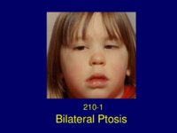 |
Bilateral Ptosis | This case, previously reported in 2007, is published courtesy of John Newsom-Davis, M.D., FRCP, FRS, CBE. Weatherall Institute of Molecular Medicine, John Radcliffe Hospital, Oxford. This patient was unusual in presenting in early childhood and the development of persistent facial muscle and tongue... | Text |
| 18 |
 |
Bilateral Ptosis | Patient is a 65 year old woman who presented with acute onset of bilateral ptosis. She awoke one day and found her eyelids half shut and she was unable to see. The lids completely shut and she came to the Massachusetts General Hospital Emergency Room and was admitted. Past History: Negative for pt... | Image/MovingImage |
| 19 |
 |
Bilateral Ptosis | The patient is a 65 year old physician who presented with intermittent drooping of his eyelids, particularly at the end of the day. He found that if he gently closed his eyes when he came to a stop in his car at a set of traffic lights, his eyelids opened more fully. He subsequently developed inte... | Image/MovingImage |
| 20 |
 |
Bilateral Ptosis | This case, previously reported in 2007, is published courtesy of John Newsom-Davis, M.D., FRCP, FRS, CBE. Weatherall Institute of Molecular Medicine, John Radcliffe Hospital, Oxford. This patient was unusual in presenting in early childhood and the development of persistent facial muscle and tongue... | Image/MovingImage |
| 21 |
 |
Bilateral Ptosis Facial Diplegia | The patient is a 47 year old attorney who was transferred from an outside hospital to the Massachusetts General Hospital (MGH) for treatmebnt of the Miller Fisher variant of the Guillian Barré syndrome (GBS). On the morning of September 14, 1993, the patient awoke feeling dizzy and he was unsteady ... | Image/MovingImage |
| 22 |
 |
Bilateral Ptosis Facial Diplegia | The patient is a 74 year old woman who one month prior to admission suffered from a non-productive cough, right ear pain, and pharyngitis treated with amoxicillin. On the day prior to admission she awoke with blurred vision and horizontal and vertical diplopia that persisted all day. The next m... | Image/MovingImage |
| 23 |
 |
Bilateral Sixth Nerve Palsy | This patient was known to be a chronic alcoholic. He presented with acute onset of unsteadiness walking which he attributed to double vision. He was unable to give an accurate history and was disoriented for time and place. Neurological examination: Significant for impaired memory and cognitive ... | Image/MovingImage |
| 24 |
 |
Bilateral Sixth Nerve Palsy | This patient has CNS B-cell lymphoma involving the dura mater. She presented with a chief complaint of "double vision and a squint is his right eye". On examination she was found to have bilateral sixth nerve palsy and paralytic esotropia. Comment: Lymphoma involving the dura is uncommon. Most p... | Image/MovingImage |
| 25 |
 |
Bilateral Sixth Nerve Palsy | The patient is a 22 year old man. Five days prior to admission (PTA) he developed a sore throat, non-productive cough and a temperature of 99.8 during the day and chills and diaphoresis at night. Four days PTA he had double vision in primary gaze, difficulty with his balance and gait, and a tinglin... | Image/MovingImage |
| 26 |
 |
Bilateral Sixth Nerve Palsy | The patient is a 70 year old Italian man with atrial fibrillation on long-term coumadin therapy. In October 1995, he developed generalized headache, horizontal double vision and his left eye deviated inwards (esotropia). A diagnosis of left sixth nerve palsy was made and attributed to microvascular ... | Image/MovingImage |
| 27 |
 |
Bilateral Sixth Nerve Palsy | This patient is a 62 year old woman with a six month history of double vision and difficulty walking. In August 1996, she first noted her right upper eyelid twitching followed by dizziness, nausea and vomiting. Soon after her voice became "shaky" and she experienced mild difficulty walking. She... | Image/MovingImage |
| 28 |
 |
Bilateral Sixth Nerve Palsy | The patient is a 60 year old woman who consulted her ophthalmologist with a chief complaint of double vision looking to the left. He diagnosed of a left sixth nerve palsy. No investigations were done. Two years later she complained of diplopia looking to the right. A diagnosis of bilateral six... | Image/MovingImage |
| 29 |
 |
Blepharospasm Round Up (Guest Lecture) | Text | |
| 30 |
 |
Brain Control of Horizontal Saccadic Eye Movements (Guest Lecture) | Text | |
| 31 |
 |
Brain MRI in Multiple Sclerosis (Guest Lecture) | Text | |
| 32 |
 |
Brainstem Cavernous Angioma | The patient is a 50 year old woman who presented in November 1977 with a transient facial droop, nystagmus, diplopia, dysarthria and vertigo. She was admitted to New England Tufts Medical Center and had an extensive workup including an electroencephalogram, first generation CT brain scan, angiogram ... | Text |
| 33 |
 |
Cavernous Angioma | The patient is a 19 year old sophomore who presented in 1983 with numbness of the left hand, involving initially just the fingers, and numbness and weakness of the right side of the face. He described the numbness in his hand as if it was "intensely asleep". The facial numbness involved the peri... | Text |
| 34 |
 |
Cavernous Sinus Meningioma | This patient is a 46 year old woman from Portugal who was admitted to the Massachusetts General Hospital in September 1986 with ophthalmoplegia of the left eye (OS) and signs of aberrant reinnervation of the third nerve. She presented, in August 1985, with an episode of diplopia. The diplopia was s... | Text |
| 35 |
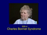 |
Charles Bonnet Syndrome | The patient is a 79 year old woman with a chief complaint of visual hallucinations. She carries a diagnosis of glaucoma and cataracts. The patient was in good health until two weeks prior to admission when she noted a black cloud in her visual field in the top central area. The cloud gradually c... | Text |
| 36 |
 |
Chest CT Thymoma (Guest Lecture) | Text | |
| 37 |
 |
Chiari I Malformation | Text | |
| 38 |
 |
Childhood Migraine Disease | Text | |
| 39 |
 |
Claudes Syndrome | The patient is a 76 year old woman who woke one morning unable to see out of her right eye because of ptosis. She came to the Massachusetts General Hospital emergency room and was admitted. Neurological Examination: Right third nerve palsy involving the pupil Ataxia of the left hand on finger-nose t... | Text |
| 40 |
 |
Clivus Chordoma | This 46 year old patient had at age 6, a tendency for the left eye to wander out. Her face photograph at that age shows an exotropia and at age 7, a year later, the exotropia was not quite as prominent. It was assumed that the exotropia was due to a non-paralytic strabismus. Past History: ... | Text |
| 41 |
 |
CNS Lymphoma | Text | |
| 42 |
 |
Congenital Horizontal Gaze Palsy | The patient is an 8 year old boy with a rare autosomal recessive disorder characterized by congenital absence of conjugate horizontal eye movements preservation of vertical gaze and convergence and progressive scoliosis (HGPPS) developing in childhood. The child was referred to Dr. Cogan with a ... | Image/MovingImage |
| 43 |
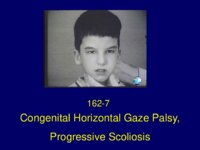 |
Congenital Horizontal Gaze Palsy Progressive Scoliosis | The patient is an 8 year old boy with a rare autosomal recessive disorder characterized by congenital absence of conjugate horizontal eye movements preservation of vertical gaze and convergence and progressive scoliosis (HGPPS) developing in childhood. The child was referred to Dr. Cogan with a ... | Text |
| 44 |
 |
Congenital Nystagmus | This young boy has oculocutaneous albinism. In 1979 he presented for evaluation of oscillations of his eyes present since birth. He had no head turn or head tremor. Diagnosis: Albinism Congenital sensory nystagmus Ocular Albinism: Infants with albinism of all types are typically slow to see, ofte... | Image/MovingImage |
| 45 |
 |
Congenital Nystagmus | This 11 year old Italian boy was referred for evaluation of nystagmus. Emanuel is the second son in a family of two and his older brother, age 13, had no visual problem. Emanuel was born at term, normal delivery, birth weight 9 pounds. Neonatal and infant development was perfectly normal. A pedia... | Image/MovingImage |
| 46 |
 |
Congenital Nystagmus - Latent Nystagmus | This boy was not recognized to have nystagmus until he accidentally discovered that he had blurred vision in one eye while pulling a sweater off over his head and blocking the vision of one eye. An ophthalmologist saw him and diagnosed latent nystagmus. He had no strabismus or any other ophthalmol... | Image/MovingImage |
| 47 |
 |
Congenital Nystagmus: Blurred vision | This 34 year old physician has had a life long problem with congenital nystagmus. He was an orphan and his adopted parents took him to see a pediatrician when he was very small. A missed diagnosis of a "lazy eye" was made. Subsequently, at the age of 9, when he complained of intermittent blurred ... | Image/MovingImage |
| 48 |
 |
Congenital Nystagmus: Eyes jump around | This 14 year old boy was born one month premature weighing 6 pounds 5 ounces. The top of his head failed to close until age 6. In addition he had only half a clavicle and stunted growth. Diagnosis: Cleidocranial dysostosis. He was referred by his endocrinologist for evaluation of difficulty ... | Image/MovingImage |
| 49 |
 |
Congenital Nystagmus: Oscillations of the Eyes | This child has had nystagmus since childhood with a head turn to the left. He was seen by his pediatrician. Diagnosis: Horizontal jerk nystagmus (congenital motor nystagmus) Congenital nystagmus Classification: The Classification of Eye Movement Abnormalities and Strabismus Working Group has recomme... | Image/MovingImage |
| 50 |
 |
Congenital Nystagmus: Oscillations of the Eyes-2 | This child was noted to have oscillations of the eyes in infancy and was given a diagnosis of congenital nystagmus. | Image/MovingImage |
