Videos, clinical notes and related presentations concerning neuro-ophthalmological and neurovisual disorders collected during Dr. Wray's work as the Director of Neuro-Visual Disorders at Massachusetts General Hospital.
Shirley H. Wray, M.D., Ph.D., FRCP, Professor of Neurology Harvard Medical School, Director, Unit for Neurovisual Disorders, Massachusetts General Hospital.
NOVEL: https://novel.utah.edu/
TO
Filters: Collection: "ehsl_novel_shw"
| Title | History | Type | ||
|---|---|---|---|---|
| 1 |
 |
Alcoholic Cerebellar Degeneration | The patient is a 72 year old woman who presented with a 4 year history of progressive difficulty with balance, frequent falls and unsteadiness walking. She required a cane to steady herself. Past History: Significant for alcohol abuse. In 1980, she came to Boston for a second opinion and was seen... | Text |
| 2 |
 |
Alexia Without Agraphia | The patient is a 69 year old left handed man with a history of hypertension, insulin dependent diabetes mellitus and atrial fibrillation. Treated with coumadin, adjusted to keep the INR between 2 and 3. On the morning of admission he awoke at 4 a.m., sat momentarily on the side of the bed and then... | Image/MovingImage |
| 3 |
 |
Alexia Without Agraphia | The patient is a 69 year old left handed man with a history of hypertension, insulin dependent diabetes mellitus and atrial fibrillation. Treated with coumadin, adjusted to keep the INR between 2 and 3. On the morning of admission he awoke at 4 a.m., sat momentarily on the side of the bed and then... | Text |
| 4 |
 |
Alzheimers Disease | The patient is a 78 year old left handed woman with a diagnosis of a left parietal infarct in 1995, bilateral carotid artery stenosis and hypertension. She was first seen in August 1997 for evaluation of involuntary movements of the lower face in the setting of rapidly progressive dementia and was... | Text |
| 5 |
 |
Amyotrophic Lateral Sclerosis (Guest Lecture) | Text | |
| 6 |
 |
Apraxia Eyelid Opening | This patient first presented to his PCP with increasing immobility. A diagnosis of Parkinson's disease was made. When his condition progressed, he was referred to the Neurology Clinic. Neuro-ophthalmological examination showed: Apraxia of eyelid opening Impaired initiation of horizontal saccades ... | Image/MovingImage |
| 7 |
 |
Apraxia of Eyelid Opening | In January 1997, This 73 year old patient was referred to the Neurovisual Clinic. At that time his speech was slurred and he stated that his eyes were his "biggest" complaint due to: 1. Impaired focusing "close up" 2. His eyes shut spontaneously much of the time 3. Bright sunlight provoked eye ... | Image/MovingImage |
| 8 |
 |
Ataxic Gait | The patient is a 72 year old woman who presented with a 4 year history of progressive difficulty with balance, frequent falls and unsteadiness walking. She required a cane to steady herself. Past History: Significant for alcohol abuse. In 1980, she came to Boston for a second opinion and was seen... | Image/MovingImage |
| 9 |
 |
Benign Essential Blepharospasm | The patient is a 60 year old estate manager with a history of retinal laser therapy, dry eyes and age related bilateral ptosis. He carries a diagnosis of hilar lymphadenopathy due to sarcoid and has had cancer of the kidney. He presented in 1995 with a 6 month history of frequent blinking and sp... | Image/MovingImage |
| 10 |
 |
Benign Essential Blepharospasm | Patient is a 55 year old woman functionally blind from severe benign essential blepharospasm. She presented with frequent blinking.continuous spasms of eye closure and great difficulty opening her eyes i.e. blepharospasm associated "apraxia of eyelid opening". Symptomatic Inquiry negative for... | Image/MovingImage |
| 11 |
 |
Benign Neonatal Ocular Flutter | Shortly after birth, this baby was noted to have "jiggling eyes" by his mother. He was in good general health and neurologically intact. Cogan and I saw the baby and Cogan made the diagnosis of neonatal ocular flutter. In 1954 Cogan first used the term "ocular flutter" to describe a rare disorder o... | Image/MovingImage |
| 12 |
 |
Bilateral Internuclear Ophthalmoplegia | The patient is a 25 year old woman who was in excellent health until 4 days prior to admission when she noted blurred vision and horizontal double vision on lateral gaze to right and left. Past History: Negative for strabismus as a child. No previous episodes of transient neurological symptoms. Fa... | Image/MovingImage |
| 13 |
 |
Bilateral Internuclear Ophthalmoplegia | This patient was seen at the Yale Eye Center at the age of 37. She had a long history of multiple sclerosis. At age 22, she had an acute attack of optic neuritis in the left eye which recovered fully within three weeks. Some months later she had a recurrent episode in the same eye, which also ... | Image/MovingImage |
| 14 |
 |
Bilateral Internuclear Ophthalmoplegia | This patient was seen at the Yale Eye Center at the age of 37. She had a long history of multiple sclerosis. At age 22, she had an acute attack of optic neuritis in the left eye which recovered fully within three weeks. Some months later she had a recurrent episode in the same eye, which also ... | Text |
| 15 |
 |
Bilateral Internuclear Ophthalmoplegia | This case was reported by Cogan DG and Wray SH. Internuclear ophthalmoplegia as an early sign of brain tumor. Neurology 1970; 20:629-633. The patient is Case 1. He is a 4 ½ year old boy whose parents noted that the right eye had been "drifting" for four months. On examination the only signific... | Image/MovingImage |
| 16 |
 |
Bilateral Internuclear Ophthalmoplegia (INO) | The patient is a 48 year old woman who presented with horizontal double vision looking to the right and to the left. She also noted blurring of vision with the image jerking horizontally back and forth. Neuro-ophthalmological examination: Visual acuity 20/20 OU corrected Visual fields, pupils, and f... | Image/MovingImage |
| 17 |
 |
Bilateral Ptosis | Patient is a 65 year old woman who presented with acute onset of bilateral ptosis. She awoke one day and found her eyelids half shut and she was unable to see. The lids completely shut and she came to the Massachusetts General Hospital Emergency Room and was admitted. Past History: Negative for pt... | Image/MovingImage |
| 18 |
 |
Bilateral Ptosis | This case, previously reported in 2007, is published courtesy of John Newsom-Davis, M.D., FRCP, FRS, CBE. Weatherall Institute of Molecular Medicine, John Radcliffe Hospital, Oxford. This patient was unusual in presenting in early childhood and the development of persistent facial muscle and tongue... | Image/MovingImage |
| 19 |
 |
Bilateral Ptosis | The patient is a 65 year old physician who presented with intermittent drooping of his eyelids, particularly at the end of the day. He found that if he gently closed his eyes when he came to a stop in his car at a set of traffic lights, his eyelids opened more fully. He subsequently developed inte... | Image/MovingImage |
| 20 |
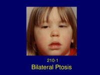 |
Bilateral Ptosis | This case, previously reported in 2007, is published courtesy of John Newsom-Davis, M.D., FRCP, FRS, CBE. Weatherall Institute of Molecular Medicine, John Radcliffe Hospital, Oxford. This patient was unusual in presenting in early childhood and the development of persistent facial muscle and tongue... | Text |
| 21 |
 |
Bilateral Ptosis Facial Diplegia | The patient is a 74 year old woman who one month prior to admission suffered from a non-productive cough, right ear pain, and pharyngitis treated with amoxicillin. On the day prior to admission she awoke with blurred vision and horizontal and vertical diplopia that persisted all day. The next m... | Image/MovingImage |
| 22 |
 |
Bilateral Ptosis Facial Diplegia | The patient is a 47 year old attorney who was transferred from an outside hospital to the Massachusetts General Hospital (MGH) for treatmebnt of the Miller Fisher variant of the Guillian Barré syndrome (GBS). On the morning of September 14, 1993, the patient awoke feeling dizzy and he was unsteady ... | Image/MovingImage |
| 23 |
 |
Bilateral Sixth Nerve Palsy | This patient was known to be a chronic alcoholic. He presented with acute onset of unsteadiness walking which he attributed to double vision. He was unable to give an accurate history and was disoriented for time and place. Neurological examination: Significant for impaired memory and cognitive ... | Image/MovingImage |
| 24 |
 |
Bilateral Sixth Nerve Palsy | The patient is a 22 year old man. Five days prior to admission (PTA) he developed a sore throat, non-productive cough and a temperature of 99.8 during the day and chills and diaphoresis at night. Four days PTA he had double vision in primary gaze, difficulty with his balance and gait, and a tinglin... | Image/MovingImage |
| 25 |
 |
Bilateral Sixth Nerve Palsy | The patient is a 70 year old Italian man with atrial fibrillation on long-term coumadin therapy. In October 1995, he developed generalized headache, horizontal double vision and his left eye deviated inwards (esotropia). A diagnosis of left sixth nerve palsy was made and attributed to microvascular ... | Image/MovingImage |
| 26 |
 |
Bilateral Sixth Nerve Palsy | The patient is a 60 year old woman who consulted her ophthalmologist with a chief complaint of double vision looking to the left. He diagnosed of a left sixth nerve palsy. No investigations were done. Two years later she complained of diplopia looking to the right. A diagnosis of bilateral six... | Image/MovingImage |
| 27 |
 |
Bilateral Sixth Nerve Palsy | This patient has CNS B-cell lymphoma involving the dura mater. She presented with a chief complaint of "double vision and a squint is his right eye". On examination she was found to have bilateral sixth nerve palsy and paralytic esotropia. Comment: Lymphoma involving the dura is uncommon. Most p... | Image/MovingImage |
| 28 |
 |
Bilateral Sixth Nerve Palsy | This patient is a 62 year old woman with a six month history of double vision and difficulty walking. In August 1996, she first noted her right upper eyelid twitching followed by dizziness, nausea and vomiting. Soon after her voice became "shaky" and she experienced mild difficulty walking. She... | Image/MovingImage |
| 29 |
 |
Blepharospasm Round Up (Guest Lecture) | Text | |
| 30 |
 |
Brain Control of Horizontal Saccadic Eye Movements (Guest Lecture) | Text | |
| 31 |
 |
Brain MRI in Multiple Sclerosis (Guest Lecture) | Text | |
| 32 |
 |
Brainstem Cavernous Angioma | The patient is a 50 year old woman who presented in November 1977 with a transient facial droop, nystagmus, diplopia, dysarthria and vertigo. She was admitted to New England Tufts Medical Center and had an extensive workup including an electroencephalogram, first generation CT brain scan, angiogram ... | Text |
| 33 |
 |
Cavernous Angioma | The patient is a 19 year old sophomore who presented in 1983 with numbness of the left hand, involving initially just the fingers, and numbness and weakness of the right side of the face. He described the numbness in his hand as if it was "intensely asleep". The facial numbness involved the peri... | Text |
| 34 |
 |
Cavernous Sinus Meningioma | This patient is a 46 year old woman from Portugal who was admitted to the Massachusetts General Hospital in September 1986 with ophthalmoplegia of the left eye (OS) and signs of aberrant reinnervation of the third nerve. She presented, in August 1985, with an episode of diplopia. The diplopia was s... | Text |
| 35 |
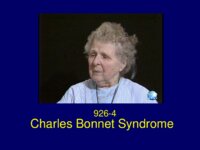 |
Charles Bonnet Syndrome | The patient is a 79 year old woman with a chief complaint of visual hallucinations. She carries a diagnosis of glaucoma and cataracts. The patient was in good health until two weeks prior to admission when she noted a black cloud in her visual field in the top central area. The cloud gradually c... | Text |
| 36 |
 |
Chest CT Thymoma (Guest Lecture) | Text | |
| 37 |
 |
Chiari I Malformation | Text | |
| 38 |
 |
Childhood Migraine Disease | Text | |
| 39 |
 |
Claudes Syndrome | The patient is a 76 year old woman who woke one morning unable to see out of her right eye because of ptosis. She came to the Massachusetts General Hospital emergency room and was admitted. Neurological Examination: Right third nerve palsy involving the pupil Ataxia of the left hand on finger-nose t... | Text |
| 40 |
 |
Clivus Chordoma | This 46 year old patient had at age 6, a tendency for the left eye to wander out. Her face photograph at that age shows an exotropia and at age 7, a year later, the exotropia was not quite as prominent. It was assumed that the exotropia was due to a non-paralytic strabismus. Past History: ... | Text |
| 41 |
 |
CNS Lymphoma | Text | |
| 42 |
 |
Congenital Horizontal Gaze Palsy | The patient is an 8 year old boy with a rare autosomal recessive disorder characterized by congenital absence of conjugate horizontal eye movements preservation of vertical gaze and convergence and progressive scoliosis (HGPPS) developing in childhood. The child was referred to Dr. Cogan with a ... | Image/MovingImage |
| 43 |
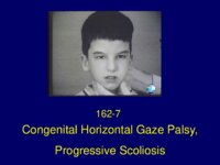 |
Congenital Horizontal Gaze Palsy Progressive Scoliosis | The patient is an 8 year old boy with a rare autosomal recessive disorder characterized by congenital absence of conjugate horizontal eye movements preservation of vertical gaze and convergence and progressive scoliosis (HGPPS) developing in childhood. The child was referred to Dr. Cogan with a ... | Text |
| 44 |
 |
Congenital Nystagmus | This young boy has oculocutaneous albinism. In 1979 he presented for evaluation of oscillations of his eyes present since birth. He had no head turn or head tremor. Diagnosis: Albinism Congenital sensory nystagmus Ocular Albinism: Infants with albinism of all types are typically slow to see, ofte... | Image/MovingImage |
| 45 |
 |
Congenital Nystagmus | This 11 year old Italian boy was referred for evaluation of nystagmus. Emanuel is the second son in a family of two and his older brother, age 13, had no visual problem. Emanuel was born at term, normal delivery, birth weight 9 pounds. Neonatal and infant development was perfectly normal. A pedia... | Image/MovingImage |
| 46 |
 |
Congenital Nystagmus - Latent Nystagmus | This boy was not recognized to have nystagmus until he accidentally discovered that he had blurred vision in one eye while pulling a sweater off over his head and blocking the vision of one eye. An ophthalmologist saw him and diagnosed latent nystagmus. He had no strabismus or any other ophthalmol... | Image/MovingImage |
| 47 |
 |
Congenital Nystagmus: Blurred vision | This 34 year old physician has had a life long problem with congenital nystagmus. He was an orphan and his adopted parents took him to see a pediatrician when he was very small. A missed diagnosis of a "lazy eye" was made. Subsequently, at the age of 9, when he complained of intermittent blurred ... | Image/MovingImage |
| 48 |
 |
Congenital Nystagmus: Eyes jump around | This 14 year old boy was born one month premature weighing 6 pounds 5 ounces. The top of his head failed to close until age 6. In addition he had only half a clavicle and stunted growth. Diagnosis: Cleidocranial dysostosis. He was referred by his endocrinologist for evaluation of difficulty ... | Image/MovingImage |
| 49 |
 |
Congenital Nystagmus: Oscillations of the Eyes | This child has had nystagmus since childhood with a head turn to the left. He was seen by his pediatrician. Diagnosis: Horizontal jerk nystagmus (congenital motor nystagmus) Congenital nystagmus Classification: The Classification of Eye Movement Abnormalities and Strabismus Working Group has recomme... | Image/MovingImage |
| 50 |
 |
Congenital Nystagmus: Oscillations of the Eyes-2 | This child was noted to have oscillations of the eyes in infancy and was given a diagnosis of congenital nystagmus. | Image/MovingImage |
| 51 |
 |
Congenital Nystagmus; Latent Nystagmus | This little girl has had nystagmus since birth. She has the classical constellation of signs, initially diagnosed as congenital motor nystagmus with: • Horizontal jerk nystagmus • A slight head turn to the right to place the eyes in the null position • Latent nystagmus • Inability to superi... | Image/MovingImage |
| 52 |
 |
Congenital Ocular Motor Apraxia | This little boy was born at term after a normal pregnancy. There was no family history of a neurological or ophthalmological disease. His mother noted from birth that he turned his head instead of his eyes and frequently tried to fix objects on either side by making a quick turn of the head past... | Image/MovingImage |
| 53 |
 |
Congenital Ocular Motor Apraxia | This 3 year old boy was referred for evaluation of inattention. He was born at term after a normal pregnancy. He had been noted, in early childhood, to have difficulty looking to the right and to the left and was slow to read. | Image/MovingImage |
| 54 |
 |
Congenital Ocular Motor Apraxia | This baby was born at term after an uneventful pregnancy and normal delivery. He was the first and only child. There was no family history of any This baby was born at term after an uneventful pregnancy and normal delivery. He was the first and only child. There was no family history of any neurol... | Image/MovingImage |
| 55 |
 |
Constructional Apraxia | Text | |
| 56 |
 |
Convergence Insufficiency | The patient is a 73 year old man with a ten year history of idiopathic Parkinson's disease characterized by difficulty in walking, generalized rigidity and a mild tremor of his hands at rest with deterioration in his handwriting. He denied any memory impairment or loss of cognitive function. He w... | Image/MovingImage |
| 57 |
 |
Convergence Insufficiency | The patient is a 58 year old man with idiopathic Parkinson's Disease for five years. He presented with an increasing shuffling gait, marked bradykinesia, mild to moderate rigidity in all four limbs and impairment in rapid movements of the hands. When he attempted to standup and walk he needed to... | Image/MovingImage |
| 58 |
 |
Convergence Insufficiency | The patient is a 73 year old man with a ten year history of idiopathic Parkinson's disease characterized by difficulty in walking, generalized rigidity and a mild tremor of his hands at rest with deterioration in his handwriting. He denied any memory impairment or loss of cognitive function. He w... | Text |
| 59 |
 |
Downbeat Nystagmus | This patient carries a diagnosis of Type I Chiari malformation. Neurological symptoms of a Chiari malformation may not develop until adolescence or adult life as in this man. The symptoms may be those of: 1. Increased intracranial pressure, mainly headache 2. Progressive cerebellar ataxia 3. Progre... | Image/MovingImage |
| 60 |
 |
Downbeat Nystagmus | In 1984, this young woman was involved in a motor vehicle accident when her car was hit from behind. She struck her head on the dash board but had no loss of consciousness. She was able to get out of the car unaided and was immediately aware of pain in her neck. She thought that she had susta... | Image/MovingImage |
| 61 |
 |
Downbeat Nystagmus | This 58 year old engineer was referred by his neurologist for evaluation of periodic episodes of difficulty focusing and blurred vision for 8 years. In 1981 whilst sightseeing in Newport, he became acutely aware of difficulty focusing and blurry vision. The symptoms lasted for twenty minutes and t... | Image/MovingImage |
| 62 |
 |
Downbeat Nystagmus | This patient carries a diagnosis of Type I Chiari malformation. In 1983 she presented with vertical double vision which persisted. Two months later, she had an acute episode of irritation of her eyes possibly due to allergy, followed two days later by difficulty in focusing, light headedness, a ... | Image/MovingImage |
| 63 |
 |
Downbeat Nystagmus | The patient is a 72 year old man who was found down and admitted as an emergency in coma. Neurological examination: Patient in coma failed to respond to painful stimuli. Downbeat nystagmus Lid nystagmus Pupils 2 mm OU responsive to light Corneal reflexes absent Oculocephalic reflex absent No resp... | Image/MovingImage |
| 64 |
 |
Downbeat Nystagmus | The patient is a 59 year old woman who carried a diagnosis of: 1. Congenital anomaly of the occipito-cervical junction with occipitalization of the C1 ring 2. Basilar invagination with Chiari Type I 3. A syrinx in the center of the cervical-thoracic spinal cord extending from C2 to T4 In 1957, at ag... | Image/MovingImage |
| 65 |
 |
Downbeat Nystagmus | This 58 year old engineer was referred by his neurologist for evaluation of periodic episodes of difficulty focusing and blurred vision for 8 years. In 1981 whilst sightseeing in Newport, he became acutely aware of difficulty focusing and blurry vision. The symptoms lasted for twenty minutes and t... | Text |
| 66 |
 |
Downbeat Nystagmus - Periodic Alternating Nystagmus | This patient carries a diagnosis of multiple sclerosis. | Image/MovingImage |
| 67 |
 |
Downbeat Nystagmus PAN | This patient carries a diagnosis of multiple sclerosis. | Text |
| 68 |
 |
Dressing Apraxia | The patient is a 72 year old right handed woman who presented in November 1995 with the sudden onset of impaired coordination of visual and motor skills following an inner right ear infection. One of her problems was difficulty sitting on a chair as she tended to place her body incorrectly. By lat... | Image/MovingImage |
| 69 |
 |
Duane's Syndrome | The patient is a 7 year old boy born two weeks premature with transposition of the major arteries of the heart, four holes in the heart, and an absent spleen. He had cardiac surgery at age 2 days and at age one year and his development was excellent thereafter. At age 6 months, it was noted that th... | Image/MovingImage |
| 70 |
 |
Elliptical Nystagmus | The patient carries a diagnosis of multiple sclerosis. | Image/MovingImage |
| 71 |
 |
Essential Palatal Tremor | The patient is a 25 year old meteorologist from Tennessee who came to Boston in the summer of 1992 to vacation with his family on the Cape. His illness started with flu-like symptoms, low grade fever from 99 to 100F, sweating and episodes of light headedness associated with occasional nausea, indige... | Text |
| 72 |
 |
Essential Palatal Tremor | The patient is a 25 year old meteorologist from Tennessee who came to Boston in the summer of 1992 to vacation with his family on the Cape. His illness started with flu-like symptoms, low grade fever from 99 to 100F, sweating and episodes of light headedness associated with occasional nausea, indige... | Image/MovingImage |
| 73 |
 |
Eyebrow Spasm | This case is published courtesy of Daniel J. Costello, M.D., Department of Neurology, Massachusetts General Hospital, Boston. The patient is a 32-year-old right-handed man with an established diagnosis of Tuberous Sclerosis Complex characterized by: - medically intractable epilepsy - developmental... | Image/MovingImage |
| 74 |
 |
Eyebrow Spasm (Guest Lecture) | This case is published courtesy of Daniel J. Costello, M.D., Department of Neurology, Massachusetts General Hospital, Boston. The patient is a 32-year-old right-handed man with an established diagnosis of Tuberous Sclerosis Complex characterized by: - medically intractable epilepsy - developmental... | Text |
| 75 |
 |
Familial Amyotrophic Lateral Sclerosis | This 58 year old woman was referred to Dr. Robert Brown in March 1995 for evaluation of slurred speech. She remained under his care until her death. On examination she had signs of a pseudobulbar palsy: Dysarthria and dysphagia Diminished palatal movement... | Text |
| 76 |
 |
Familial Myasthenia Gravis | In 1999 Dr. A. G. Engel published a monograph on Myasthenia Gravis and Myasthenic Disorders with a chapter devoted to Congenital Myasthenic Syndromes. Dr. Engel systematically defined and classified hereditary and congenital myasthenic syndromes, delineated on the basis of their electrophysiologi... | Image/MovingImage |
| 77 |
 |
Familial Nystagmus | The mother and her infant son both have conjugate horizontal pendular nysagmus. Diagnosis: Congenital sensory nystagmus Familial Infantile Nystagmus Syndrome (INS) The little boy is a bright active child with normal development. He has pendular nystagmus in central gaze without a head turn or h... | Image/MovingImage |
| 78 |
 |
Fascicular Third Nerve Palsy | The patient is a 76 year old woman who woke one morning unable to see out of her right eye because of ptosis. She came to the Massachusetts General Hospital emergency room and was admitted. Neurological Examination: Right third nerve palsy involving the pupil Ataxia of the left hand on finger-nose t... | Image/MovingImage |
| 79 |
 |
Fisher's One and a Half Syndrome | The patient is a 62 year old right handed man, status post myocardial infarction in 1989 and on Coumadin. In 1993 he presented with a history of three separate TIAs 1. Instantaneous perioral tingling and/or numbness lasting less than 1 minute. 2. Episodic numbness of the right hand and foot lasti... | Image/MovingImage |
| 80 |
 |
Fisher's One and a Half Syndrome | The patient is a 56 year old woman who was seen by a Stroke Consult on the cardiac unit because she had difficulty opening her left eye and diplopia looking to the right. On examination she had ptosis OS and "a partial ophthalmoplegia" sparing the pupil. Diagnosis Partial left third nerve palsy.... | Image/MovingImage |
| 81 |
 |
Fisher's One and a Half Syndrome | This 28 year old woman had severe multiple sclerosis with a spastic paraparesis due to a lesion of the spinal cord. She was admitted to the Intensive Care Unit complaining of dizziness and double vision. Neuro-ophthalmologic examination: Visual acuity 20/30 OU Pupils, visual fields and fundi normal ... | Image/MovingImage |
| 82 |
 |
Fisher's One and a Half Syndrome | This young patient presented with double vision and was found to have on examination the classical findings of Fisher's one-and-a-half syndrome which are: • Right internuclear ophthalmoplegia on gaze left with adduction weakness OD • Right horizontal gaze paresis with gaze evoked nystagmus • F... | Image/MovingImage |
| 83 |
 |
Fisher's One and a Half Syndrome | This young man was seen in the emergency room of his local hospital following the onset of severe headache, mild confusion and diplopia. Non-contrast CT brain scan showed: A right pontine hemorrhage He was transferred to the Massachusetts General Hospital ICU. Ocular Motility: Esotropia of the r... | Image/MovingImage |
| 84 |
 |
Fisher's One and a Half Syndrome | This 44 year old woman presented in 1973 with an acute attack of optic neuritis in the right eye that fully recovered after a course of ACTH therapy. In 1991, 18 years later, she developed unsteadiness of gait, "walking like a chicken", stiff legs that jerked spontaneously in bed at night, and num... | Image/MovingImage |
| 85 |
 |
Fourth Nerve Palsy | The patient is a 32 year old, left handed chemistry teacher who presented with intermittent vertical double vision. In August 1992 she noted, particularly late in the evening when reading in bed, vertical double vision. The images were one on top of the other and on occasions one image was slig... | Image/MovingImage |
| 86 |
 |
Fourth Nerve Palsy | The patient is a 64 year old engineer who noted whilst driving around Thanksgiving time double vision with one image on top of the other. Vertical double vision persisted and he was referred to the Massachusetts General Hospital for evaluation. Past History: 1991 Hyperthyroidism with Graves' Dise... | Image/MovingImage |
| 87 |
 |
Frontotemporal Dementia | The patient is a 68 year old right handed retired air conditioner repair man who presented with impaired balance and slow walking. For about one year he had noted difficulty lifting his feet high enough when climbing the stairs. From that time on, his movements slowed and worsened so that he had ... | Text |
| 88 |
 |
Gaucher's Disease | This little boy has Gaucher's disease. Gaucher's disease is an autosomal recessive disorder, linked to chromosome 1q21, due to glucocerebroside β-glucosidase deficiency. There are three phenotypic variances of Gaucher's disease. Type I is the most common and lacks neurological features. Type 2 is... | Image/MovingImage |
| 89 |
 |
Global Supranuclear Paralysis of Vertical Gaze | This case was presented to the Clinical Eye Movement Society at the American Neurological Association Meeting in October 2007. The patient is a healthy, 36 year old Lieutenant Commander in the Coast Guard who was last seen perfectly well at 2 a.m. on the day of admission. He awoke in the morning ... | Image/MovingImage |
| 90 |
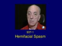 |
Hemifacial Spasm | The patient is a 72 year old man with myopia, childhood exotropia, progressive age related ptosis and right hemifacial spasm. Hemifacial spasm (HS) most often begins insidiously in the orbicularis oculi muscle in the early stages, as in this man. He presented with a 2 year history of involuntary t... | Text |
| 91 |
 |
Hemifacial Spasm | The patient is a 72 year old man with myopia, childhood exotropia, progressive age related ptosis and right hemifacial spasm. Hemifacial spasm (HS) most often begins insidiously in the orbicularis oculi muscle in the early stages, as in this man. He presented with a 2 year history of involuntary t... | Image/MovingImage |
| 92 |
 |
Hemifacial Spasm: Eyelid Twitching | This patient is an atypical case of hemifacial spasm because he is a young college student only 29 years of age. Hemifacial spasm usually develops in the fifth and sixth decade of life and affects women more than men. It often begins insidiously in the orbicularis oculi muscle in the early stage... | Image/MovingImage |
| 93 |
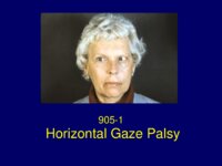 |
Horizontal Gaze Palsy | This 56 year old woman with known adenocarcinoma of the breast presented with the recent onset of horizontal diplopia and deviation of her left eye inwards. Her oncologist referred her for a neuro-ophthalmic evaluation. This 56 year old woman with known adenocarcinoma of the breast presented with... | Text |
| 94 |
 |
Information Processing in the Visual System: David H. Hubel, Nobel Laureate Physiology or Medicine 1981 | David H. Hubel is the John Enders University Professor of Neurobiology at Harvard Medical School. Born in Canada of American parents, he grew up in Montreal, graduated from McGill Medical School, and received training in neurology at the Montreal Neurological Institute and Johns Hopkins Hospital. ... | Image/MovingImage |
| 95 |
 |
Internuclear Ophthalmoplegia in Childhood (Guest Lecture) | Text | |
| 96 |
 |
Jaw Winking | This young boy was born at full term after a normal pregnancy with congenital unilateral ptosis of the left eyelid. His mother noticed that when he was sucking on a bottle the ptotic eyelid opened and closed. A diagnosis of the Marcus-Gunn jaw winking phenomenon was made. The first case of thi... | Image/MovingImage |
| 97 |
 |
Jaw Winking | This little boy was born with congenital ptosis of the left eyelid, which "flicked up and down" when his jaw moved. The pediatrician made a diagnosis of the Marcus-Gunn jaw winking phenomenon was made. The first case of this unusual synkinesis between the pterygoid muscles and the levator musc... | Image/MovingImage |
| 98 |
 |
Latent Nystagmus | This is one of the first cases of latent nystagmus that I saw with Dr. Cogan in the 1970's. The presence of latent nystagmus was unknown to this patient, a little girl who was found to have latent nystagmus by the ophthalmologist at school trying to test the vision of each eye separately. When she... | Image/MovingImage |
| 99 |
 |
Lateropulsion | This 60 year old patient has Wallenberg's syndrome due to infarction of the left dorsolateral medulla. Wallenberg's syndrome is the best recognized syndrome involving the vestibular nuclei and adjacent structures. Unilateral infarcts affecting the vestibular nuclei may produce an oculomotor imbalanc... | Image/MovingImage |
| 100 |
 |
Lateropulsion | This 44 year old woman has a diagnosis of Multiple Sclerosis. | Image/MovingImage |
