Videos, clinical notes and related presentations concerning neuro-ophthalmological and neurovisual disorders collected during Dr. Wray's work as the Director of Neuro-Visual Disorders at Massachusetts General Hospital.
Shirley H. Wray, M.D., Ph.D., FRCP, Professor of Neurology Harvard Medical School, Director, Unit for Neurovisual Disorders, Massachusetts General Hospital.
NOVEL: https://novel.utah.edu/
TO
Filters: Collection: "ehsl_novel_shw"
| Title | History | Type | ||
|---|---|---|---|---|
| 26 |
 |
Migraine / PET Study | In December 1994 the New England Journal of Medicine published a remarkable paper Bilateral Spreading Cerebral Hypoperfusion during Spontaneous Migraine Headache. Roger P. Woods, Marco Iacoboni and John C. Mazziotta. which is reproduced in part, and accompanied by a video illustration. Courtesy of J... | Image/MovingImage |
| 27 |
 |
Paroxysmal Skew Deviation | In 1970 I saw this unique case with Dr. Cogan. The patient is a 60 year old hypertensive woman with a ten year history of intermittent vertical double vision and oscillopsia. Neurological examination between attacks of diplopia showed: Visual acuity 20/25 OU Visual fields, pupils and fundus exami... | Image/MovingImage |
| 28 |
 |
Fisher's One and a Half Syndrome | This young man was seen in the emergency room of his local hospital following the onset of severe headache, mild confusion and diplopia. Non-contrast CT brain scan showed: A right pontine hemorrhage He was transferred to the Massachusetts General Hospital ICU. Ocular Motility: Esotropia of the r... | Image/MovingImage |
| 29 |
 |
Migraine Visual Aura | The patient is a 73 year old retired teacher who was referred in 1993 for a second opinion regarding treatment of episodic visual hallucinations. As a school boy in junior school, he began to experience transient episodes of a spot appearing in the right lower homonymous quadrant of his field of vi... | Image/MovingImage |
| 30 |
 |
Cavernous Angioma | The patient is a 19 year old sophomore who presented in 1983 with numbness of the left hand, involving initially just the fingers, and numbness and weakness of the right side of the face. He described the numbness in his hand as if it was "intensely asleep". The facial numbness involved the peri... | Text |
| 31 |
 |
Unilateral Internuclear Ophthalmoplegia | The patient is a 19 year old sophomore who presented in 1983 with numbness of the left hand, involving initially just the fingers, and numbness and weakness of the right side of the face. He described the numbness in his hand as if it was "intensely asleep". The facial numbness involved the peri... | Image/MovingImage |
| 32 |
 |
Migraine Visual Aura: A Discussion with Nobel Laureate David H. Hubel | I am greatly indebted to the Nobel Laureate, David Hubel for his permission to publish his description of his migraine aura. The recording was made fortuitously at the time that I invited David to the Unit for Neuro-Visual Disorders to record an audio clip describing the experiments in the cat that... | Image/MovingImage |
| 33 |
 |
Migraine Visual Aura: A Personal Account | I am greatly indebted to the Nobel Laureate, David Hubel for his permission to publish his description of his migraine aura. The recording was made fortuitously at the time that I invited David to the Unit for Neuro-Visual Disorders to record an audio clip describing the experiments in the cat that... | Image/MovingImage |
| 34 |
 |
Third Nerve Palsy; Aberrant Reinnervation | The patient is a 48 year old man from Saudi Arabia who came to the Massachusetts General Hospital for a second opinion and hopefully for surgical correction of ptosis of the left eye (OS). He had a long standing left third nerve palsy due to compression of the nerve trunk by a cavernous sinus mening... | Image/MovingImage |
| 35 |
|
Unilateral Horizontal Gaze Palsy | The patient is a 62 year old woman with known hypertension. She was referred by her PCP because she could not move her eyes fully and look left. Neuro-ophthalmological examination: Visual acuity, fields, pupils and fundi normal Ocular Motility: • Slight head turn to the left • Conjugate dev... | Image/MovingImage |
| 36 |
 |
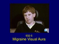
Migraine Visual Aura | The patient is a 9 year old right handed boy who developed headaches in 1993 at the age of 8. At that time he told his mother that he had bad headaches starting at the back of the head, usually bioccipital, spreading over the top of the head to his forehead. The headaches were short in duration la... | Image/MovingImage |
| 37 |
 |
Duane's Syndrome | The patient is a 7 year old boy born two weeks premature with transposition of the major arteries of the heart, four holes in the heart, and an absent spleen. He had cardiac surgery at age 2 days and at age one year and his development was excellent thereafter. At age 6 months, it was noted that th... | Image/MovingImage |
| 38 |
 |
Supranuclear Paralysis of Upgaze | This young child presented with headache and unsteadiness. He was found to have obstructive hydrocephalus, aqueduct stenosis and a medulloblastoma. The constellation of clinical eye signs localized to the Dorsal Midbrain and included: • Supranuclear paralysis of upgaze (saccadic and pursuit move... | Image/MovingImage |
| 39 |
 |
Migraine Visual Aura | The patient is a 9 year old right handed boy who developed headaches in 1993 at the age of 8. At that time he told his mother that he had bad headaches starting at the back of the head, usually bioccipital, spreading over the top of the head to his forehead. The headaches were short in duration la... | Text |
| 40 |
 |
Overview of TDP 43 (Guest Lecture) | Text | |
| 41 |
 |
Third Nerve Palsy | This patient is a 46 year old woman from Portugal who was admitted to the Massachusetts General Hospital in September 1986 with ophthalmoplegia of the left eye (OS) and signs of aberrant reinnervation of the third nerve. She presented, in August 1985, with an episode of diplopia. The diplopia was s... | Image/MovingImage |
| 42 |
 |
Unilateral Horizontal Gaze Palsy | This 56 year old woman with known adenocarcinoma of the breast presented with the recent onset of horizontal diplopia and deviation of her left eye inwards. Her oncologist referred her for a neuro-ophthalmic evaluation. This 56 year old woman with known adenocarcinoma of the breast presented with... | Image/MovingImage |
| 43 |
 |
Upbeat Nystagmus | The patient, a 36 year old Italian, presented in October 1967, at the age of 27, with acute dizziness and ataxia. He was evaluated in Rome. A pneumoencephalogram showed hydrocephalus, attributed to arachnoiditis, and a ventriculo-atrial shunt was placed. Three months post shunt placement he ... | Image/MovingImage |
| 44 |
 |
Cavernous Sinus Meningioma | This patient is a 46 year old woman from Portugal who was admitted to the Massachusetts General Hospital in September 1986 with ophthalmoplegia of the left eye (OS) and signs of aberrant reinnervation of the third nerve. She presented, in August 1985, with an episode of diplopia. The diplopia was s... | Text |
| 45 |
 |
Downbeat Nystagmus | This 58 year old engineer was referred by his neurologist for evaluation of periodic episodes of difficulty focusing and blurred vision for 8 years. In 1981 whilst sightseeing in Newport, he became acutely aware of difficulty focusing and blurry vision. The symptoms lasted for twenty minutes and t... | Image/MovingImage |
| 46 |
 |
Palinopsia | The patient is a healthy 59 year old woman who presented in 1978 with transiet visual symptoms. The first visual disturbance occurred in December 1978 when suddenly she noted: • Fluttering of vision in the left eye (OS) • The appearance of a central black spot • Around the edge of the blac... | Image/MovingImage |
| 47 |
 |
Third Nerve Palsy | This patient is a 58 year old woman from Peru who, in 1975, developed intermittent headaches and right retro-orbital eye pain. She was seen by several ophthalmologists in South America who were unable to make a diagnosis. In March 1977 she awoke one morning with vertical diplopia most marked on... | Image/MovingImage |
| 48 |
 |
Parasellar Meningioma | This patient is a 58 year old woman from Peru who, in 1975, developed intermittent headaches and right retro-orbital eye pain. She was seen by several ophthalmologists in South America who were unable to make a diagnosis. In March 1977 she awoke one morning with vertical diplopia most marked on... | Text |
| 49 |
 |
Downbeat Nystagmus | This 58 year old engineer was referred by his neurologist for evaluation of periodic episodes of difficulty focusing and blurred vision for 8 years. In 1981 whilst sightseeing in Newport, he became acutely aware of difficulty focusing and blurry vision. The symptoms lasted for twenty minutes and t... | Text |
| 50 |
 |
Upbeat Nystagmus | The patient, a 36 year old Italian, presented in October 1967, at the age of 27, with acute dizziness and ataxia. He was evaluated in Rome. A pneumoencephalogram showed hydrocephalus, attributed to arachnoiditis, and a ventriculo-atrial shunt was placed. Three months post shunt placement he ... | Text |