Home
Browse
Ask Us
Chat
Harmful Language Statement
Log in
NOVEL - Neil R. Miller Collection
Advanced Search
About
The Neil R. Miller Collection covers a broad range of neuro-ophthalmologic conditions and syndromes in videos, images, and lecture presentations.
Year
1996
1997
1998
1999
2000
2001
2002
2003
2004
2005
2006
2007
2008
2009
2010
2011
2012
2013
2014
2015
2016
2017
2018
2019
2020
2021
2022
2023
2024
TO
1996
1997
1998
1999
2000
2001
2002
2003
2004
2005
2006
2007
2008
2009
2010
2011
2012
2013
2014
2015
2016
2017
2018
2019
2020
2021
2022
2023
2024
Type
Image
601
Image/MovingImage
135
Text
1
Format
image/jpeg
642
video/mp4
135
application/pdf
8
Collection
NOVEL - Neil R. Miller Collection
785
Filters:
Collection:
"ehsl_novel_nrm"
601
-
700
of
785
<
1
2
3
4
5
6
7
8
>
Gallery view
Number of results to display per page
10
25
50
100
200
Sort by Relevance
Sort by Title A-Z
Sort by Title Z-A
Sort by Date Ascending
Sort by Date Descending
Sort by Last Modified Ascending
Sort by Last Modified Descending
Title
Date
Type
601
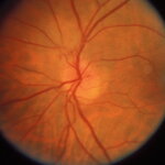
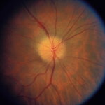
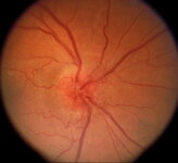
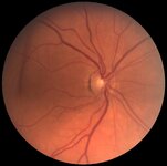
Nonarteritic Anteriior Ischemic Optic Neuropathy in an Eye with Buried Drusen
2024-06
Image
602
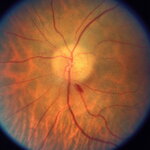
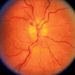
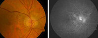
Nonarteritic Anterior Ischemic Optic Neuropathy
2018-02-08
Image
603
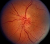
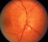
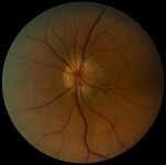
Nonarteritic Anterior Ischemic Optic Neuropathy Associated with Amiodarone Use
2024-06-28
Image
604
Nonarteritic Anterior Ischemic Optic Neuropathy Associated with Amiodarone Use
2024-06-28
Image
605
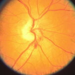
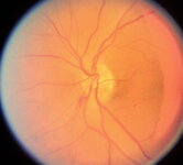
Nonarteritic Anterior Ischemic Optic Neuropathy Associated with Amiodarone Use
2024-06-28
Image
606
Nonarteritic Anterior Ischemic Optic Neuropathy Associated with Amiodarone Use
2024-06-28
Image
607
Nonarteritic Anterior Ischemic Optic Neuropathy Associated with Amiodarone Use
2024-06-28
Image
608
Nonarteritic Anterior Ischemic Optic Neuropathy Associated with Amiodarone Use
2024-06-28
Image
609
Nonarteritic Anterior Ischemic Optic Neuropathy Associated with Amiodarone Use
2024-06-28
Image
610
Nonarteritic Anterior Ischemic Optic Neuropathy Associated with Amiodarone Use
2024-06-28
Image
611
Nonarteritic Anterior Ischemic Optic Neuropathy Associated with Amiodarone Use
2024-06-28
Image
612
Nonarteritic Anterior Ischemic Optic Neuropathy Associated with Amiodarone Use
2024-06-28
Image
613
Nonarteritic Anterior Ischemic Optic Neuropathy Associated with Amiodarone Use
2024-06-28
Image
614
Nonarteritic Anterior Ischemic Optic Neuropathy Associated with Amiodarone Use
2024-06-28
Image
615
Nonarteritic Anterior Ischemic Optic Neuropathy Following Intraocular Avastin Injection for AMD
Image
616
Nonarteritic Anterior Ischemic Optic Neuropathy in an Eye with Previously Buried Drusen, Now Visible as Optic Atrophy Develops
2024-06
Image
617
Nonspecific Visual Field Defects in a Patient with CADASIL
2024-07-10
Image
618
Normal Optic Disc with 0.3 Cup/Disc Ratio in an Eye with Contralateral Arteritic Anterior Ischemic Optic Neuropathy
2024-06
Image
619
OCT in a Patient with Nonarteritic Anterior Ischemic Optic Neuropathy in the Left Eye
2018-02-16
Image
620
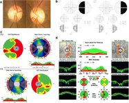
OCT Showing Band Atrophy in a Patient with an Optic Chiasmal Syndrome
2024-07
Image
621
OCT Showing Band Atrophy in a Patient with an Optic Chiasmal Syndrome
2024-07
Image
622
OCT Showing Bilateral Papilledema
2024-06-28
Image
623
Ocular Bobbing
2024-05
Image/MovingImage
624
Ocular Flutter
2024-06
Image/MovingImage
625
Ocular Flutter
2024-06
Image/MovingImage
626
Ocular Flutter
2024-06
Image/MovingImage
627
Ocular Flutter in a Child With an Intracranial Arachnoid Cyst Thought to be Responsible for the Abnormal Eye Movements but in Whom an Evaluation Revealed an Underlying Neuroblastoma
2024-06
Image/MovingImage
628
Ocular Motility Disturbance in a Patient With Progressive Supranuclear Palsy
2024-06
Image/MovingImage
629
Ocular Motor Dysfunction in a Child With Spinocerebellar Atrophy Type 7
2024-06
Image/MovingImage
630
Ocular Myasthenia Gravis in a Child
2024-05
Image/MovingImage
631
Ocular Neuromyotonia
2024-05
Image/MovingImage
632
Ocular Neuromyotonia
2024-05
Image/MovingImage
633
Oculomotor Nerve Palsy With Cyclic Spasms
2024-06
Image/MovingImage
634
Oculomotor Nerve Palsy With Cyclic Spasms
2024-06
Image/MovingImage
635
Oculopalatal Tremor
2024-05
Image/MovingImage
636
Oculopalatal Tremor
2024-05
Image/MovingImage
637
Oculopalatal Tremor
2024-05
Image/MovingImage
638
Oculopalatal Tremor
2024-06
Image/MovingImage
639
One-and-a-half Syndrome
2024-05
Image/MovingImage
640
Opsoclonus
2024-05
Image/MovingImage
641
Opsoclonus
2024-05
Image/MovingImage
642
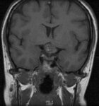
Optic Chiasm Damage Following Excision of a Cavernous Angioma
2024-07
Image
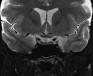
643
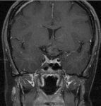
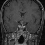
Optic Chiasm: Normal Appearance on Coronal MRI
2024-06-28
Image
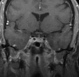
644
Optic Chiasm: Normal Appearance on Coronal MRI
2024-07
Image
645
Optic Chiasm: Normal Appearance on MRI
2024-07
Image
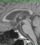
646
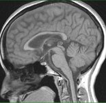
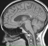
Optic Chiasm: Normal Appearance on Sagittal MRI
2024-07
Image
647
Optic Chiasm: Normal Appearance on Sagittal MRI
2024-07
Image
648
Optic Chiasm: Normal Appearance on Sagittal MRI
2024-07
Image
649
Optic Chiasmal Cavernoma
2024-07
Image
650
Optic Chiasmal Cavernoma
2024-07
Image
651
Optic Chiasmal Cavernoma
2024-07
Image
652
Optic Chiasmal Syndrome
2024-07
Image
653
Optic Disc with No Cup (Disc at Risk) in a Patient with NAION in the Fellow Eye
2018-07
Image
654
Optic Disc with no Cup (Disc at Risk) in a Patient with Nonarteritic Anterior Ischemic Optic Neuropathy in the Fellow Eye
2018-02-08
Image
655
Optic Disc with No Cup (Disc at Risk) in the Eye of a Patient with Nonarteritic Anterior Ischemic Optic Neuropathy in the Fellow Eye
2018-02
Image
656
Optic Nerve Cupping - 3
Image
657
Optic Nerve Cupping - 4
Image
658
Optic Nerve Cupping - 5
Image
659
Optic Nerve Cupping - 6
Image
660
Optic Nerve Cupping - 7
Image
661
Optic Nerve Cupping - 8
Image
662
Optic Nerve Cupping - a
Image
663
Optic Nerve Cupping - MR - 1
Image
664
Optic Nerve Cupping - MR - 4
Image
665
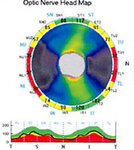
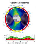
Optic Nerve Cupping - OCT - 2016
2016
Image
666
Optic Nerve Cupping - OD
Image
667
Optic Nerve Cupping - OS
Image
668
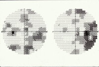
Optic Nerve Cupping - VF - 1
Image
669
Optic Nerve Cupping - VF - 1a
Image
670
Optic Nerve Cupping - VF - 1b
Image
671
Optic Nerve Cupping - VF - 2
Image
672
Optic Nerve Sheath Decompression: Nasal Approach (Narrated)
2024-06
Image/MovingImage
673
Optic Nerve Sheath Decompression: Nasal Route
2024-06
Image/MovingImage
674
Optic Nerve Sheath Fenestration Technique
2024-06
Image/MovingImage
675
Optokinetic Nystagmus
2024-05
Image/MovingImage
676
Optokinetic Nystagmus (Symmetrical)
2024-05
Image/MovingImage
677
Palatal Myoclonus in a Patient After a Pontine Stroke
2024-06
Image/MovingImage
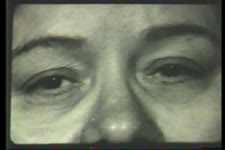
678
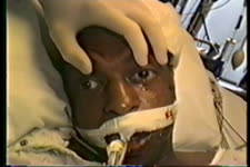
Papilledema in a Patient with Cryptococcal Meningitis in the Setting of AIDS
1996-12-13; 1998-11-11
Image
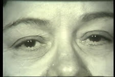
679
Papilledema in a Patient with Cryptococcal Meningitis in the Setting of AIDS
1996-12-13; 1998-11-11
Image
680
Paraneoplastic Ocular Motor Dysfunction in a Patient With Prostate Cancer
2024-06
Image/MovingImage
681
Parinaud Syndrome
2024-06
Image/MovingImage
682
Parinaud Syndrome
2024-06
Image/MovingImage
683
Partially Thrombosed Right-sided Intracavernous Aneurysm
2024-07-10
Image
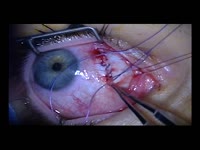
684
Patient Immediately Following Successful Transvenous Embolization of a Left-sided Direct Carotid-cavernous Sinus Fistula Via the Superior Ophthalmic Vein
2024-07-10
Image
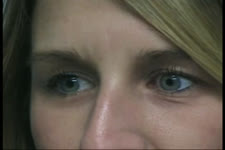
685
Patient with Giant Right-sided Intracavernous Aneurysm, Showing Ipsilateral Proptosis and Orbital Fullness. Rightward Gaze
2024-07-10
Image
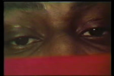
686
Patient with Giant Right-sided Intracavernous Aneurysm, Showing Moderate Fight Proptosis and Orbital Swelling
2024-07-10
Image
687
Patient with Left-sided Proptosis and Orbital Swelling from an Ipsilateral Direct Carotid-cavernous Sinus Fistula Prior to Transvenous Embolization
2024-07-10
Image
688
Patient with Right-sided Giant Intracavernous Aneurysm During Attempted Leftward Gaze
2024-07-10
Image
689
Patient with Right-sided Intracavernous Aneurysm on Attempted Downgaze
2024-07-10
Image
690
Patient with Right-sided Intracavernous Aneurysm Showing Right Orbital Fullness and Suggestion of a Horner Syndrome
2024-07-10
Image
691
Pendular Nystagmus (Binocular)
2024-05
Image/MovingImage
692
Pendular Nystagmus (Monocular)
2024-05
Image/MovingImage
693
Pendular Nystagmus (Monocular)
2024-05
Image/MovingImage
694
Pendular Oscillations
2024-05
Image/MovingImage
695
Periodic Alternating Gaze Deviation
2024-05
Image/MovingImage
696
Periodic Alternating Nystagmus
2024-05
Image/MovingImage
697
Periodic Alternating Nystagmus
2024-05
Image/MovingImage
698
Periodic Alternating Nystagmus
2024-05
Image/MovingImage
699
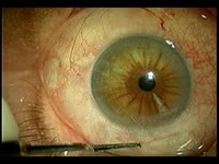
Persistent Post-cataract Pupillary Mydriasis
2024-07-10
Image
700
Post-papilledema Retino-choroidal Striae and Optic Atrophy
2024-07
Image
601
-
700
of
785
<
1
2
3
4
5
6
7
8
>