Home
Browse
Ask Us
Chat
Harmful Language Statement
Log in
NOVEL - Neil R. Miller Collection
Advanced Search
About
The Neil R. Miller Collection covers a broad range of neuro-ophthalmologic conditions and syndromes in videos, images, and lecture presentations.
Year
1996
1997
1998
1999
2000
2001
2002
2003
2004
2005
2006
2007
2008
2009
2010
2011
2012
2013
2014
2015
2016
2017
2018
2019
2020
2021
2022
2023
2024
TO
1996
1997
1998
1999
2000
2001
2002
2003
2004
2005
2006
2007
2008
2009
2010
2011
2012
2013
2014
2015
2016
2017
2018
2019
2020
2021
2022
2023
2024
Type
Image
601
Image/MovingImage
135
Text
1
Format
image/jpeg
642
video/mp4
135
application/pdf
8
Collection
NOVEL - Neil R. Miller Collection
785
Filters:
Collection:
"ehsl_novel_nrm"
201
-
300
of
785
<
1
2
3
4
5
6
7
8
>
Gallery view
Number of results to display per page
10
25
50
100
200
Sort by Relevance
Sort by Title A-Z
Sort by Title Z-A
Sort by Date Ascending
Sort by Date Descending
Sort by Last Modified Ascending
Sort by Last Modified Descending
Title
Date
Type
201
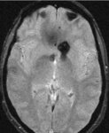
Cavernoma in Region of Chiasm (SWI MRI)
2024-07
Image
202
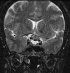
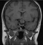
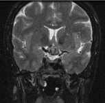
Cavernoma of the Optic Chiasm Causing Chiasmal Apoplexy
2024-07
Image
203
Cavernoma of the Optic Chiasm Causing Chiasmal Apoplexy
2024-07
Image
204
Cavernoma of the Optic Chiasm Causing Chiasmal Apoplexy
2024-07
Image
205
Cavernoma of the Optic Chiasm Causing Chiasmal Apoplexy
2024-07
Image
206
Cavernoma of the Optic Chiasm Causing Chiasmal Apoplexy
2024-07
Image
207
Cavernoma of the Optic Chiasm Causing Chiasmal Apoplexy
2024-07
Image
208
Cavernous Angioma in the Optic Chiasm in a Patient with Familial Cavernomas (SWI)
2024-06-28
Image
209
Cavernous Sinus Thrombosis
2024-07-10
Image
210
CCF - Surgery - SOV9
2024-07-10
Image
211
Central Retinal Artery Occlusion - 1
Image
212
Central Retinal Artery Occlusion - 1a
Image
213
Central Retinal Artery Occlusion - 2
Image
214
Central Retinal Artery Occlusion - 3
Image
215
Central Retinal Artery Occlusion - normal cao
Image
216
Central Retinal Vein Occlusion - 1a
Image
217
Central Retinal Vein Occlusion - 1b
Image
218
Central Retinal Vein Occlusion - 2a
Image
219
Central Retinal Vein Occlusion - 2b
Image
220
Central Retinal Vein Occlusion - 3a
Image
221
Central Retinal Vein Occlusion - 3b
Image
222
Central Retinal Vein Occlusion - 4a
Image
223
Central Retinal Vein Occlusion - 4b
Image
224
Central Retinal Vein Occlusion - 5
Image
225
Central Retinal Vein Occlusion - 6
Image
226
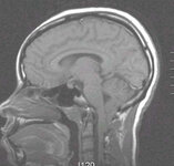
Chiari Malformation
2024-06
Image/MovingImage
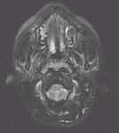
227
Chiari Malformation - 01
2024-07
Image
228
Chiari Malformation - 02
2024-07
Image
229
Chiari Malformation - 03
2024-07
Image
230
Chiari Malformation - 04
2024-07
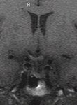
Image
231
Chiari Malformation - 05
2024-07
Image
232
Chiari Malformation - 06
2024-07
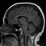
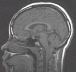
Image
233
Chiari Malformation - a
2024-07
Image
234
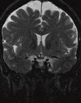
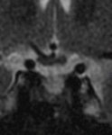
Chiari Malformation in a Patient who Presented with Papilledema (MRI)
2024-07
Image
235
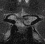
Chiari Malformation in a Patient who Presented with Papilledema (MRI)
2024-07
Image
236
Chiari Malformation in a Patient who Presented with Papilledema (MRI)
2024-07
Image
237
Chiari Malformation with Papilledema - 02
2024-07
Image
238
Chiari Malformation with Papilledema - OD
2024-07
Image
239
Chiari Malformation with Papilledema - OS
2024-07
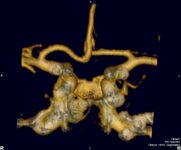
Image
240
Chiasm Anatomy - 001
2024-07
Image
241
Chiasm Anatomy - Birdbeak
2024-07
Image
242
Chiasm Anatomy - Chiasmal Adenohypophysis - 1
2024-07
Image
243
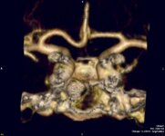
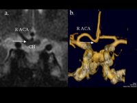
Chiasm Anatomy - Chiasmal Adenohypophysis - 2
2024-07
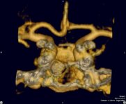
Image
244
Chiasm Anatomy - Chiasmal Germinoma - 1
2024-07
Image
245
Chiasm Anatomy - Chiasmal Germinoma - 2
2024-07
Image
246
Chiasm Anatomy - Chiasmal Infarction
2024-07
Image
247
Chiasm Anatomy - Chiasmal Neuritis - 1
2024-07
Image
248
Chiasm Anatomy - Chiasmal Neuritis - 2
2024-07
Image
249
Chiasm Anatomy - Germinoma - 01
2024-07
Image
250
Chiasm Anatomy - Hydrocephalus - 002
2024-07
Image
251
Chiasm Anatomy - Hydrocephalus - 006
2024-07
Image
252
Chiasm Anatomy - Hydrocephalus - 02
2024-07
Image
253
Chiasm Anatomy - Infarction - 001
2024-07
Image
254
Chiasm Anatomy - Infarction - 002
2024-07
Image
255
Chiasm Anatomy - Infarction - 01
2024-07
Image
256
Chiasm Anatomy - Infarction - 03
2024-07
Image
257
Chiasm Anatomy - Infarction - 04
2024-07
Image
258
Chiasm Anatomy - Pit Carcinoma 2
2024-07
Image
259
Chiasmal Compression - Vascular - 010
2024-07
Image
260
Chiasmal Compression - Vascular - 08
2024-07
Image
261
Chiasmal Compression - Vascular - 09
2024-07
Image
262
Chiasmal Compression - Vascular - CTA
2024-07
Image
263
Chiasmal Compression - Vascular - g
2024-07
Image
264
Chiasmal Compression - Vascular - h
2024-07
Image
265
Chiasmal Compression - Vascular - i
2024-07
Image
266
Chiasmal Compression - Vascular - j
2024-07
Image
267
Chiasmal Compression - Vascular - k
2024-07
Image
268
Chiasmal Compression - Vascular - KineticOD
2024-07
Image
269
Chiasmal Compression - Vascular - KineticOS
2024-07
Image
270
Chiasmal Compression - Vascular - l
2024-07
Image
271
Chiasmal Compression - Vascular - m
2024-07
Image
272
Chiasmal Compression - Vascular - OCSOD1
2024-07
Image
273
Chiasmal Compression - Vascular - OCSOD2
2024-07
Image
274
Chiasmal Compression - Vascular - OCSOS1
2024-07
Image
275
Chiasmal Compression - Vascular - VF
2024-07
Image
276
Chiasmal Compression by a Vascular Loop
2024-07
Image
277
Chiasmal Compression from a Dilated Third Ventricle in a Patient with Severe Hydrocephalus (MRI)
2024-07
Image
278
Chiasmal Compression from a Vascular Loop
2024-07
Image
279
Chiasmal PNET - 3
2024-07
Image
280
Chiasmal PNET - 8
2024-07
Image
281
Cholesterol Granuloma - 08
2024-07
Image
282
Choriocarcinoma Primary - 1a
2024-07
Image
283
Choriocarcinoma Primary - 1b
2024-07
Image
284
Choriocarcinoma Primary - 1c
2024-07
Image
285
Choriocarcinoma Primary - 2
2024-07
Image
286
Choriocarcinoma Primary - 2a
2024-07
Image
287
Choriocarcinoma Primary - 2b
2024-07
Image
288
Choriocarcinoma Primary - 2c
2024-07
Image
289
Choriocarcinoma Primary - 2d
2024-07
Image
290
Choroidal Folds - 3
2024-07
Image
291
Choroidal Hypoperfusion in a Patient with CADASIL
2024-07-10
Image
292
Chronic Progressive External Ophthalmoplegia - 0001
Image
293
Chronic Progressive External Ophthalmoplegia - 0002
Image
294
Chronic Progressive External Ophthalmoplegia - 0003
Image
295
Chronic Progressive External Ophthalmoplegia - 0004
Image
296
Chronic Progressive External Ophthalmoplegia - 0005
Image
297
Chronic Progressive External Ophthalmoplegia - 0006
Image
298
Chronic Progressive External Ophthalmoplegia - 0007
Image
299
Chronic Progressive External Ophthalmoplegia - 0008
Image
300
Chronic Progressive External Ophthalmoplegia - 0009
Image
201
-
300
of
785
<
1
2
3
4
5
6
7
8
>