John A. Moran Eye Center Neuro-Ophthalmology Collection: A variety of lectures, videos and images relating to topics in Neuro-Ophthalmology created by faculty at the Moran Eye Center, University of Utah, in Salt Lake City.
NOVEL: https://novel.utah.edu/
TO
Filters: Collection: "ehsl_novel_jmec"
1 - 25 of 18
| Title | Description | Type | ||
|---|---|---|---|---|
| 1 |
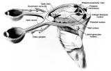 | Anatomy of the Pupillary Light Reflex Pathway | Anatomy of the pupillary light reflex pathway. (Miller NR: Walsh And Hoyt's Clinical Neuro-Ophthalmology, p 421. Vol 2, 4th ed. Baltimore: Williams & Wilkins, 1985, with permission.) | Image |
| 2 |
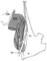 | The Course of the Postganglionic Segment of the Oculosympathetic Fibers from the Internal Carotid Artery | The course of the postganglionic segment of the oculosympathetic fibers from the internal carotid artery (ICA) to the orbit is depicted as a dotted line. Note that they briefly join the abducens nerve (cranial nerve VI) before joining the nasociliary branch of the of the ophthalmic division of the t... | Image |
| 3 |
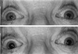 | Enhanced Mydriasis in Response to Hydroxyamphetamine | Enhanced mydriasis in response to hydroxyamphetamine in a 77-year-old woman with a long-standing, preganglionic, right-sided Horner's syndrome that occurred following cervical neck dissection for thoracic outlet syndrome 30 years earlier. Miosis of the right pupil is apparent in room light (top). Th... | Image |
| 4 |
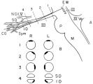 | Location of Pupillomotor Fibers | Location of pupillomotor fibers are depicted as dark regions on cross-sections of the right (R) and left (L) oculomotor nerve at various locations along its course, including its emergence from the brain stem in the interpeduncular fossa (1), the midsubarachnoid segment (2), the level of the dorsum ... | Image |
| 5 |
 | The Normal Pupillary Light Reflex | The normal pupillary light reflex is initiated following exposure to light. After a brief latency, both the right (solid line) and left (broken line) pupil constrict, then undergo a small amount of redilation (escape), followed by oscillations (hippus) if the light is sustained. Hippus is not a path... | Image |
| 6 |
 | B-scan Technique | This video describes and demonstrates the B-scan examination technique for examination of the eye using ultrasonography. | Image/MovingImage |
| 7 |
 | Ultrasonography: Immersion Technique | This video describes and demonstrates the immersion technique for examination of the eye using ultrasonography. | Image/MovingImage |
| 8 |
 | Ultrasonography Techniques | This video describes and demonstrates the various techniques for examination of the eye using ultrasonography, including A-scan, B-scan and immersion. | Image/MovingImage |
| 9 |
 | A-scan Technique | This video describes and demonstrates the A-scan examination technique for examination of the eye using ultrasonography. | Image/MovingImage |
| 10 |
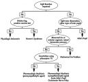 | Flow Chart for Sorting Out Anisocoria - Direct Light Reaction of the Pupil | Flow chart for sorting out anisocoria based initially on the integrity of the direct light reaction of the pupil. | Image |
| 11 |
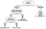 | Flow Chart for Sorting Out Anisocoria - Bright Light and Darkness | Flow chart for sorting out anisocoria based initially on how it is influenced by bright light and darkness. | Image |
| 12 |
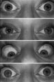 | Pupil Signs in a 32-year-old Woman with Right-sided Adie's Pupil | Pupil signs in a 32-year-old woman with right-sided Adie's pupil. The right pupil is larger than the left pupil (top), reacts poorly to direct light stimulation (second panel), and better in response to near stimulation (third panel). The right pupil also shows a supersensitive response 30 minutes a... | Image |
| 13 |
 | Third Nerve Palsy, Pupil Involving | Example of patient with third nerve palsy. Left eye shows pupilary involvement. Left eye doesn't immediately duct, but abducts well, with impaired superduction. Secondary and primary deviations are demonstrated. Anisocoria is more prominent when light is on, showing a parasympathetic defect to the p... | Image/MovingImage |
| 14 |
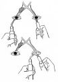
 | Dilation Lag | Two examples of dilation lag (Horner's syndrome). In the first example, the right pupil dilates much faster than the left pupil when the light is turned out. In the second example, the left pupil dilates much faster than the right pupil when the light is turned out. Discussion of methods of document... | Image/MovingImage |
| 15 |
 | Assessment of an Afferent Pupillary Defect When Only One Iris is Functional | Assessment of an afferent pupillary defect when only one iris is functional. In this example, a right-sided parasellar tumor is compressing both the optic and oculomotor nerves, causing an optic neuropathy and a pupil-involving third crainial nerve palsy. The pupil on the affected side has both an a... | Image |
| 16 |
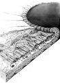 | Structures of the iris | Structures of the iris. The a indicates the anterior border layer that terminates at the pigmentary ruff of the pupillary border (b). The c indicates the iris sphincter muscle, which is oriented circumferentially within the stroma and located deep to the anterior border layer; d indicates vessels th... | Image |
| 17 |
 | Silent Sinus Syndrome | Silent sinus syndrome (SSS) is characterized by spontaneous and progressive unilateral enophthalmos. | Text |
| 18 |
 | The Electroretinogram and Electro-oculogram: Clinical Applications | The global or full-field electroretinogram (ERG) is a mass electrical response of the retina to photic stimulation. The ERG is a test used worldwide to assess the status of the retina in eye diseases in human patients and in laboratory animals used as models of retinal disease. | Text |
1 - 25 of 18