John A. Moran Eye Center Neuro-Ophthalmology Collection: A variety of lectures, videos and images relating to topics in Neuro-Ophthalmology created by faculty at the Moran Eye Center, University of Utah, in Salt Lake City.
NOVEL: https://novel.utah.edu/
TO
Filters: Collection: "ehsl_novel_jmec"
1 - 25 of 3
| Title | Curriculum | Description | Subject | Collection | ||
|---|---|---|---|---|---|---|
| 1 |
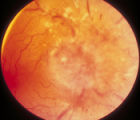 |
3-59a - Glioma | curriculum_fellow; KBDcompinfilglioma | This 45-year-old man presented with vision loss in his right eye; his examination showed severe disc swelling in this eye and vision loss on visual field testing (3-59a). MRI with fat saturation and enhancement and MRI with T2 signals also confirm an enlarged optic nerve. (3-59c) Excisional biopsy o... | Glioma | Neuro-Ophthalmology Virtual Education Library - The Moran Eye Center Neuro-Ophthalmology Collection: https://novel.utah.edu/Moran/ |
| 2 |
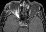 |
3-59c - Glioma | curriculum_fellow; KBDcompinfilglioma | This 45-year-old man presented with vision loss in his right eye; his examination showed severe disc swelling in this eye and vision loss on visual field testing (3-59a). MRI with fat saturation and enhancement and MRI with T2 signals also confirm an enlarged optic nerve. (3-59c) Excisional biopsy o... | Glioma | Neuro-Ophthalmology Virtual Education Library - The Moran Eye Center Neuro-Ophthalmology Collection: https://novel.utah.edu/Moran/ |
| 3 |
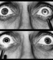 |
Right-sided Relative Afferent Pupillary Defect | curriculum_fellow; KBDrapd; KBDmeasurerapd; IC-C9d-relative-afferent-pupillary-defect; IC-D5fiii-afferent-pupillary-defect | Right-sided relative afferent pupillary defect in a man with optic nerve glioma. When the unaffected left eye is stimulated by light, both pupils constrict (top). When the light is then swung over to the affected right eye, both pupils dilate (bottom). This indicates that pupillomotor conduction thr... | Pupil Disorders; Relative Afferent Pupillary Defect; RAPD; Afferent Pupillary Defect | Neuro-Ophthalmology Virtual Education Library - The Moran Eye Center Neuro-Ophthalmology Collection: https://novel.utah.edu/Moran/ |
1 - 25 of 3