John A. Moran Eye Center Neuro-Ophthalmology Collection: A variety of lectures, videos and images relating to topics in Neuro-Ophthalmology created by faculty at the Moran Eye Center, University of Utah, in Salt Lake City.
NOVEL: https://novel.utah.edu/
TO
Filters: Collection: "ehsl_novel_jmec"
| Title | Description | Type | ||
|---|---|---|---|---|
| 176 |
 |
Optic Disc: Anatomy, Variants, Unusual discs | Discussion of viewing the optic disc. Includes development of direct ophthalmoscope. Covers normal optic disc and nerve fiber; nerve fiber loss and defects; cilioretinal arteries; venous anomolies; papilledema; pseudopapilledema; myopic disc; hyperoptic disc; little red discs; megallopapilla; myelin... | Text |
| 177 |
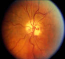 |
Shunt Vessel Meningioma | RETINO-CHOROIDAL (OPTO-CILIARY) COLLATERAL VESSELS: (also known as Retinal-choroidal venous collaterals, opticociliary veins or ciliary shunt vessels) Retino-choroidal collaterals are potential telangiectatic connections between the retina and choroidal circulation. Although sometimes called "shunts... | Image |
| 178 |
 |
Duane's Retraction Syndrome Type 1: Lid Retraction | Example of patients with Duane's Retraction Syndrome, Type 1. Description of components of Duane's Syndrome: limitation of abduction, variable limitation of adduction, and palpebral fissure narrowing and globe retraction with attempted adduction. Type 1 includes limited or absent abduction with norm... | Image/MovingImage |
| 179 |
 |
The 3 Step Test: Looking for a 4th Nerve Palsy | Description of the three step test (3 step test) used when looking for a 4th nerve palsy. | Text |
| 180 |
 |
Normal Light Reflex without RAPD | This clip demonstrates the examination of the Relative Afferent Pupillary Defect (RAPD.) Demonstration of gauging the size of the pupil in light, testing light reflexes, swinging flashlight test for optic nerve abnormality. | Image/MovingImage |
| 181 |
 |
See-saw Nystagmus | 7-year-old female whose mother noticed her eyes "bouncing" for 2 months. Visual acuity 20/70 OD and 20/40 OS, reduced color vision OU, and no afferent pupillary defect. See-saw nystagmus documented with videography. Manual perimetry revealed a complete right homonymous hemianopia. MRI revealed a lar... | Image/MovingImage |
| 182 |
 |
Cochet-Bonnet Esthesiometer (similar to Von Frey Hair) Exam | The Cochet-Bonnet esthesiometer , similar to Von Frey Hair testing, has been used in studies to quantify sensory changes in the trigeminal system, especially the cornea. The device uses a thin fiber to test sensation. The shorter the fiber, the more sensation is felt. The monofilament is applied per... | |
| 183 |
 |
Oculopalatal Myoclonus | Oculopalatal myoclonus (OPM) Rhythmic oscillations of eyes and palate. Occurred after specific brainstem injury from stroke, following stenting. Related PowerPoint Presentation: http://content.lib.utah.edu/u?/EHSL-Moran-Neuro-opth,129 Disease/Diagnosis: Oculopalatal myoclonus. | Image/MovingImage |
| 184 |
 |
Oculopalatal Myoclonus (PPT) | Oculopalatal myoclonus (OPM) Rhythmic oscillations of eyes and palate. Occurred after specific brainstem injury from stroke, following stenting. Related Video: http://content.lib.utah.edu/u?/EHSL-Moran-Neuro-opth,128 Disease/Diagnosis: Oculopalatal myoclonus | Image/MovingImage |
| 185 |
 |
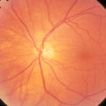
2-53a - Venous Pulsations | On the disc, look for spontaneous venous pulsations. Spontaneous venous pulsations can be seen in the large trunks of veins at the level of the disc margin. They are normally present and seen in 37-90% of normals -- depending on the experience of the examiner and the shape of the disc. The spontaneo... | Image |
| 186 |
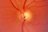 |
2-53b - Venous Pulsations | On the disc, look for spontaneous venous pulsations. Spontaneous venous pulsations can be seen in the large trunks of veins at the level of the disc margin. They are normally present and seen in 37-90% of normals -- depending on the experience of the examiner and the shape of the disc. The spontaneo... | Image |
| 187 |
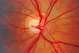 |
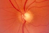
2-7a - Disc Anatomy | The optic disc appearance is determined by: the size of the eye, the size of the scleral canal, how the nerve is inserted into the globe, the appearance of the lamina cribrosa, where myelination stops, and what is left behind in normal development. Even though this is a disc with a very large cup, i... | Image |
| 188 |
 |
Spiral and Stellate Visual Fields Non-physiologic Variants | Description of testing the spiral and stellate visual fields. | |
| 189 |
 |
2-4a - Disc Anatomy | The optic disc appearance is determined by: the size of the eye, the size of the scleral canal, how the nerve is inserted into the globe, the appearance of the lamina cribrosa, where myelination stops, and what is left behind in normal development. Even though this is a disc with a very large cup, i... | Image |
| 190 |
 |
Clover-leaf Visual Field Defects | Description of clover-leaf visual field defects. | |
| 191 |
 |
Documenting the Neuro-ophthalmic Patient: External Photography | Description of documenting the neuro-ophthalmic patient using external photography. This covers pupils and extra ocular muscles. | |
| 192 |
 |
Tunnel Vision on Tangent Screen Testing | Description of tunnel vision and tangent screen testing. | |
| 193 |
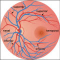 |
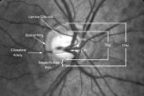
2-37a - Vascular Features | When looking at the disc, the central retinal artery and vein should be visible. The central retinal artery is usually slightly narrower than the vein. When the central retinal artery goes though the lamina cribrosa, the artery becomes smaller because of diminution of the muscular layer and loss of ... | Image |
| 194 |
 |
2-37b - Vascular Features | When looking at the disc, the central retinal artery and vein should be visible. The central retinal artery is usually slightly narrower than the vein. When the central retinal artery goes though the lamina cribrosa, the artery becomes smaller because of diminution of the muscular layer and loss of ... | Image |
| 195 |
 |
Near Reflex and Accomodation | Description of testing the near reflex and accomodation. | |
| 196 |
 |
Tangent Screen Testing Visual Field | Description of tangent screen testing. | |
| 197 |
 |
Benign Episodic Unilateral Mydriasis | Presentation covering benign episodic mydriasis. | Text |
| 198 |
 |
Silent Sinus Syndrome | Silent sinus syndrome (SSS) is characterized by spontaneous and progressive unilateral enophthalmos. | |
| 199 |
 |
Fluoresein Angiography | Comprehensive description of using fluoresein angiography in examinations. | |
| 200 |
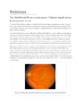 |
The Multifocal Electroretinogram: Clinical Applications | The most important development in ERGs is the multifocal ERG (mfERG). Erich Sutter adapted the mathematical sequences called binary m-sequences creating a program that can extract hundreds of focal ERGs from a single electrical signal. This system allows assessment of ERG activity in small areas of ... | Text |