John A. Moran Eye Center Neuro-Ophthalmology Collection: A variety of lectures, videos and images relating to topics in Neuro-Ophthalmology created by faculty at the Moran Eye Center, University of Utah, in Salt Lake City.
NOVEL: https://novel.utah.edu/
TO
| Title | Description | Type | ||
|---|---|---|---|---|
| 151 |
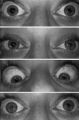 |
Pupil Signs in a 32-year-old Woman with Right-sided Adie's Pupil | Pupil signs in a 32-year-old woman with right-sided Adie's pupil. The right pupil is larger than the left pupil (top), reacts poorly to direct light stimulation (second panel), and better in response to near stimulation (third panel). The right pupil also shows a supersensitive response 30 minutes a... | Image |
| 152 |
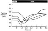 |
Pupillogram Demonstrating Paradoxical Pupillary Constriction to Darkness | Pupillogram demonstrating paradoxical pupillary constriction to darkness in four patients with congenital achromatopsia. Note that the pupils initially constrict when the light is extinguished. (Price MJ, Thompson HS, Judisch GF et al: Pupillary constriction to darkness. Br J Ophthalmol 1981;69:205-... | Image |
| 153 |
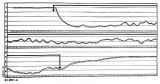 |
Pupillogram of a Healthy Young Subject | Pupillogram of a healthy young subject showing continuous pupillary oscillations of both pupils when light is sustained, indicated by the dark arrow at the top of the recording. Note that the oscillations of the pupils are synchronous and demonstrate variable amplitude and frequency. This pattern of... | Image |
| 154 |
 |
RAPD Present | This clip demonstrates the technique used to determine that Relative Afferent Pupillary Defect (RAPD) is present in a patient. | Image/MovingImage |
| 155 |
 |
Rebound Nystagmus | Example of a patient with rebound nystagmus, where the oscillations alternate direction as the patient shifts gaze in different directions. Discussion of relationship to disease and disorders of the cerebellum, including degenerations of the cerebellum, infarction, and demyelination. | Image/MovingImage |
| 156 |
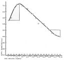 |
Relationship Between Age and Pupil Size | Relationship between age and pupil size, determined using an infrared flash photograph technique with subjects placed in darkness for 3 minutes. The numbers above the abscissa indicate the number of subjects tested in each age range. (Reprinted with permission of Loewenfeld IE: "Simple, central" ani... | Image |
| 157 |
 |
Retinal Fluorescein Angiography | This slide set provides a brief description of Retinal Fluorescein Angiography. First introduced in 1960, sodium fluorescein, a dye, is administered through an angiocatheter (3-5cc) by a nurse or technician. The dye reaches the central retinal artery after passing through the heart and lungs. | Text |
| 158 |
 |
Retinitis Pigmentosa Disease of Rods | Discussion of retinitis pigmentosa which is a retinal/choroidal degeneration caused by various genetic defects. | Text |
| 159 |
 |
Retino-choroidal Vessels or Optociliary Veins or Ciliary Shunt | Overview of retino-choroidal collaterals, which are potential telangiectatic connections between the retina and choroidal circulation. Although sometimes called "shunts", these collaterals are between the retinal venous circulation and the choroidal venous circulation. | Text |
| 160 |
 |
Retraction Nystagmus | Patient with retraction nystagmus (no audio) | Image/MovingImage |
| 161 |
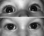 |
Right-sided Pseudo-Horner's Syndrome | Right-sided pseudo-Horner's syndrome in an 8-month-old infant referred because her mother had noted a larger pupil on the left for a few months and her pediatrician thought the right upper lid was droopy. Both pupils reacted normally to light and darkness, the degree of anisocoria was similar in bot... | Image |
| 162 |
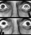 |
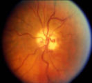
Right-sided Relative Afferent Pupillary Defect | Right-sided relative afferent pupillary defect in a man with optic nerve glioma. When the unaffected left eye is stimulated by light, both pupils constrict (top). When the light is then swung over to the affected right eye, both pupils dilate (bottom). This indicates that pupillomotor conduction thr... | Image |
| 163 |
 |
Rotary Downbeat | Patient with rotary downbeat nystagmus (no audio) | Image/MovingImage |
| 164 |
 |
Rotary Nystagmus | Example of a patient with rotary nystagmus, showing occasional counterclockwise rotary movements of both eyes. Seen more in intrinsic disorders of the brainstem. | Image/MovingImage |
| 165 |
 |
Sector Palsies and Light-Near Dissociation | Example of patient with bilateral Adie's pupils. Exam is performed with a slit-lamp. Shows iris stroma and focal segments of iris sphincter that retain their contractilty. Suggests post-ganglionic parasympathetic denervation. | Image/MovingImage |
| 166 |
 |
See-saw Nystagmus | Example of a patient with see-saw nystagmus, showing how one eye elevates as the other depresses, with the elevating eye intorting as the depressing eye extorts. Shows vertical oscillations with pendular waveforms. Suggests a large structural lesion in the pericellar region (associated with bi-tempo... | Image/MovingImage |
| 167 |
 |
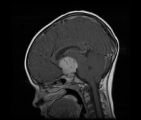
See-saw Nystagmus | 7-year-old female whose mother noticed her eyes "bouncing" for 2 months. Visual acuity 20/70 OD and 20/40 OS, reduced color vision OU, and no afferent pupillary defect. See-saw nystagmus documented with videography. Manual perimetry revealed a complete right homonymous hemianopia. MRI revealed a lar... | Image/MovingImage |
| 168 |
 |
See-saw Nystagmus MRI 1 | MRI; See-saw Nystagmus | Image |
| 169 |
 |
See-saw Nystagmus MRI 2 | MRI; See-saw Nystagmus | Image |
| 170 |
 |
Shaken Baby Syndrome | Text | |
| 171 |
 |
Shunt Vessel Meningioma | RETINO-CHOROIDAL (OPTO-CILIARY) COLLATERAL VESSELS: (also known as Retinal-choroidal venous collaterals, opticociliary veins or ciliary shunt vessels) Retino-choroidal collaterals are potential telangiectatic connections between the retina and choroidal circulation. Although sometimes called "shunts... | Image |
| 172 |
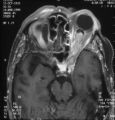 |
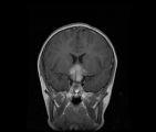
Shunt Vessel Meningioma - MRI | Meningiomas block venous egress and open potential venous channels known as retinochoroidal (optociliary) collateral vein. This meningioma extends from the back of the globe through the optic canal. | Image |
| 173 |
 |
Spasm of the Near Reflex | Example of patient with spasm of the near reflex and voluntary nystagmus. Discussion of similar-looking conditions (e.g. six nerve palsy, limitation of abduction, lateral rectus muscle problems) and how to tell them apart from spasm of the near reflex by observing the myosis evoked by the near respo... | Image/MovingImage |
| 174 |
 |
Spasmus Nutans | Example of patient with spasmus nutans. Discussion of characteristics of this disorder, such as dissociated or monocular nystagmus, abnormal head position, and to-and-fro head oscillation. Sometimes an eccentric gaze is seen as well (as in patient). Patient has a monocular horizontal nystagmus in th... | Image/MovingImage |
| 175 |
 |
Spasmus Nutans | Example of patient with spasmus nutans. | Image/MovingImage |