John A. Moran Eye Center Neuro-Ophthalmology Collection: A variety of lectures, videos and images relating to topics in Neuro-Ophthalmology created by faculty at the Moran Eye Center, University of Utah, in Salt Lake City.
NOVEL: https://novel.utah.edu/
TO
Filters: Collection: "ehsl_novel_jmec"
| Title | Description | Type | ||
|---|---|---|---|---|
| 151 |
 |
See-saw Nystagmus | Example of a patient with see-saw nystagmus, showing how one eye elevates as the other depresses, with the elevating eye intorting as the depressing eye extorts. Shows vertical oscillations with pendular waveforms. Suggests a large structural lesion in the pericellar region (associated with bi-tempo... | Image/MovingImage |
| 152 |
|
Transillumination - Lisch Nodules | Demonstration of transillumination of the Lisch nodules on a patient with neurofibromatosis. Shows how Lisch nodules that were not very visible in slit-lamp examination are better seen with transillumination, which may therefore be useful in detecting Lisch nodules earlier in children where they are... | Image/MovingImage |
| 153 |
 |
Cochet-Bonnet Esthesiometer (similar to Von Frey Hair) Exam | The Cochet-Bonnet esthesiometer , similar to Von Frey Hair testing, has been used in studies to quantify sensory changes in the trigeminal system, especially the cornea. The device uses a thin fiber to test sensation. The shorter the fiber, the more sensation is felt. The monofilament is applied per... | |
| 154 |
 |
Internuclear Ophthalmoplegia (2 Examples) | Two examples of patients with internuclear ophthalmoplegia. First patient has a right internuclear ophthalmoplegia. Patient had subacute bacterial endocarditis with a bacterial abscess in the brain stem. Ductions and gaze to the right look good, but when gazing to the left, the right eye does not ad... | Image/MovingImage |
| 155 |
 |
Third Nerve Palsy, Pupil Involving | Example of patient with third nerve palsy. Left eye shows pupilary involvement. Left eye doesn't immediately duct, but abducts well, with impaired superduction. Secondary and primary deviations are demonstrated. Anisocoria is more prominent when light is on, showing a parasympathetic defect to the p... | Image/MovingImage |
| 156 |
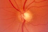 |
2-53a - Venous Pulsations | On the disc, look for spontaneous venous pulsations. Spontaneous venous pulsations can be seen in the large trunks of veins at the level of the disc margin. They are normally present and seen in 37-90% of normals -- depending on the experience of the examiner and the shape of the disc. The spontaneo... | Image |
| 157 |
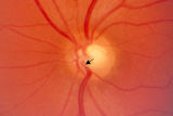 |
2-53b - Venous Pulsations | On the disc, look for spontaneous venous pulsations. Spontaneous venous pulsations can be seen in the large trunks of veins at the level of the disc margin. They are normally present and seen in 37-90% of normals -- depending on the experience of the examiner and the shape of the disc. The spontaneo... | Image |
| 158 |
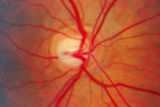 |
2-7a - Disc Anatomy | The optic disc appearance is determined by: the size of the eye, the size of the scleral canal, how the nerve is inserted into the globe, the appearance of the lamina cribrosa, where myelination stops, and what is left behind in normal development. Even though this is a disc with a very large cup, i... | Image |
| 159 |
 |
Spasmus Nutans | Example of patient with spasmus nutans. Discussion of characteristics of this disorder, such as dissociated or monocular nystagmus, abnormal head position, and to-and-fro head oscillation. Sometimes an eccentric gaze is seen as well (as in patient). Patient has a monocular horizontal nystagmus in th... | Image/MovingImage |
| 160 |
 |
Spiral and Stellate Visual Fields Non-physiologic Variants | Description of testing the spiral and stellate visual fields. | |
| 161 |
 |
Superior Oblique Myokymia | Example of patients with superior oblique myokymia, a saccadic intrusion. First patient is seen to have intermittent, intorting movements with superimposed slight vertical deviations in right eye. Discussion of disorder as benign, but frequently disabling, as patients experience episodes of diplopia... | Image/MovingImage |
| 162 |
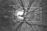 |
2-4a - Disc Anatomy | The optic disc appearance is determined by: the size of the eye, the size of the scleral canal, how the nerve is inserted into the globe, the appearance of the lamina cribrosa, where myelination stops, and what is left behind in normal development. Even though this is a disc with a very large cup, i... | Image |
| 163 |
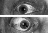 |
Aberrant Regeneration of the Right Pupil | Aberrant regeneration of the right pupil in a man with a large intracavernous sinus meningioma causing a pupil-involving, incomplete third cranial nerve palsy. His pupil is round when he gazes straight ahead (top). When he tries to rotate the eye medially, the pupil constricts, but a segment of the ... | Image |
| 164 |
 |
Before Tensilon | Example of patient with myasthenia gravis. Demonstration of baseline examination, followed by administration of 2mg of tensilon, which is a test dose. Procedure for administration of tensilon test is described, including variations. Patient is then shown after being given 4mg of tensilon, with very ... | Image/MovingImage |
| 165 |
 |
Clover-leaf Visual Field Defects | Description of clover-leaf visual field defects. | |
| 166 |
 |
Dilation Lag | Two examples of dilation lag (Horner's syndrome). In the first example, the right pupil dilates much faster than the left pupil when the light is turned out. In the second example, the left pupil dilates much faster than the right pupil when the light is turned out. Discussion of methods of document... | Image/MovingImage |
| 167 |
 |
Documenting the Neuro-ophthalmic Patient: External Photography | Description of documenting the neuro-ophthalmic patient using external photography. This covers pupils and extra ocular muscles. | |
| 168 |
 |
Duane's Retraction Syndrome Type 3 | Example of a patient with Type 3 Duane's Retraction Syndrome, as well as bilateral Duane's Syndrome. Shows limitation of abduction in both eyes and adduction in the left eye. Also shows side-view of globe retraction in abduction. | Image/MovingImage |
| 169 |
 |
Facial Myokymia Unilateral | Example of patient with facial myokymia, a disorder of the seventh nerve, probably due to brain stem involvement. Patient has multiple sclerosis. Discussion of characteristics, such as continuous, undulating, contractions in the distribution of the seventh nerve, and a spreading of these movements t... | Image/MovingImage |
| 170 |
 |
Hemifacial Spasm | Example of patients with hemifacial spasm. First patient has a sequela of Bell's palsy, and is seen to have mainly clonic movements around the eye, with occasional tonic movements around the mouth. Second patient has a cerebellopontine angle epidurmoid tumor, and is seen to have movements around the... | Image/MovingImage |
| 171 |
 |
How to Measure the RAPD | This clip demonstrates the examination technique for measuring the Relative Afferent Pupillary Defect (RAPD). Demonstration of balancing an afferent papillary defect using filters in a patient with a resolving optic neuritis and an afferent papillary defect on the left. | Image/MovingImage |
| 172 |
 |
Opsoclonus | Example of patients with opsoclonus, a saccadic abnormality. Discussion of characteristics of opsoclonus, such as involuntary, rapid, brief, random, conjugate saccades. Discussion of possible causes, including brain stem encephalitis (as in first patient), a paraneoplastic effect, tumors, and drug t... | Image/MovingImage |
| 173 |
 |
Parinaud's Syndrome | Two examples of patients with Parinaud's syndrome, a dorsal midbrain syndrome. Discussion of hallmarks of this syndrome, including convergence retraction nystagmus, vertical gaze palsies, light-near dissociation, and Collier's Sign. Discussion of age-dependent disorders associated with this syndrome... | Image/MovingImage |
| 174 |
 |
Spasm of the Near Reflex | Example of patient with spasm of the near reflex and voluntary nystagmus. Discussion of similar-looking conditions (e.g. six nerve palsy, limitation of abduction, lateral rectus muscle problems) and how to tell them apart from spasm of the near reflex by observing the myosis evoked by the near respo... | Image/MovingImage |
| 175 |
 |
Tunnel Vision on Tangent Screen Testing | Description of tunnel vision and tangent screen testing. |