John A. Moran Eye Center Neuro-Ophthalmology Collection: A variety of lectures, videos and images relating to topics in Neuro-Ophthalmology created by faculty at the Moran Eye Center, University of Utah, in Salt Lake City.
NOVEL: https://novel.utah.edu/
TO
Filters: Collection: "ehsl_novel_jmec"
| Title | Description | Type | ||
|---|---|---|---|---|
| 126 |
 |
Upbeat Nystagmus | Example of a patient with upbeat nystagmus. Shows vertical jerk nystagmus with fast phases in the up direction. Localizes to brain stem, and occurs with strokes, demyelination, and tumors. | Image/MovingImage |
| 127 |
 |
Vestibular Nystagmus | Discussion of vestibular nystagmus. Seen with peripheral disorders and central disorders, and in two varieties: spontaneous and positional. Horizontal jerk with small amplitude. | Image/MovingImage |
| 128 |
 |
Voluntary Nystagmus | Example of patient with voluntary nystagmus. Discussion of how a lack of uniform, patterned movement of the eyes along with associated lid movements suggests that activity is voluntary. | Image/MovingImage |
| 129 |
 |
Wall-Eyed Bilateral Internuclear Ophthalmoplegia (WEBINO) | Example of patient with horizontal binocular diplopia. Demonstration of exam, which shows alternating exotropia in cover test. As patient follows object, right eye does not pass the midline as the object moves to the left, while left eye go slightly past the midline, but does not abduct completely. ... | Image/MovingImage |
| 130 |
 |
Aberrant Regeneration of Third Nerve, Bilaterally (1 degree OD, 2 Digrees OS) | Example of patient with bilateral aberrancy of the third nerve. Shows lids popping up (synkinetic) with adduction. Patient had bilateral internal carotid artery aneurisms with third nerve compression. | Image/MovingImage |
| 131 |
 |
Convergence Retraction Nystagmus (Parinaud's Syndrome) | Examples of patients with convergence retraction nystagmus. Shows saccadic oscillations in patients looking upwards and following downwards moving targets. Also shows a side-view of the retracting movements of the globes. | Image/MovingImage |
| 132 |
|
Latent Nystagmus | Example of a patient with latent nystagmus. Demonstrates a lack of oscillations in forward gaze, followed by the occlusion of each eye, showing how this generates a jerking oscillation in the non-occluded eye away from the occluded eye. | Image/MovingImage |
| 133 |
 |
Levator Disinsertion | Example of patient with levator disinsertion, a lid disorder. Patient is pregnant and wears poorly fitting contacts. Discussion of characteristics, such as lid ptosis (shown in the left eye of patient), but with full levator function. | Image/MovingImage |
| 134 |
 |
Light-near Dissociation | Example of patient with Argyll Robertson pupil with neurosyphilis. Shows a lack of pupillary response to light and some pupillary response to nearness of finger. | Image/MovingImage |
| 135 |
 |
Ocular Flutter | Two examples of patients, the first with rotary, flutter-like movements, but not ocular flutter, and the second with genuine ocular flutter. Discussion of difference between ocular flutter and nystagmus, and how to elicit ocular flutter. | Image/MovingImage |
| 136 |
 |
Optic Disc: Anatomy, Variants, Unusual discs | Discussion of viewing the optic disc. Includes development of direct ophthalmoscope. Covers normal optic disc and nerve fiber; nerve fiber loss and defects; cilioretinal arteries; venous anomolies; papilledema; pseudopapilledema; myopic disc; hyperoptic disc; little red discs; megallopapilla; myelin... | Text |
| 137 |
 |
Paradoxical Constriction of Pupils to Darkness (Flynn Phenomenon) | Example of patients both with and without paradoxical constriction of pupils. Observed in many congenital retinal disorders, such as achromatopsia, congenital stationary night-blindness, and Leber's congenital amaurosis. Sometimes seen in optic nerve disorders, such as dominant optic atrophy. | Image/MovingImage |
| 138 |
 |
Periodic Alternating Nystagmus | Example of a patient with periodic alternating nystagmus, showing an alternation between left-beats and right-beats as the patient maintains forward gaze. Nystagmus maintain horizontal direction regardless of position of gaze. | Image/MovingImage |
| 139 |
|
Progressive Supranuclear Palsy | Example of patient with progressive supranuclear palsy. Discussion of difference between saccadic movement in supranuclear palsy and nystagmus. Shows saccadic intrusions in forward gaze, pursuit, saccades, and doll's head maneuver. | Image/MovingImage |
| 140 |
 |
Rebound Nystagmus | Example of a patient with rebound nystagmus, where the oscillations alternate direction as the patient shifts gaze in different directions. Discussion of relationship to disease and disorders of the cerebellum, including degenerations of the cerebellum, infarction, and demyelination. | Image/MovingImage |
| 141 |
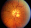 |
Shunt Vessel Meningioma | RETINO-CHOROIDAL (OPTO-CILIARY) COLLATERAL VESSELS: (also known as Retinal-choroidal venous collaterals, opticociliary veins or ciliary shunt vessels) Retino-choroidal collaterals are potential telangiectatic connections between the retina and choroidal circulation. Although sometimes called "shunts... | Image |
| 142 |
 |
The 3 Step Test: Looking for a 4th Nerve Palsy | Description of the three step test (3 step test) used when looking for a 4th nerve palsy. | Text |
| 143 |
 |
Aberrant Regeneration of the Seventh Nerve | Examples of patients with aberrant regeneration of the seventh nerve. First example is a patient with contractions around the mouth and dimpling, demonstrated with slow and rapid eye blinking. Second example shows contraction around nose with eye blink. | Image/MovingImage |
| 144 |
 |
Binocular Pendular Nystagmus | Example of a patient with binocular pendular nystagmus. Patient has somewhat dissociated nystagmus, with nystagmus seen more prominently in the left eye. Patient shows an occasional jerk nystagmus to the right in the right eye. Left eye oscillations are mostly pendular. | Image/MovingImage |
| 145 |
 |
Congenital Nystagmus | Example of patients with congenital nystagmus. First patient's nystagmus are mostly jerk and not pendular. Second patient's nystagmus are mostly pendular. Both patients show a uniform horizontal oscillation. Second patient also shows differences in frequency of oscillations depending on gaze, includ... | Image/MovingImage |
| 146 |
 |
Monocular Pendular Nystagmus | Example of a patient with monocular pendular nystagmus, with discussion of situations in which this condition is seen: acquired disorder of the visual-sensory pathway, and acquired disorder of the brain stem (e.g. multiple sclerosis). | Image/MovingImage |
| 147 |
 |
Normal Light Reflex without RAPD | This clip demonstrates the examination of the Relative Afferent Pupillary Defect (RAPD.) Demonstration of gauging the size of the pupil in light, testing light reflexes, swinging flashlight test for optic nerve abnormality. | Image/MovingImage |
| 148 |
|
Ocular Lateropulsion (Wallenberg's Syndrome) | Example of patient with ocular lateropulsion. Patient also has central Horner syndrome and nystagmus in right gaze. When shifting gaze back to forward, eyes overshoot their mark. Eyes laterally deviate to the right upon opening. | Image/MovingImage |
| 149 |
 |
Ocular Myasthenia | Example of patient with myasthenia gravis. Demonstration of tensilon test. Patient shown to have bilateral ptosis, bilateral duction deficits, and left hypertropia. Discussion of techniques to observe subtle changes, such as bringing in a neutral observer or taking still photographs. Shows split-scr... | Image/MovingImage |
| 150 |
 |
Sector Palsies and Light-Near Dissociation | Example of patient with bilateral Adie's pupils. Exam is performed with a slit-lamp. Shows iris stroma and focal segments of iris sphincter that retain their contractilty. Suggests post-ganglionic parasympathetic denervation. | Image/MovingImage |