John A. Moran Eye Center Neuro-Ophthalmology Collection: A variety of lectures, videos and images relating to topics in Neuro-Ophthalmology created by faculty at the Moran Eye Center, University of Utah, in Salt Lake City.
NOVEL: https://novel.utah.edu/
TO
Filters: Collection: "ehsl_novel_jmec"
| Title | Description | Type | ||
|---|---|---|---|---|
| 101 |
 |
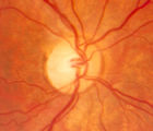
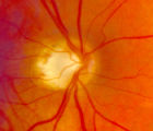
Normal Optic Disc | Overview of the structure and function of the normal optic disc. | Text |
| 102 |
 |
Optic Disc Pallor Pseudo and Real | Discussion of the causes of optic disc pallor. | Text |
| 103 |
 |
Papilledema 2013 | Discussion of papilledema, the swelling due to increased pressure. | Text |
| 104 |
 |
Pupil Exam | Demonstration of pupil examination. | Text |
| 105 |
 |
Retinitis Pigmentosa Disease of Rods | Discussion of retinitis pigmentosa which is a retinal/choroidal degeneration caused by various genetic defects. | Text |
| 106 |
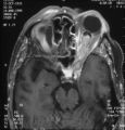 |
Shunt Vessel Meningioma - MRI | Meningiomas block venous egress and open potential venous channels known as retinochoroidal (optociliary) collateral vein. This meningioma extends from the back of the globe through the optic canal. | Image |
| 107 |
 |
Stages of Papilledema | Text | |
| 108 |
 |
Stereoacuity Testing | Demonstration of examination for stereoacuity. | Text |
| 109 |
 |
Superior Oblique Myokymia | Close-up video of a patient with superior oblique myokymia (no audio.) | Image/MovingImage |
| 110 |
 |
Tilted Discs | Short PowerPoint discussion of tilted discs with illustrations and images. | |
| 111 |
 |
Ultrasonography Techniques | This video describes and demonstrates the various techniques for examination of the eye using ultrasonography, including A-scan, B-scan and immersion. | Image/MovingImage |
| 112 |
 |
Wall-Eyed Bilateral Internuclear Ophthalmoplegia (WEBINO) | Example of patient with Wall-Eyed Bilateral Internuclear Ophthalmoplegia. Patient is led through instructions for direction and distance of gaze. | Image/MovingImage |
| 113 |
 |
Internuclear Ophthalmoplegia (2 Examples) | Two examples of patients with internuclear ophthalmoplegia. First patient has a right internuclear ophthalmoplegia. Patient had subacute bacterial endocarditis with a bacterial abscess in the brain stem. Ductions and gaze to the right look good, but when gazing to the left, the right eye does not ad... | Image/MovingImage |
| 114 |
 |
Third Nerve Palsy, Pupil Involving | Example of patient with third nerve palsy. Left eye shows pupilary involvement. Left eye doesn't immediately duct, but abducts well, with impaired superduction. Secondary and primary deviations are demonstrated. Anisocoria is more prominent when light is on, showing a parasympathetic defect to the p... | Image/MovingImage |
| 115 |
 |
Spasmus Nutans | Example of patient with spasmus nutans. Discussion of characteristics of this disorder, such as dissociated or monocular nystagmus, abnormal head position, and to-and-fro head oscillation. Sometimes an eccentric gaze is seen as well (as in patient). Patient has a monocular horizontal nystagmus in th... | Image/MovingImage |
| 116 |
 |
Superior Oblique Myokymia | Example of patients with superior oblique myokymia, a saccadic intrusion. First patient is seen to have intermittent, intorting movements with superimposed slight vertical deviations in right eye. Discussion of disorder as benign, but frequently disabling, as patients experience episodes of diplopia... | Image/MovingImage |
| 117 |
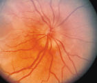 |
3-33b - Papilledema Stages | Grading Papilledema: Stage 2 = Elevation of the disc margin 360 degrees. Since the blood vessels at the disc margin are not swollen or obscured, this disc could be mistaken for pseudo-papilledema. | Image |
| 118 |
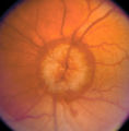 |
3-36a - Papilledema Stages | Grading Papilledema: Stage 5 Stage 5 = Dome-shaped appearance with all vessels being obscured. (Sometimes called "champagne cork" swelling--because of its dome shape.) | Image |
| 119 |
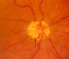 |
3-64a - Shunt Vessels (CRVO) | This man with a chronic CRVO and retino-choroidal collaterals developed AION and his collaterals disappeared. CRVO with retinochoroidal collaterals is almost always associated with multiple peripheral dot and blot hemorrhages as well as nerve fiber layer infarcts of various ages. Notice the retino-c... | Image |
| 120 |
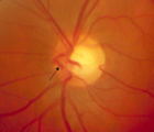 |
3-65 - Shunt Vessels (Glaucoma) | Chronic end-stage glaucoma produces high pressure that interferes with venous drainage from the disc and broad smooth venous collaterals drain the disc centrifugally to the disc margin where they drain. | Image |
| 121 |
 |
4-35 - Cupped Optic Nerve | Atrophic Glaucoma Atrophic glaucomatous discs show thinning of the neuro-retinal rim, "saucerization" (which is shallow cupping), evidence of peripapillary atrophy, and pallor of the very narrow neuroretinal rim. Notice that there is severe atrophy of the nerve fiber layer. | Image |
| 122 |
 |
4-52b - Dominant Optic Neuropathy | A son presented with bilateral optic atrophy of unknown etiology after he failed a school visual exam. When looking for dominant optic atrophy, look at the parents. Mother was examined to find similar kind of atrophy. 4-52a mother, 4-52b son. | Image |
| 123 |
 |
4-60a - Dominant Optic Neuropathy | A son presented with bilateral optic atrophy of unknown etiology after he failed a school visual exam. When looking for dominant optic atrophy, look at the parents. Mother was examined to find similar kind of atrophy. 4-60a mother, 4-60b son. | Image |
| 124 |
 |
Anterior Ischemic Optic Neuropathy | PPT describing Anterior Ischemic Optic Neuropathy (AION). Covers clinical signs, such as monocular vision loss, swollen nerve, and visual field defects, as well as risk factors. | Text |
| 125 |
 |
Bilateral Asynchronous Blepharospasm with Facial and Cervical Dystonia | Bilateral Asynchronous Blepharospasm with Facial and Cervical Dystonia. | Image/MovingImage |