John A. Moran Eye Center Neuro-Ophthalmology Collection: A variety of lectures, videos and images relating to topics in Neuro-Ophthalmology created by faculty at the Moran Eye Center, University of Utah, in Salt Lake City.
NOVEL: https://novel.utah.edu/
TO
Filters: Collection: "ehsl_novel_jmec"
| Title | Description | Type | ||
|---|---|---|---|---|
| 101 |
 |
Measuring Visual Acuity | Demonstration on self of visual acuity exam, using a standard card. | Image/MovingImage |
| 102 |
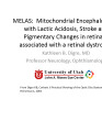 |
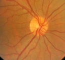
MELAS and RP | MELAS; Mitochondrial Encephalopathy with Lactic Acidosis, Stroke and Pigmentary Changes in retina-associated with a retinal dystrophy. This 53 year old man had seizures, encephalopathy and lactic acidosis typical of MELAS. His fundus examination showed granularity and some slight pigmentary changes ... | Text |
| 103 |
 |
Ocular Myotonia | Example of patient with ocular myotonia. Patient is led through instructions for direction of gaze and opening and closing of eyes. Right eye is shown to be stuck in position after held gaze to the left and right, with very slow relaxation back into forward gaze. | Image/MovingImage |
| 104 |
 |
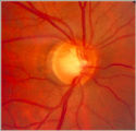
Tour of the Fundus | This clip demonstrates the funduscopic examination technique. | Image/MovingImage |
| 105 |
 |
3-31b - Papilledema Stages | Grading Papilledema: Stage 0 GRADING PAPILLEDEMA GRADING PAPILLEDEMA We grade papilledema in order to tell us how severe it is. The most sensible grading scheme has been provided by Lars Frisén. STAGE 0: This woman had documented increased intracranial pressure of 340 mm water. Very little if any ... | Image |
| 106 |
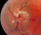 |
3-35a - Papilledema Stages | Grading Papilledema: Stage 4 Stage 4 = Complete obliteration of the cup and complete obscuration of at least some vessels on the surface of the disc. There may be small dilated capillaries on the disc that resemble telangiectasia. It is not the NFL infarcts or hemorrhages but the obscuration of the ... | Image |
| 107 |
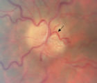 |
3-66a - Shunt Vessels (Post-papilledema) | The retino-choroidal collaterals seen with chronic papilledema begin with a "Hairnet" of telangiectasias that gradually winnow down to one or more large collateral tortuous draining channel. The presence of these vessels is evidence of long standing disc swelling. When the CSF pressure is lowered, t... | Image |
| 108 |
 |
3-66d - Shunt Vessels (Post-papilledema) | The retino-choroidal collaterals seen with chronic papilledema begin with a "Hairnet" of telangiectasias that gradually winnow down to one or more large collateral tortuous draining channel. The presence of these vessels is evidence of long standing disc swelling. When the CSF pressure is lowered, t... | Image |
| 109 |
 |
Abducting (Dissociated) Nystagmus | Example of a patient with abducting (dissociated) nystagmus. Patient has a subtle internuclear ophthalmoplegia. Right eye has right-beating jerk nystagmus, with smaller oscillations in the left eye. Disease/Diagnosis: Abducting Nystagmus | Image/MovingImage |
| 110 |
 |
Brun's Nystagmus | Observation of patient with Brun's Nystagmus. Shows patient gazing to the right and the nystagmus beating in the direction of the gaze. | Image/MovingImage |
| 111 |
 |
Cogan's Lid Twitch | Example of a patient with Cogan's lid twitch, with discussion of how to detect it in an exam. | Image/MovingImage |
| 112 |
 |
Congenital Ocular Motor Apraxia | Two examples of congenital ocular motor apraxia. Patients have trouble initiating saccades, and compensate with head movement. Discussion of how to distinguish this condition from simply not seeing well. | Image/MovingImage |
| 113 |
 |
Downbeat Nystagmus | Example of patients with downbeating jerk nystagmus. Demonstrates how oscillations grow more prominent when the patient gazes down or laterally. Discusses some causes, including Arnold-Chiari malformation, infarction, and demyelination. | Image/MovingImage |
| 114 |
 |
Fourth Nerve Palsy | Demonstration of examination of patient who experienced blurry vision and pain in the left eye. Demonstrates checking of eye movements, focusing on object while each eye is covered and uncovered, turning head both ways and repeating. Shows limitation of depression in adduction of left eye, left hype... | Image/MovingImage |
| 115 |
 |
How to Use the Direct Ophthalmoscope in an Exam | Demonstration of using the direct ophthalmoscope to examine the optic disc. Covers hand placement , which eye to use, and distance from patient. | Image/MovingImage |
| 116 |
 |
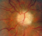
Notching of the Neuro-retinal Rim | The neuro-retinal rim becomes thinner; in particular the rim superotemporally and inferortemporally may develop a notch which is usually superior or inferior and rarely nasal or temporal. These notches are believed to be due to focal ischemic damage to the neuro-retinal rim. Glaucoma with Notching a... | Image |
| 117 |
 |
Physiologic (End-Gaze) Nystagmus | Demonstration of physiological nystagmus, where oscillations do not represent pathology, but occur when the patient's gaze is drawn too far laterally. | Image/MovingImage |
| 118 |
 |
Pulsating Exophthalmos | Example of a patient with neurofibromatosis with an absent sphenoid wing. Shows left eye pulsating back and forth with the pulse from front and side views. | Image/MovingImage |
| 119 |
 |
RAPD Present | This clip demonstrates the technique used to determine that Relative Afferent Pupillary Defect (RAPD) is present in a patient. | Image/MovingImage |
| 120 |
 |
Retino-choroidal Vessels or Optociliary Veins or Ciliary Shunt | Overview of retino-choroidal collaterals, which are potential telangiectatic connections between the retina and choroidal circulation. Although sometimes called "shunts", these collaterals are between the retinal venous circulation and the choroidal venous circulation. | Text |
| 121 |
|
Rotary Nystagmus | Example of a patient with rotary nystagmus, showing occasional counterclockwise rotary movements of both eyes. Seen more in intrinsic disorders of the brainstem. | Image/MovingImage |
| 122 |
 |
Square Wave Jerks | Example of patient with square wave jerks. Discussion of difference between square wave jerks (saccadic oscillations) and horizontal nystagmus. | Image/MovingImage |
| 123 |
 |
Testing the Visual Fields | Demonstration of various methods of testing visual fields, including counting fingers, motion, and color of several objects. | Image/MovingImage |
| 124 |
 |
Tour of the Direct Ophthalmoscope | This clip describes the parts and operation of the ophthalmoscope as an ocular examination tool. Includes adjustment of aperture size and adjustment of lenses. | Image/MovingImage |
| 125 |
|
Transillumination - Ciliary Body Neurofibromas | Example of transillumination on a patient with neurofibromatosis, but without Lisch nodules. Shows suspected neurofibromas in the ciliary body. | Image/MovingImage |