John A. Moran Eye Center Neuro-Ophthalmology Collection: A variety of lectures, videos and images relating to topics in Neuro-Ophthalmology created by faculty at the Moran Eye Center, University of Utah, in Salt Lake City.
NOVEL: https://novel.utah.edu/
TO
Filters: Collection: "ehsl_novel_jmec"
| Title | Description | Type | ||
|---|---|---|---|---|
| 201 |
 |
Spontaneous Venous Pulsations | This clips shows a spontaneous venous pulsation viewed during an ocular examination. | Image/MovingImage |
| 202 |
 |
Square Wave Jerks | Example of patient with square wave jerks. Discussion of difference between square wave jerks (saccadic oscillations) and horizontal nystagmus. | Image/MovingImage |
| 203 |
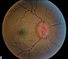 |
Stage 2 - Papilledema | Image | |
| 204 |
 |
Stages of Papilledema | Text | |
| 205 |
 |
Stargardt's Disease | Discussion of Stargardt's disease, an inherited maculopathy which frequently presents with a loss of central vision. | Text |
| 206 |
 |
Stereoacuity Testing | Demonstration of examination for stereoacuity. | Text |
| 207 |
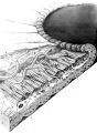 |
Structures of the iris | Structures of the iris. The a indicates the anterior border layer that terminates at the pigmentary ruff of the pupillary border (b). The c indicates the iris sphincter muscle, which is oriented circumferentially within the stroma and located deep to the anterior border layer; d indicates vessels th... | Image |
| 208 |
 |
Superior Oblique Myokymia | Close-up video of a patient with superior oblique myokymia (no audio.) | Image/MovingImage |
| 209 |
 |
Superior Oblique Myokymia | Example of patients with superior oblique myokymia, a saccadic intrusion. First patient is seen to have intermittent, intorting movements with superimposed slight vertical deviations in right eye. Discussion of disorder as benign, but frequently disabling, as patients experience episodes of diplopia... | Image/MovingImage |
| 210 |
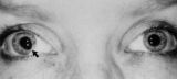 |
Tadpole-shaped Pupil | Tadpole-shaped pupil in a 20-year-old women with frequent episodes of blurred vision and achiness of the right eye lasting several minutes. The patient took a photograph of her eyes during an attack to document the peaked, segmental dilation of her right pupil (black arow). (Thompson HS, Zackon DH, ... | Image |
| 211 |
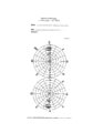 |
Tangent Screen Recording Chart | The tangent screen recording chart. | Text |
| 212 |
 |
Tangent Screen Testing Visual Field | Description of tangent screen testing. | Text |
| 213 |
 |
Test Duane | Image/MovingImage | |
| 214 |
 |
Testing the Visual Fields | Demonstration of various methods of testing visual fields, including counting fingers, motion, and color of several objects. | Image/MovingImage |
| 215 |
 |
Third Nerve Palsy | Patient with third nerve palsy (no audio) | Image/MovingImage |
| 216 |
 |
Third Nerve Palsy, Pupil Involving | Example of patient with third nerve palsy. Left eye shows pupilary involvement. Left eye doesn't immediately duct, but abducts well, with impaired superduction. Secondary and primary deviations are demonstrated. Anisocoria is more prominent when light is on, showing a parasympathetic defect to the p... | Image/MovingImage |
| 217 |
 |
Tilted Discs | Short PowerPoint discussion of tilted discs with illustrations and images. | Text |
| 218 |
 |
Tour of the Direct Ophthalmoscope | This clip describes the parts and operation of the ophthalmoscope as an ocular examination tool. Includes adjustment of aperture size and adjustment of lenses. | Image/MovingImage |
| 219 |
 |
Tour of the Fundus | This clip demonstrates the funduscopic examination technique. | Image/MovingImage |
| 220 |
 |
Transillumination - Ciliary Body Neurofibromas | Example of transillumination on a patient with neurofibromatosis, but without Lisch nodules. Shows suspected neurofibromas in the ciliary body. | Image/MovingImage |
| 221 |
|
Transillumination - Lisch Nodules | Demonstration of transillumination of the Lisch nodules on a patient with neurofibromatosis. Shows how Lisch nodules that were not very visible in slit-lamp examination are better seen with transillumination, which may therefore be useful in detecting Lisch nodules earlier in children where they are... | Image/MovingImage |
| 222 |
|
Transillumination Ocular Melanoma | Video describing condition. | Image/MovingImage |
| 223 |
 |
Trigeminal Nerve Exam | Explanation of a trigeminal nerve exam. | Image/MovingImage |
| 224 |
 |
Tunnel Vision on Tangent Screen Testing | Description of tunnel vision and tangent screen testing. | Text |
| 225 |
 |
Ultrasonography Techniques | This video describes and demonstrates the various techniques for examination of the eye using ultrasonography, including A-scan, B-scan and immersion. | Image/MovingImage |
| 226 |
 |
Ultrasonography: Immersion Technique | This video describes and demonstrates the immersion technique for examination of the eye using ultrasonography. | Image/MovingImage |
| 227 |
 |
Unilateral Blepharospasm | Example of patient with unilateral blepharospasm. | Image/MovingImage |
| 228 |
 |
Upbeat Nystagmus | Example of a patient with upbeat nystagmus. Shows vertical jerk nystagmus with fast phases in the up direction. Localizes to brain stem, and occurs with strokes, demyelination, and tumors. | Image/MovingImage |
| 229 |
 |
Vestibular Nystagmus | Example of patient with vestibular nystagmus. Patient is led through instructions for direction of gaze. Shown also with Frenzel goggles. | Image/MovingImage |
| 230 |
 |
Vestibular Nystagmus | Discussion of vestibular nystagmus. Seen with peripheral disorders and central disorders, and in two varieties: spontaneous and positional. Horizontal jerk with small amplitude. | Image/MovingImage |
| 231 |
 |
Visually Evoked Potentials | Detailed explanation of visually evoked potentials. The terms visually evoked potential (VEP), visually evoked response (VER) and visually evoked cortical potential (VECP) are equivalent. They refer to electrical potentials, initiated by brief visual stimuli, which are recorded from the scalp overl... | Text |
| 232 |
 |
Voluntary Nystagmus | Example of patient with voluntary nystagmus. Discussion of how a lack of uniform, patterned movement of the eyes along with associated lid movements suggests that activity is voluntary. | Image/MovingImage |
| 233 |
 |
Wall-Eyed Bilateral Internuclear Ophthalmoplegia (WEBINO) | Example of patient with horizontal binocular diplopia. Demonstration of exam, which shows alternating exotropia in cover test. As patient follows object, right eye does not pass the midline as the object moves to the left, while left eye go slightly past the midline, but does not abduct completely. ... | Image/MovingImage |
| 234 |
 |
Wall-Eyed Bilateral Internuclear Ophthalmoplegia (WEBINO) | Example of patient with Wall-Eyed Bilateral Internuclear Ophthalmoplegia. Patient is led through instructions for direction and distance of gaze. | Image/MovingImage |
| 235 |
 |
The Wall-Eyed Potato Farmer | Young man presenting with apparent episodic neurologic evants that initially was thought to be multiple sclerosis, but as time went on, he had progressive changes in his neurologic exam and in his imaging findings. Brain biopsy revealed Gliomatosis cerebri. Anatomy: Brain Stem; Pons; Midbrain. Patho... | Text |
| 236 |
 |
Why Don't You See Double? | This presentation was given at the Neurology Grand Rounds in Fall 2011 at the University of Utah. A number of Duane Syndrome cases are covered. Related video can be found in this collection at: Duane's Syndrome Type I: http://content.lib.utah.edu/u?/EHSL-Moran-Neuro-opth,130 Duane's Syndrome Type I... | Text |