John A. Moran Eye Center Neuro-Ophthalmology Collection: A variety of lectures, videos and images relating to topics in Neuro-Ophthalmology created by faculty at the Moran Eye Center, University of Utah, in Salt Lake City.
NOVEL: https://novel.utah.edu/
TO
| Title | Description | Type | ||
|---|---|---|---|---|
| 1 |
 |
Basal Encephaloceles | Text | |
| 2 |
 |
Mimics of Atrophy | Text | |
| 3 |
 |
Macula | Overview of the structure and viewing of the macula. | Text |
| 4 |
 |
Hydroxychloroquine Maculopathy (Plaquenil) | An overview of Chloroquine Maculopathy. | Text |
| 5 |
 |
Nutritional Amblyopia | Example of patient with amblyopia with nutritional causes. | Text |
| 6 |
 |
Optic Nerve Tumors Benign and Malignant | Discussion of optic nerve tumors including meningioma and glioma. | Text |
| 7 |
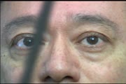
 |
Shaken Baby Syndrome | Text | |
| 8 |
 |
Stargardt's Disease | Discussion of Stargardt's disease, an inherited maculopathy which frequently presents with a loss of central vision. | Text |
| 9 |
 |
Amsler Grid Testing | Demonstration of Amsler Grid examination. | Text |
| 10 |
 |
Basic Headache | Presentation covering an overview of headache and migraine. | Text |
| 11 |
 |
Color Vision Testing | Demonstration of color vision examination. | Text |
| 12 |
 |
Cone Dystrophy | PPT covering Cone Dystrophy - An inherited degeneration that presents between 10 - 30 years of age. Symptoms are decreased visual acuity, poor color vision, and sometimes light sensitivity. | Text |
| 13 |
 |
Exophthalmometry | Demonstration of exophthalmometry examination. | Text |
| 14 |
 |
Leber's Hereditary Optic Neuropathy | Images and visual fields from a boy with acute visual loss. | Text |
| 15 |
 |
Normal Optic Disc | Overview of the structure and function of the normal optic disc. | Text |
| 16 |
 |
Optic Disc Pallor Pseudo and Real | Discussion of the causes of optic disc pallor. | Text |
| 17 |
 |
Papilledema 2013 | Discussion of papilledema, the swelling due to increased pressure. | Text |
| 18 |
 |
Pupil Exam | Demonstration of pupil examination. | Text |
| 19 |
 |
Retinitis Pigmentosa Disease of Rods | Discussion of retinitis pigmentosa which is a retinal/choroidal degeneration caused by various genetic defects. | Text |
| 20 |
 |
Stages of Papilledema | Text | |
| 21 |
 |
Stereoacuity Testing | Demonstration of examination for stereoacuity. | Text |
| 22 |
 |
Anterior Ischemic Optic Neuropathy | PPT describing Anterior Ischemic Optic Neuropathy (AION). Covers clinical signs, such as monocular vision loss, swollen nerve, and visual field defects, as well as risk factors. | Text |
| 23 |
 |
Fusional Vergence Amplitudes | Demonstration of fusional vergence amplitudes examination. Incluudes: a. Convergence Amplitudes b. Divergence Amplitudes c. Vertical Ampitudes | Text |
| 24 |
 |
Herpes Zoster Ophthalmicus with Third Nerve Palsy | Images showing presentation of Herpes Zoster (Zoster Ophthalmicus). | Text |
| 25 |
 |
Normal Eye Movements | This is an examination of a person with normal eye movements. Notice the patient has normal excursions. He has normal pursuit and saccades (horizontally and vertically). | Text |