|
|
Title | Date | Type |
| 1 |
 |
30 | 2021 | Image |
| 2 |
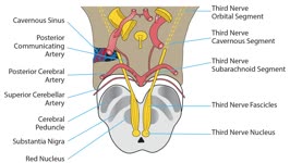 |
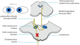
3rd Fascicle Lesion | 2021 | Image/MovingImage |
| 3 |
 |
3rd Superior Division Lesion Patient | 2021 | Image/MovingImage |
| 4 |
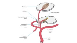 |
3rd Top of the Basilar | 2021 | Image/MovingImage |
| 5 |
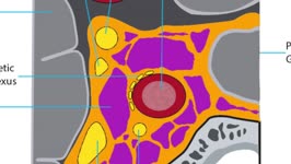 |
4th Nerve Cavernous Lesion | 2021 | Image/MovingImage |
| 6 |
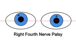 |
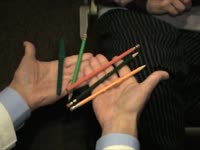
4th Nerve Palsy Blue Eyes | 2021 | Image/MovingImage |
| 7 |
|
4th Nerve Palsy Blue Eyes Plus Double Maddox Rod (animated) | 2021 | Image/MovingImage |
| 8 |
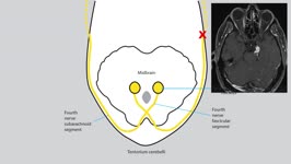 |
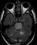
4th Tentorium Meningioma | 2021 | Image/MovingImage |
| 9 |
 |
6-16 | 2021 | Image |
| 10 |
 |
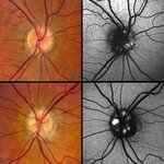
6-16 Non-Arteritic | 2021 | Image |
| 11 |
 |
6-4 Prime | 2021 | Image |
| 12 |
 |
6th Nerve Fasicle Lesion Plus Blue Eyes | 2021 | Image/MovingImage |
| 13 |
 |
6th Nerve Orbital Negative | 2021 | Image |
| 14 |
 |
80 Infiltrative Orbital Negative | 2021 | Image |
| 15 |
 |
81-b | 2021 | Image |
| 16 |
|
81-b Glaucoma | 2021 | Image |
| 17 |
|
81-b Glaucoma (again) | 2021 | Image |
| 18 |
 |
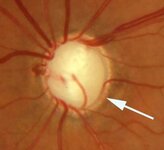
81-b Glaucoma with Arrow | 2021 | Image |
| 19 |
 |
81-b Glaucoma with Arrow (cropped) | 2021 | Image |
| 20 |
|
81-b Glaucoma with Arrow (cropped, again) | 2021 | Image |
| 21 |
 |
9-27B | 2021 | Image |
| 22 |
 |
9-27C | 2021 | Image |
| 23 |
 |
Absent Physiologic Cup (cropped) | 2021 | Image |
| 24 |
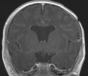 |
Absent Septum Pellucidum | 2021 | Image |
| 25 |
 |
Absent Septum Pellucidum with Arrow | 2021 | Image |
| 26 |
|
Absent Septum Pellucidum with Arrow (again) | 2021 | Image |
| 27 |
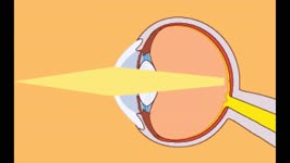 |
Accommodation | 2021 | Image/MovingImage |
| 28 |
|
Accommodation and Presbyopia | 2021 | Image/MovingImage |
| 29 |
 |
Acquired 4th Nerve Palsy | 2021 | Image/MovingImage |
| 30 |
 |
Acquired Brown Superior Oblique Tendon Sheath Syndrome | 2021-09 | Image/MovingImage |
| 31 |
 |
Acquired Ocular Motor Apraxia in Hypoxic-Ischemic Encephalopathy | 2021-09 | Image/MovingImage |
| 32 |
 |
Acquired Pendular Nystagmus | 2021-09 | Image/MovingImage |
| 33 |
 |
Acquired Pendular Nystagmus | 2021 | |
| 34 |
|
Acquired Pendular Nystagmus Fragment (Sept 2) | 2021 | Image/MovingImage |
| 35 |
 |
Acquired Right Fourth Nerve Palsy | 2021-09 | Image/MovingImage |
| 36 |
 |
Acute Comitant Esotropia | 2021-09 | Image/MovingImage |
| 37 |
 |
Acute Peripheral Vestibulopathy | 2021-09 | Image/MovingImage |
| 38 |
 |
Acute Peripheral Vestibulopathy | 2021 | |
| 39 |
 |
Acute Peripheral Vestibulopathy (converted) | 2021 | Image/MovingImage |
| 40 |
 |
Afferant LN Dissoc | 2021 | Image/MovingImage |
| 41 |
 |
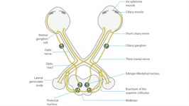
Afferant Parasympathetic Loop | 2021 | Image/MovingImage |
| 42 |
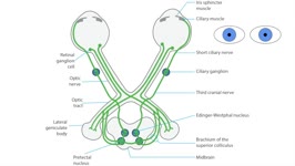
|
Afferant Parasympathetic Loop (revised) | 2021 | Image/MovingImage |
| 43 |
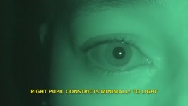 |
Afferent Light Near Dissociation | 2021-09 | Image/MovingImage |
| 44 |
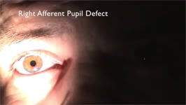 |
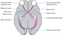
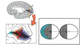
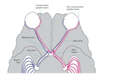
Afferent Pupil Defect (no audio) | 2021 | Image/MovingImage |
| 45 |
 |
Aicardi | 2021 | Image |
| 46 |
 |
Alternating Skew Deviation | 2021-09 | Image/MovingImage |
| 47 |
 |
Alternating Skew Deviation and Gaze Alignment | 2021-09 | Image/MovingImage |
| 48 |
 |
Altitudinal Disc Pallor (cropped) | 2021 | Image |
| 49 |
 |
Altitudinal Lesion | 2021 | Image/MovingImage |
| 50 |
 |
Altitudinal Lesion | 2021 | Image/MovingImage |
| 51 |
 |
Amsler Grid Testing | 2021-09 | Image/MovingImage |
| 52 |
 |
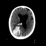
Aneurysm (CT) | 2021 | Image |
| 53 |
 |
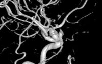
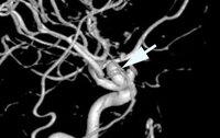
Aneurysm, Angio with Arrow | 2021 | Image |
| 54 |
 |
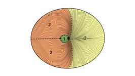
Animated Arcuate Bundle | 2021 | Image/MovingImage |
| 55 |
 |
Animated Arcuate Bundle | 2021-09 | Image/MovingImage |
| 56 |
 |
Animated MP Bundle | 2021 | Image/MovingImage |
| 57 |
 |
Animated MP Bundle | 2021 | Image/MovingImage |
| 58 |
|
Animated Nasal Radial Bundle | 2021 | Image/MovingImage |
| 59 |
 |
Anisocoria | 2021 | Image |
| 60 |
 |
Anisocoria Overview | 2021 | |
| 61 |
 |
Anisocoria with logo | 2021 | Image |
| 62 |
 |
Anomalous Vasculature Optic Disc (cropped) | 2021 | Image |
| 63 |
|
Anomalous Vasculature Optic Disc (cropped, again) | 2021 | Image |
| 64 |
 |
Anterior and Middle Visual Cortex Lesion With Field | 2021 | Image/MovingImage |
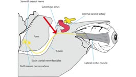
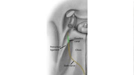
| 65 |
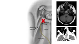
 |
Apraclonidine Bottle | 2021 | Image |
| 66 |
 |
Apraxia of Lid Opening (cropped) | 2021 | Image |
| 67 |
 |
Aqueductal Stenosis | 2021 | Image |
| 68 |
 |
Arteritic Ischemic Optic Neuropathy (OU) | 2021 | Image |
| 69 |
 |
Asymmetric Papilledema (OU) | 2021 | Image |
| 70 |
 |
Atrophic Disc | 2021 | Image |
| 71 |
 |
Atrophic Disc with Cataract (cropped) | 2021 | Image |
| 72 |
 |
Atrophic EOMs | 2021 | Image |
| 73 |
|
Atrophic EOMs with Arrows | 2021 | Image |
| 74 |
 |
Atrophic EOMs, CPEO, CT | 2021 | Image |
| 75 |
 |
Atrophic Papilledema | 2021 | Image |
| 76 |
 |
Autopsy Alzheimer | 2021 | Image |
| 77 |
 |
AV with Fistula Arrow | 2021 | Image |
| 78 |
 |
Axial Enhancing Optic Nerve (MRI) | 2021 | Image |
| 79 |
 |
Axial Enhancing Optic Nerve MRI with Arrow | 2021 | Image |
| 80 |
 |
Balint Biparietal Infarcts | 2021 | Image |
| 81 |
 |
Balint Hall | 2021 | Image/MovingImage |
| 82 |
 |
Balint-Holmes Syndrome | 2021-09 | Image/MovingImage |
| 83 |
 |
Bihemispheric Innervation | 2021 | Image |
| 84 |
 |
Bilateral 4th Nerve Trauma | 2021 | Image/MovingImage |
| 85 |
 |
Bilateral End Organ Lesion | 2021 | Image |
| 86 |
 |
Bilateral Hom | 2021 | Image/MovingImage |
| 87 |
 |
Bilateral Occipital Infarctions (CT) | 2021 | Image |
| 88 |
 |
Bilateral Opthalmoplegia (Third and Sixth Cranial Nerve Palsies) | 2021-09 | Image/MovingImage |
| 89 |
 |
Bilateral Orbital Inflamation | 2021 | Image |
| 90 |
 |
Bilateral Orbital Inflammation in GCA with Arrows | 2021 | Image |
| 91 |
 |
Bilateral Pontine Infarction | 2021 | Image |
| 92 |
 |
Bilateral Restricted Diffusion Optic Nerves | 2021 | Image |
| 93 |
 |
Bilateral Restricted Diffusion Optic Nerves with Arrows | 2021 | Image |
| 94 |
|
Bilateral Restricted Diffusion Optic Nerves with Arrows (new image) | 2021 | Image |
| 95 |
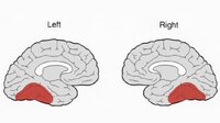 |
Bilateral Temporal Legion | 2021 | Image |
| 96 |
 |
Biparietal Abscesses Balint | 2021 | Image |
| 97 |
 |
Biparietal Infarcts (DWI) | 2021 | Image |
| 98 |
|
Biparietal Infarcts (DWI) 1 | 2021 | Image |
| 99 |
|
Biparietal Infarcts (DWI) 2 | 2021 | Image |
| 100 |
 |
Bitemporal - 1 | 2021 | Image |
| 101 |
 |
Bitemporal Hemorrhage | 2021 | Image |
| 102 |
 |
Bithalamic Infarcts with Arrows | 2021 | Image |
| 103 |
 |
Blepharospasm (cropped) | 2021 | Image |
| 104 |
 |
Blinking Fourth Nerve Cavernous Segment | 2022 | Image/MovingImage |
| 105 |
 |
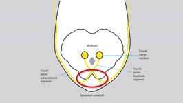
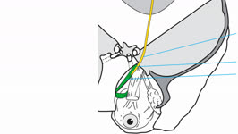
Blinking Fourth Nerve Fascicular Segment | 2022 | Image/MovingImage |
| 106 |
 |
Blinking Fourth Nerve Nucleus | 2022 | Image/MovingImage |
| 107 |
 |
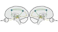
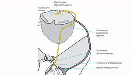
Blinking Fourth Nerve Pathway | 2022 | Image/MovingImage |
| 108 |
 |
Blinking Fourth Nerve Subarchnoid Segment | 2022 | Image/MovingImage |
| 109 |
 |
Blinking Fourth Orbital Segment | 2021 | Image/MovingImage |
| 110 |
 |
Blinking Superior Oblique Muscle | 2022 | Image/MovingImage |
| 111 |
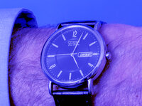 |
Blue | 2021 | Image |
| 112 |
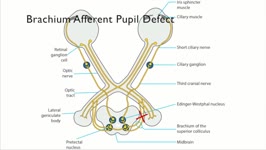 |
Brachium Lesion | 2021 | Image/MovingImage |
| 113 |
 |
Brain Hemorrhage | 2021 | Image |
| 114 |
 |
Carotid Aneurysm | 2021 | Image |
| 115 |
 |
Carotid Aneurysm 3D | 2021 | Image |
| 116 |
|
Carotid Aneurysm 3D (again) | 2021 | Image |
| 117 |
 |
Carotid Aneurysm with Arrow | 2021 | Image |
| 118 |
 |
Carotid Cavernous Fistula | 2021-09 | Image/MovingImage |
| 119 |
 |
Carotid Fasicula | 2021 | Image |
| 120 |
 |
Carotoid | 2021 | Image |
| 121 |
 |
Cartoon Downgaze | 2021 | Image/MovingImage |
| 122 |
 |
Cataract | 2021 | Image |
| 123 |
 |
Caudal Quadrigeminal Glioma with Arrow | 2021 | Image |
| 124 |
 |
Cavernoma Medulla with Arrow | 2021 | Image |
| 125 |
 |
Cavernous 6th Lesion | 2021 | Image |
| 126 |
 |
Cavernous Horner Syndrome | 2021 | Image/MovingImage |
| 127 |
 |
Cavernous Sixth Nerve Activated Coronal With Lesion | 2021 | Image/MovingImage |
| 128 |
 |
Cavernous Sympathetics Activated | 2021 | Image/MovingImage |
| 129 |
 |
Cerebellar Atrophy | 2021 | Image |
| 130 |
 |
Cerebellar Atrophy in SCA with Arrow (MRI) | 2021 | Image |
| 131 |
 |
Cerebellar Hem with Arrow | 2021 | Image |
| 132 |
 |
Cerebral Metamorphopsia | 2021 | Image |
| 133 |
 |
Cerebral Polyopia | 2021 | Image |
| 134 |
 |
Chiasmal Sorting | 2021 | Image/MovingImage |
| 135 |
 |
Choriorentinitis | 2021 | Image |
| 136 |
 |
Choriorentinitis (cropped) | 2021 | Image |
| 137 |
|
Choriorentinitis (cropped, again) | 2021 | Image |
| 138 |
 |
Chronic Orbital Myositis | 2021-09 | Image/MovingImage |
| 139 |
 |
Chronic Progressive External Ophthalmoplegia | 2021 | Image/MovingImage |
| 140 |
 |
Chronic Progressive External Ophthalmoplegia | 2021-09 | Image/MovingImage |
| 141 |
 |
Classic Hemisphereic Signs of MS (MRI) | 2021 | Image |
| 142 |
|
Classic Hemisphereic Signs of MS (MRI, again) | 2021 | Image |
| 143 |
 |
Clear Margins Elevated Optic Disc (cropped) | 2021 | Image |
| 144 |
 |
Close Up Relieving Diplopia (figure b) | 2021 | Image |
| 145 |
 |
Coarse Visual Snow | 2021 | Image |
| 146 |
 |
Coloboma (cropped) | 2021 | Image |
| 147 |
|
Coloboma (cropped, again) | 2021 | Image |
| 148 |
|
Coloboma -1 (cropped) | 2021 | Image |
| 149 |
 |
Coloboma Optic Disc | 2021 | Image |
| 150 |
 |
Coloboma with Arrow (cropped, new image) | 2021 | Image |
| 151 |
 |
COMA Hamm (MRI) | 2021 | Image |
| 152 |
|
COMA Hamm (MRI, again) | 2021 | Image |
| 153 |
 |
Comet Tail | 2021 | Image |
| 154 |
 |
Comitant and Incomitant Disorders of Ocular Alignment | 2021-08 | Image/MovingImage |
| 155 |
 |
Complete Right Internuclear Ophthalmoplegia | 2021-09 | Image/MovingImage |
| 156 |
 |
Completely Excavated Optic Disc Cropped | 2021 | Image |
| 157 |
 |
Compressive Optic Disc Pallor | 2021 | Image |
| 158 |
 |
Compressive Optic Neuropathy (MRI) | 2021 | Image |
| 159 |
 |
Confrontation | 2021-09 | Image/MovingImage |
| 160 |
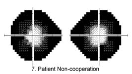 |
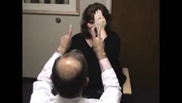
Constricted Fields of Deliberate Non-Cooperation | 2021-09 | Image/MovingImage |
| 161 |
 |
Convergence and Retraction in Dorsal Midbrain Syndrome | 2021-09 | Image/MovingImage |
| 162 |
 |
Convergence-Retraction Eye Movements | 2021-09 | Image/MovingImage |
| 163 |
 |
Coronal Atrophy (MRI, R, SOM) | 2021 | Image |
| 164 |
 |
CPEO (no audio) | 2021 | Image/MovingImage |
| 165 |
 |
Cranio Coronal with Arrow (MRI) | 2021 | Image |
| 166 |
 |
Cranio Cyst | 2021 | Image |
| 167 |
 |
Crescent Sparing (Sep 2) | 2021 | Image/MovingImage |
| 168 |
 |
CRVO | 2021 | Image |
| 169 |
 |
Cup - 2 | 2021 | Image |
| 170 |
 |
Cupless Optic Disc | 2021 | Image |
| 171 |
 |
Cupless Optic Disc (cropped) | 2021 | Image |
| 172 |
 |
De Morsier with Arrows (MRI) | 2021 | Image |
| 173 |
 |
Decompensated Left Superior Oblique Muscle Dysfunction | 2021-09 | Image/MovingImage |
| 174 |
 |
Decompensated SOM | 2021 | Image/MovingImage |
| 175 |
 |
Diabetic papillopathy | 2022 | |
| 176 |
 |
Diabetic Papillopathy (OU), a brighter image | 2021 | Image |
| 177 |
 |
Diabetic Papillopathy (OU), a brighter image with arrows | 2021 | Image |
| 178 |
 |
Dilated SOV Cortez with Arrow | 2021 | Image |
| 179 |
 |
Dim | 2021 | Image |
| 180 |
 |
Diplopia | 2021 | Image |
| 181 |
 |
Diplopia | 2021 | |
| 182 |
 |
Direct Fistula with Arrow (SOV) | 2021 | Image |
| 183 |
 |
Direct Fistula with Arrow on Cavernous Sinus | 2021 | Image |
| 184 |
 |
Direct Fistula with Arrow on Inferior Petrosal Sinus | 2021 | Image |
| 185 |
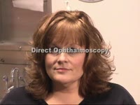 |
Direct Ophthalmoscopy | 2021-09 | Image/MovingImage |
| 186 |
 |
Displaced Pituitary Bright Spot | 2021 | Image |
| 187 |
 |
Displaced Pituitary Bright Spot with Arrow | 2021 | Image |
| 188 |
|
Displaced Pituitary Bright Spot with Arrow, again | 2021 | Image |
| 189 |
 |
Dissection | 2021 | Image |
| 190 |
|
Dissection MRA (MRI) | 2021 | Image |
| 191 |
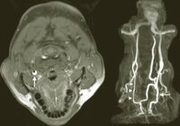 |
Dissection MRA (MRI), again | 2021 | Image |
| 192 |
 |
Dissociated Vertical Deviation | 2021-09 | Image/MovingImage |
| 193 |
 |
Distal Optic Nerve Lesion | 2021 | Image/MovingImage |
| 194 |
 |
Dominant Small Caliber | 2021 | Image |
| 195 |
 |
Dorellos Canal (animated) | 2021 | Image/MovingImage |
| 196 |
 |
Dorellos Canal Lesion | 2021 | Image/MovingImage |
| 197 |
 |
Dorellos Canal, Blinking | 2021 | Image/MovingImage |
| 198 |
 |
Dorsal Midbrain Lesion | 2021 | Image |
| 199 |
 |
Dorsal Midbrain Lesion, NMO with Arrow | 2021 | Image |
| 200 |
 |
Dorsal Midbrain Syndrome in CSF Shunt Malfunction | 2021-09 | Image/MovingImage |