The Emory Eye Center Neuro-Ophthalmology Collection contains a variety of lectures, videos and images relating to the discipline of neuro-ophthalmology created by faculty at Emory University in Atlanta, GA.
NOVEL: https://novel.utah.edu/
TO
Filters: Collection: "ehsl_novel_eec"
1 - 25 of 3
| Title | Description | Creator | ||
|---|---|---|---|---|
| 1 |
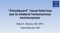 |
Checkboard Visual Field Loss Due to Bilateral Homonymous Hemianopsias | A 70-year-old man with vascular risk factors was seen for assessment of sudden visual field loss in both eyes. His examination showed: visual acuity: 20/20 OD and 20/25 OS; pupils: equal with no relative afferent pupillary defect; color vision: 14/14 plates correct OU; anterior segment exam: normal ... | Rahul A. Sharma, MD, MPH; Valérie Biousse, MD |
| 2 |
 |
Occipital Hemorrhagic Infarction Secondary to Bacterial Endocarditis-Congruent Homonymous Scotomatous Hemianopic Defect | MRI features of occipital hemorrhage secondary to bacterial endocarditis Figure 1 : Humphrey visual fields showing a congruent homonymous scotomatous hemianopic defect Figure 2 : axial gradient echo T2*w brain MRI Figure 3 : axial T1w and T2w brain MRI Figure 4 : axial postcontrast T1w brain MRI | Samuel Bidot, MD; Amit M. Saindane, MD; Valérie Biousse, MD |
| 3 |
 |
Retrograde Trans-Synaptic Degeneration from a Longstanding Occipital Lobe Tumor | This is an illustrated guide that (1) discusses the localization of paracentral homonymous hemianopic scotomatous visual field defects and (2) discusses the concept of trans-synaptic retrograde degeneration. A 43-year-old woman was assessed for longstanding blurred vision in both eyes. Her examinati... | Rahul A. Sharma, MD, MPH; Valérie Biousse, MD |
1 - 25 of 3