The Emory Eye Center Neuro-Ophthalmology Collection contains a variety of lectures, videos and images relating to the discipline of neuro-ophthalmology created by faculty at Emory University in Atlanta, GA.
NOVEL: https://novel.utah.edu/
TO
Filters: Collection: "ehsl_novel_eec"
| Title | Description | Creator | ||
|---|---|---|---|---|
| 1 |
 |
Vitreomacular Traction Syndrome (VMTS) | Vitreomacular traction (VMT) syndrome is characterized by an abnormal vitreous adherence to the macula due to incomplete posterior vitreous detachment (PVD). The resulting traction produces morphological changes of the macula. Patients may complain of decreased visual acuity, photopsia, micropsia, o... | Kirstyn Taylor; Sachin Kedar, MD |
| 2 |
 |
Vitreopapillary Traction Syndrome (VPT) | Vitreopapillary traction (VPT) is characterized by abnormal vitreous adherence to the optic disc due to a fibrocellular membrane or incomplete posterior vitreous detachment (PVD). Traction on the optic disc can result in elevation of the disc, blurred margins, and rarely peripapillary hemorrhage. Pa... | Kirstyn Taylor; Sachin Kedar, MD |
| 3 |
 |
Compressive Optic Neuropathy from Cavernous Hemangioma | A 42-year-old man was seen for assessment of progressive blurring of vision in his left eye (OS) over several weeks. His examination showed a left optic neuropathy: visual acuity: 20/20 OD and 20/20-2 OS; pupils: 1+ left relative afferent pupillary defect; color vision: 14/14 plates correct OU. Ther... | Rahul A. Sharma, MD, MPH; Aaron M. Yeung, MD; Valérie Biousse, MD |
| 4 |
 |
Giant Cell Arteritis: Temporal Artery Anatomy and Histology | Gross anatomy and histology of the normal superficial temporal artery.; Histopathology of the superficial temporal artery involved by active and healed GCA; Summary of the main histopathologic findings in GCA | Samuel Bidot, MD; Valérie Biousse, MD |
| 5 |
 |
Anatomy of the Orbits on CT and MRI | MRI and CT imaging of the orbit. | Valérie Biousse, MD |
| 6 |
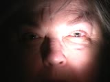 |
Left RAPD | Video clip displaying pupillary examination and RAPD measurement. | Valérie Biousse, MD |
| 7 |
 |
Enophthalmos from Breast Cancer Metastasis to the Orbit | Right painful ophthalmoplegia with right enophthalmos secondary to breast cancer metastasis to the right orbit. | Valérie Biousse, MD |
| 8 |
 |
Ophthalmic Artery Aneurysm | Slideshow describing ophthalmic artery aneurysm with MRI imaging. | Valérie Biousse, MD |
| 9 |
 |
Right Lateral Mediullary Syndrome (Wallenberg Syndrome) With Lateropulsion and Ocular Tilt Reaction | A 55-year old man presented with acute onset right-sided facial numbness, left-sided body numbness, vertigo, right ptosis, and binocular vertical diplopia. External examination showed right ptosis and miosis indicating a right Horner syndrome (Figure 1). He had gaze-evoked nystagmus only on right g... | Jonathan A. Micieli, MD; Valérie Biousse, MD |
| 10 |
 |
Malignant Hypertension With Bilateral Optic Nerve Edema | This is a case of malignant hypertension and severe hypertensive retinopathy. A 30-year-old woman with headache and vision loss in the left eye was found to have a markedly elevated blood pressure of 205/100. CT head without contrast showed acute hemorrhage in the right temporal-occipital junction a... | Rahul A. Sharma, MD, MPH; Michael Dattilo, MD, PhD; Valérie Biousse, MD |
| 11 |
 |
Toxoplasmic Chorioretinitis with Unilateral Disc Edema | A 53-year-old man had a history of high myopia and a seronegative spondyloarthropathy treated with immunosuppressive agents. He presented with mild, painless vision loss in his right eye. His examination showed findings of a right anterior optic neuropathy: visual acuity: 20/20 OD (right eye), 20/20... | Rahul A. Sharma, MD, MPH; Nancy J. Newman, MD; Valérie Biousse, MD |
| 12 |
 |
Occipital Infarction with Incomplete Congruent Homonymous Hemianopia | CT appearance of a remote occipital infarction. Congruent homonymous hemianopia. | Samuel Bidot, MD; Amit M. Saindane, MD; Valérie Biousse, MD |
| 13 |
 |
Checkboard Visual Field Loss Due to Bilateral Homonymous Hemianopsias | A 70-year-old man with vascular risk factors was seen for assessment of sudden visual field loss in both eyes. His examination showed: visual acuity: 20/20 OD and 20/25 OS; pupils: equal with no relative afferent pupillary defect; color vision: 14/14 plates correct OU; anterior segment exam: normal ... | Rahul A. Sharma, MD, MPH; Valérie Biousse, MD |
| 14 |
 |
Neurosarcoidosis | This is an illustrated guide to the clinical diagnosis of neurosarcoidosis. Sarcoidosis is a chronic systemic inflammatory disorder characterized by non-caseating granulomas. It can affect multiple organ systems including the lungs, skin, orbit, and brain. When there is central nervous system (CNS) ... | Bryce Buchowicz, MD; Valérie Biousse, MD |
| 15 |
 |
Vitreopapillary Traction | A 64-year-old woman was referred for bilateral optic disc edema. Examination of her optic nerves showed indistinct margins at the nasal aspect of both eyes (Figure 1). Humphrey 24-2 SITA-Fast visual fields showed non-specific depressed points in both eyes (Figure 2). Optical coherence tomography (... | Jonathan A. Micieli, MD; Valérie Biousse, MD |
| 16 |
 |
Clinical Features of Neuroretinitis | A 13-year-old girl was seen for assessment of blurred vision and optic disc edema in her right eye. Her examination showed: best-corrected visual acuity of hand motion OD and 20/25 OS; pupils: no relative afferent pupil defect; color vision: 0/14 plates OD and 14/14 plates OS; humphrey visual fields... | Rahul A. Sharma, MD, MPH; Jason H. Peragallo, MD; Valérie Biousse, MD |
| 17 |
 |
Superior Segmental Optic Nerve Hypoplasia | This is a case of superior segmental optic nerve hypoplasia in a woman with a history of maternal diabetes. A 25 year-old woman noticed a visual field defect in her right eye. Her examination showed: visual acuity: 20/20 OD, 20/20 OS; pupils: trace relative afferent pupillary defect OD; color visi... | Naa Naamuah M. Tagoe, MBChB, FWACS, FGCS; Rahul A. Sharma, MD, MPH; Valérie Biousse, MD; Nancy J. Newman, MD |
| 18 |
 |
Iris Transillumination | Single case of iris transillumination in a patient with albinism. Figure 1 : Anterior segment photograph demonstrating reddish hue to iris in albinism Figure 2 : Slit lamp photograph with retroillumination demonstrating iris transillumination Figure 3 : Slit lamp photograph with retroillumination... | Jason Peragallo, MD; Valérie Biousse, MD |
| 19 |
 |
Ocular Fundus Examination | Review of various techniques of ocular fundus examination, including direct ophthalmoscopy, binocular indirect ophthalmoscopy, slit lamp binocular indirect ophthalmoscopy, and fundus photography. Advantages and disadvantages of each technique are discussed. | Devin D. Mackay, MD; Valérie Biousse, MD |
| 20 |
 |
Pulsatile Proptosis Profile | See the PowerPoint description for Pulsatile Proptosis From Sphenoid Wing Hypoplasia in Neurofibromatosis Type 1 at: http://content.lib.utah.edu/cdm/ref/collection/ehsl-eec/id/64 See also Pulsatile Proptosis Full Face video: http://content.lib.utah.edu/cdm/ref/collection/ehsl-eec/id/14 | Valérie Biousse, MD |
| 21 |
 |
Large Right Hypophyseal Aneurysm Causing a Junctional Scotoma | Right, multi-lobulated superior hypophyseal artery aneurysm measuring 1.6 x 1.2 x 2.2 cm with 6 mm neck causing a right junctional scotoma . Images from a brain CT with contrast, a brain CT angiography with contrast, cerebral angiogram, Humphrey visual fields and ocular fundus photographs are includ... | Laurel N. Vuong, MD; Valérie Biousse, MD |
| 22 |
 |
Rathke's Cleft Cyst Apoplexy with Junctional Scotoma | MRI features of Rathke's cleft cyst apoplexy. - Figure 1 : Humphrey visual fields at initial presentation - Figure 2 : Brain MRI without contrast at initial presentation - Figure 3 : Brain MRI with contrast at initial presentation - Figure 4 : Postoperative Humphrey visual fields | Samuel Bidot, MD; Amit M. Saindane, MD; Valérie Biousse, MD |
| 23 |
 |
Classic Pathology Findings in Giant Cell Arteritis | An 80-year-old Caucasian woman presented with a 10 day history of headaches, intermittent binocular diplopia, and jaw pain. Temporal artery biopsy confirmed a diagnosis of giant cell arteritis. Pathology findings were classic for giant cell arteritis with numerous inflammatory cells in the tunica me... | Andre Aung, MD; Corrina Azarcon, MD; Wael A. Alsakran, MD; Valérie Biousse, MD |
| 24 |
 |
Myelin Oligodendrocyte Glycoprotein (MOG) - Antibody Optic Neuritis | This is an illustrated guide to the clinical diagnosis of myelin oligodendrocyte glycoprotein (MOG)-antibody optic neuritis. Myelin oligodendrocyte glycoprotein (MOG) is a glycoprotein on the surface of myelin and is found exclusively in the central nervous system (CNS). MOG likely mediates a comple... | Bryce Buchowicz, MD; Valérie Biousse, MD |
| 25 |
 |
Optic Disc Pseudoedema Due to Leber Hereditary Optic Neuropathy | A 23-year-old woman developed sequential painless central vision loss in both eyes (right eye 5 months ago and left eye 2 months ago). Her examination showed bilateral optic neuropathies: visual acuity: 20/300 eccentrically OU (no improvement with pinhole); pupils: equal and reactive with no relativ... | Rahul A. Sharma, MD, MPH; Valérie Biousse, MD |
