The Health Education Assets Library (HEAL) is a collection of over 22,000 freely available digital materials for health sciences education. The collection is now housed at the University of Utah J. Willard Marriott Digital Library.
TO
Filters: Collection: "ehsl_heal"
1 - 25 of 9
| Title | Description | Subject | Collection | ||
|---|---|---|---|---|---|
| 1 |
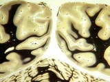 | Visual cortex or calcarine cortex | Visual cortex or calcarine cortex. At the level of the occiput, close up - afip series. Coronal plane. Photograph. Multimedia. | Brain; Visual cortex; Central nervous system; Anatomy | Slice of Life |
| 2 |
| Visual cortex or calcarine cortex | Visual cortex or calcarine cortex. At the level of the occiput, close up - afip series. Coronal plane. Photograph. Multimedia. | Visual cortex; Brain; Central nervous system; Anatomy | Slice of Life |
| 3 |
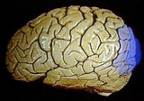 | Visual cortex, areas 17,18,19 | Visual cortex, areas 17,18,19. Lateral occipital gyri, graphic overlay on lateral hemisphere primary and association visual cortex. Photograph. Multimedia. | Visual cortex; Brain; Occipital lobe; Central nervous system; Anatomy | Slice of Life |
| 4 |
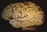 | Visual cortex, primary | Visual cortex, primary. Graphic overlay, lateral surface, occipital pole. Photograph. Multimedia. | Visual cortex; Brain; Central nervous system; Anatomy | Slice of Life |
| 5 |
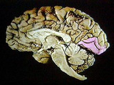 | Visual cortex, primary | Visual cortex, primary. Graphic overlay. Sagittal plane. Photograph. Multimedia. | Visual Cortex; Brain; Central Nervous System; Anatomy | Slice of Life |
| 6 |
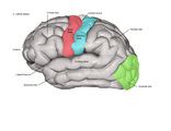 | Sensory Cortex, Visual Cortex, Auditory Cortex and Motor Cortex - Lateral (Labeled) | Sensory, visual, auditory, and motor cortices. | Central Sulcus; Sensory Area; Lateral Fissure; Auditory Area | Royal College of Surgeons in Ireland Illustrations |
| 7 |
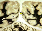 | Calcarine fissure, line of gennari visible, striate cortex | Calcarine fissure, line of gennari visible, striate cortex. AFIP series. Coronal plane. Photograph. Multimedia. | Visual cortex; Brain; Central nervous system; Anatomy | Slice of Life |
| 8 |
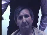 | Cranial Nerve Exam: Abnormal Examples: Cranial Nerve 2 - Visual Fields | The patient's visual fields are being tested with gross confrontation. A right sided visual field deficit for both eyes is shown. This is a right hemianopia from a lesion behind the optic chiasm involving the left optic tract, radiation or striate cortex. NeuroLogic Exam has been supported by a gran... | Cranial Nerve Examination | NeuroLogic Exam: An Anatomical Approach |
| 9 |
| Cranial Nerve Exam: Abnormal Examples: Cranial Nerve 2 - Visual Fields (x2) | The patient's visual fields are being tested with gross confrontation. A right sided visual field deficit for both eyes is shown. This is a right hemianopia from a lesion behind the optic chiasm involving the left optic tract, radiation or striate cortex. NeuroLogic Exam has been supported by a gran... | Cranial Nerve Examination | NeuroLogic Exam: An Anatomical Approach |
1 - 25 of 9