The Health Education Assets Library (HEAL) is a collection of over 22,000 freely available digital materials for health sciences education. The collection is now housed at the University of Utah J. Willard Marriott Digital Library.
Filters: Collection:"ehsl_heal"
| Title | Description | Subject | Collection | ||
|---|---|---|---|---|---|
| 1 | 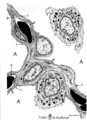 | Air-blood barrier in the lung (mammals) | Scheme electron microscopy. (1, ↓) Represents type I pneumocytes lining alveolar spaces (A). Cell (2) represents a free alveolar macrophage. The type II pneumocyte (3) is adherent to type I pneumocyte extensions (note junctional connection), and contains multilamellar bodies (surfactant). A myofib... | Pneumocyte type I ; Pneumocyte type II | Poja Histology Collection - Respiratory System Subset |
| 2 | 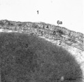 | The air-blood barrier in the lung (rat) | Electron microscopy (high magnification). The alveolar space (1) and the capillary space (2) (with part of an erythrocyte, 3) are separated from each other respectively by cytoplasm of endothelial cell (5), the common basal lamina (6) with a lamina densa (6a) and the type I alveolar cell (4, pneumo... | Pneumocyte I; Alveolar cell type I | Poja Histology Collection - Respiratory System Subset |
| 3 |  | Alveolar cell type II (pneumocyte II) in an alveolus (dog) | Electron microscopy. At (X) the alveolar space, the bulging alveolar cell type II shows the characteristic multilamellar bodies (*, cytosomes) that contain precusor material of surfactant. The lamellar bodies are responsible for the vacuolated appearance of these cells, and they give rise to surfact... | Pneumocyte II; Alveolar cell type II | Poja Histology Collection - Respiratory System Subset |
| 4 | 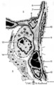 | Alveolar cells in the lung (mammals) | Scheme electron microscopy. (5) alveolar space; (6) type I Pneumocyte; (7) basal lamina; (8) myofibroblast; (9) collagen and elastin fibers; (10) mesothelial cell of the visceral pleura; (11) capillary with erythrocyte; (12) endothelial cell lining the capillary; (13) type II pneum... | Pneumocyte type I ; Pneumocyte type I I | Poja Histology Collection - Respiratory System Subset |
| 5 | 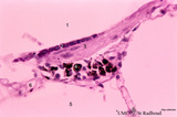 | Alveolar duct in the lung (mouse) | Stain: PAS and hematoxylin. Part of an alveolar duct lumen (1) that shows bronchiolar characteristics such as cuboidal epithelium (2) covering a bundle of smooth muscle (3) and connective tissue containing macrophages (4) with black pigment deposits. Note PAS-positivity of these cuboidal cells (Clar... | Alveolar ducts; Clara cells | Poja Histology Collection - Respiratory System Subset |
| 6 |  | Alveolar macrophage in lung (rat) | Electron microscopy. A wandering alveolar macrophage (1) migrates through an alveolar pore from an alveolar space (A1) into another (A2). Note the dark lysosomal structures and abundant organelles in its cytoplasm. The thin lining type I alveolar cell is hardly discernable (thin arrows →). Interst... | Pneumocyte I; Alveolar macrophages | Poja Histology Collection - Respiratory System Subset |
| 7 | 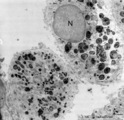 | Alveolar macrophages in lung (human, adult) | Electron microscopy. Two macrophages in the alveolar space (N = nucleus). Note the heterogeneity of many lysosomal structures containing among others carbon particles and the abundancy of organelles. | Alveolar macrophages | Poja Histology Collection - Respiratory System Subset |
| 8 | 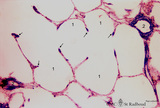 | Alveolar sac in the lung (human) | Stain: Azan. Characteristic alveolar tips (arrows) of neighbouring thin-walled alveoli (1). The tips contain elastin masked by collagen (blue-stained). At (2) small pulmonary arteries. | Alveolar tips | Poja Histology Collection - Respiratory System Subset |
| 9 | 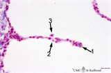 | Alveolar septum in the lung (human, high magnification) | Stain: Azan. Two neighboring alveoli separated by a septum. (1) points to the tip containing bluish-pink elastin and collagen. The nucleus of a squamous alveolar cell (type I pneumocyte) is indicated by (2), and a free alveolar macrophage by (3). | Alveolar tip; Pneumocyte I | Poja Histology Collection - Respiratory System Subset |
| 10 | 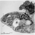 | The alveolar tip in lung tissue (human, adult) | Electron microscopy. The lung tip is covered by a flattened alveolar cell type I (↓1) and a similar neighboring bulging one with nucleus (2). In the alveolar tip amorph elastin (5) appears more electron-dense than the collagen fibers (Col) and is interwoven with it. Small foci of calcifications a... | Alveolar tip; Elastoblast | Poja Histology Collection - Respiratory System Subset |
| 11 | 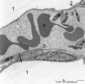 | The alveolus and air-blood barrier in the lung (rat) | Electron microscopy. The alveolus (1) is lined by a thin extension (2) of the alveolar epithelial cell type I (2), the pneumocyte I and the thin endothelium (3) of the capillary filled with erythrocytes (4) and blood platelets (5). The thin-walled air-blood barrier (↔) consists of the transition f... | Pneumocyte I; Alveolar cell type I | Poja Histology Collection - Respiratory System Subset |
| 12 | 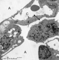 | Alveolus in lung (dog) | Electron microscopy. (A) indicate alveolar space; (C) indicate capillary with erythrocyte. Drawing-pin represents endothelium. (1) type I alveolar cell; (↓) indicate thin cytoplasm of pneumocyte I. (2) type II alveolar cell. (3) alveolar macrophage. (*) are spots of elastin, intermingled with coll... | Pneumocyte I; Pneumocyte II; Alveolar cells; Alveolar macrophage | Poja Histology Collection - Respiratory System Subset |
| 13 | 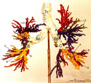 | Arbor bronchialis, the bronchial tree (human, adult) | Resin corrosion cast of the lower trachea and bronchial tree (posterior aspect). The lobar and segmental bronchi and their main branches are coloured, different colours indicate areas supplied by different segmental bronchi. White-coloured lower trachea (Tr) divides into two principal bronchi (Bp)... | Segmental bronchi ; Macroscopy | Poja Histology Collection - Respiratory System Subset |
| 14 | 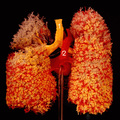 | The bronchial tree and branches of the pulmonary artery (human, adult, posterior aspect) | Resin corrosion cast of left and right lung. Posterior aspect of lower trachea (1, yellow) with two principal bronchi (yellow) with red-stained pulmonary artery (2) and its branching. The cast of both lung lobes reveals especially the intricate divisions and branching of the bronchial tree (yellow) ... | Macroscopy | Poja Histology Collection - Respiratory System Subset |
| 15 | 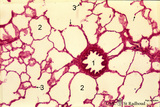 | Bronchiolus and alveoli in lung (human, adult) | Stain: Hematoxylin and eosin. (1) indicates a bronchiolus with respiratory epithelium and muscle fibers in its wall. Alveolar spaces (2) are separated by alveolar cell types that lined thin septa with capillaries. The septa end in tips and knobs (3). | Bronchiolus; Alveolar septa; Pneumocyte I; Pneumocyte II; Alveolar cell types | Poja Histology Collection - Respiratory System Subset |
| 16 | 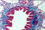 | Bronchiolus in the lung (human, adult) | Stain: Azan. The lumen of the bronchiolus is lined with one layer of ciliated epithelium (1) and is folded due to the contraction of smooth muscle fibers (2) in the wall. Note the rich cellularity (3) in the stromal surrounding of the bronchiolus. Alveolar space (4). (5) pulmonary artery branches. | Ciliated epithelium; Bronchiolus | Poja Histology Collection - Respiratory System Subset |
| 17 |  | Bronchiolus in the lung (human, adult) | Stain: Azan. The lumen is lined with one layer of ciliated epithelium (1) without goblet cells. Patches of smooth muscle fibers (2) are present. Notice small bronchial arteries (*). Arrows (↓) indicate sites of carbon accumulations between the alveoli (3). | Bronchiolus; Ciliated epithelium | Poja Histology Collection - Respiratory System Subset |
| 18 | 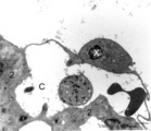 | Brush cell in lung (rat) | Electron microscopy. In the proximal as well as in the terminal airways a special type of cell the so-called brush(-border) cell could be observed. In this picture this cell (X) is located in the alveolar space. At (*) the brush border, the dense cytoplasm contains many organelles. (C) is a capillar... | Brush cells | Poja Histology Collection - Respiratory System Subset |
| 19 | 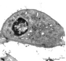 | Brush cell in lung (rat) | Electron microscopy. In the proximal as well as in the terminal airways a special type of cell can be observed, the so-called brush (-border) cell. In this picture this cell is located in the alveolar space. At (1) the brush border of thick plump microvilli. In the cytoplasm a Gogi area, mitochondri... | Brush cell | Poja Histology Collection - Respiratory System Subset |
| 20 | 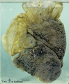 | Bullous emphysema of the lung (honeycomb lung, human, adult) | Macroscopy of the left lung showing large apical bullae (3) due to total destruction of all alveoli leaving remnants of the covering pleura. (2) indicates small bullae of foci of destroyed lobules with permanent enlargement of the air spaces and (1) points to spots of anthracosis in less-affected lu... | Macroscopy; Honeycomb lung; Interstitial fibrosis; Hamman-Rich Syndrome | Poja Histology Collection - Respiratory System Subset |
| 21 | 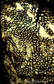 | Capillary system of a lung alveolus (cat) | Stain: specimen injected with India ink via the pulmonary system. In an en face view of the alveolar wall the black- and granular-stained capillary plexus is well shown. The larger vessels represent branches of the pulmonary arteriole. | Poja Histology Collection - Respiratory System Subset | |
| 22 | 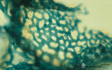 | Capillary system of an alveolus (cat) | Stain: specimen injected with trypan blue in gelatin via the pulmonary system. In a tangential view the blue-stained dense capillary plexus of one alveolus is well shown. | Poja Histology Collection - Respiratory System Subset | |
| 23 | 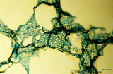 | Capillary system of lung alveoli (cat) | Stain: specimen injected with trypan blue in gelatin via the pulmonary system. In a tangential view the blue-stained dense capillary plexus surrounding each alveolus is well depicted. | Poja Histology Collection - Respiratory System Subset | |
| 24 | 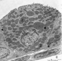 | Clara cell in lung (gerbil) | Electron microscopy. In this species a non-ciliated Clara cell (1 = nucleus) contains numerous electron-gray secretion granules (2) of varying sizes, smaller more dense granules tend to localize in the apex of the cell. Between the granules agranular endoplasmic reticulum. Irregular blunt microvill... | Respiratory bronchiolus ; Clara cells; Secretory granules | Poja Histology Collection - Respiratory System Subset |
| 25 | 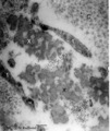 | Detail of elastin in an alveolar tip in lung tissue (human, adult) | Electron microscopy. Note the lace-like pattern of lumps of amorph elastin (X) that appears more electron-dense than the collagen fibers (C). At this high magnification cross-sectioned microfibrils (that contain among others fibrillin) are present in close association (*) with different areas of ela... | Elastin-associated proteins; Elastoblast; Myofibroblast | Poja Histology Collection - Respiratory System Subset |