The Health Education Assets Library (HEAL) is a collection of over 22,000 freely available digital materials for health sciences education. The collection is now housed at the University of Utah J. Willard Marriott Digital Library.
TO
Filters: Collection: "ehsl_heal"
| Title | Description | Subject | Collection | ||
|---|---|---|---|---|---|
| 1 |
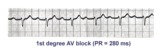 | 1st degree AV block | The normal PR interval is 0.12 - 0.20 sec, or 120 -to- 200 ms. 1st degree AV block is defined by PR intervals greater than 200 ms. This may be caused by drugs, such as digoxin; excessive vagal tone; ischemia; or intrinsic disease in the AV junction or bundle branch system. | Knowledge Weavers ECG | |
| 2 |
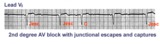 | 2nd degree AV block with junctional escapes and captures | Second degree AV block is present; conducted beats are identified by those QRS's that terminate shorter cycles than the junctional escape cycle; i.e., the 3rd and probably the 4th QRS's are captures; the other QRS's are junctional escapes. | Knowledge Weavers ECG | |
| 3 |
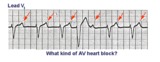
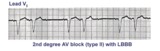 | 2nd degree AV block, mobitz type II, with LBBB | The wide QRS complexes in lead V1 indicates LBBB. 2nd degree AV block, Mobitz II is suggested by the two fixed PR intervals prior to the nonconducted P wave. The location of the block is most likely in the right bundle, because Mobitz II is usually a sign of bilateral bundle branch disease. | Knowledge Weavers ECG | |
| 4 |
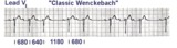 | 2nd degree AV block, type I | The 3 rules of classic AV Wenckebach are: 1. decreasing RR intervals until pause; 2. the pause is less than preceding 2 RR intervals; and 3. the RR interval after the pause is greater than the RR interval just prior to pause. Unfortunately, there are many examples of atypical forms of Wenckebach wh... | Wenckebach AV Block | Knowledge Weavers ECG |
| 5 |
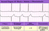 | 2nd degree AV block, type I (Wenckebach) | 2nd degree AV block, type I (Wenckebach) | Knowledge Weavers ECG | |
| 6 |
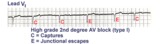 | 2nd degree AV block, type I with escapes and captures | Often in the setting of 2nd degree AV block the pauses caused by nonconducted P waves are long enough to enable escape pacemakers from the junction or ventricles to take over. This example illustrates junctional escapes, labled E and captures, labled C. Note that the PR intervals for the captures ... | Knowledge Weavers ECG | |
| 7 |
 | 2nd degree AV block, type I, with accelerated junctional escapes and a ladder diagram | The ladder diagram illustrates a Wenckebach type AV block by the increasing PR intervals before the blocked P wave. After the blocked P wave, however, a rev-ed up junctional pacemaker terminates the pause. Note that the junctional beats have a slightly different QRS morphology from the sinus beats... | Knowledge Weavers ECG | |
| 8 |
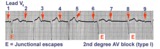 | 2nd degree AV block, type I, with junctional escapes | Junctional escapes are passive, protective events whenever the heart rate slows below that of the escape mechanism. In this example of 2nd degree AV block, type I, the escapes occur following the non-conducted P waves. Arrows indicate the position of the P waves. Note that the escape beats have a... | Knowledge Weavers ECG | |
| 9 |
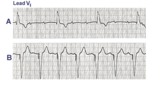 | 3rd degree AV block rx'ed with a ventricular pacemaker | In A the ECG shows complete or 3rd degree AV block with a left ventricular escape rhythm, as evidenced by the upright QRS morphology. In B the artificial right ventricular pacemaker rhythm is shown. | Knowledge Weavers ECG | |
| 10 |
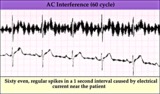 | 60 cycle artifact - marquette | 60 cycle artifact - marquette | Knowledge Weavers ECG | |
| 11 |
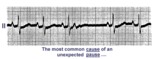 | A nonconducted PAC causes an unexpected pause | Unexpected pauses in rhythm have several causes, the most frequent being a nonconducted PAC. In this example the nonconducted PAC is seen in the ST segment of the pause. Note the change in the ST-T compared to the other ST-T waves. | Knowledge Weavers ECG | |
| 12 |
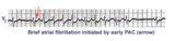 | A PAC initiates paroxysmal atrial fibrillation | The arrow indicates slight alteration of the ST-T wave by a PAC. The PAC, in turn, falls during the vulnerable period of atrial repolarization and initiates atrial fibrillation. Similar but more catastrophic events happen in the ventricles when PVC's occur during the vulnerable period, i.e. R-on-T... | Knowledge Weavers ECG | |
| 13 |
 | A very subtle 1st degree AV block | Where are the P waves??? They are hiding in the T waves as indicated by the arrows. How do we know? The PVC unmasked the sinus P wave, and now it is seen in the pause following the PVC. The PR interval is, therefore, about 500 ms. | Knowledge Weavers ECG | |
| 14 |
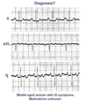 | A very subtle atrial tachycardia with 2:1 block | Although at first glance this looks like normal sinus rhythm at 95 bpm. On closer look, there are 2 P waves for every QRS; the atrial rate is 190 bpm. Note the hidden P in the T waves. This rhythm is likely due to digitalis intoxication, as are the GI symptoms. | Knowledge Weavers ECG | |
| 15 |
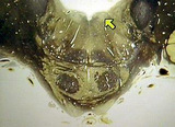
 | Abducens nerve | Abducens nerve. Yellow nerve added to digitized image from graphic output. Photograph. Multimedia. | Abducens nerve; Central nervous system; Cranial nerves; Anatomy | Slice of Life |
| 16 |
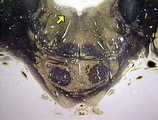
 | Abducens nerve, intracranial portion passing through tegmentum | Abducens nerve, intracranial portion passing through tegmentum. Transverse plane. Photograph. Multimedia. | Abducens nerve; Tegmentum mesencephali; Pons; Central nervous system; Anatomy | Slice of Life |
| 17 |
 | Abducens nucleus | Abducens nucleus. Transverse plane. Photograph. Multimedia. | Slice of Life | |
| 18 |
 | Abducens nucleus VI | Abducens nucleus VI. Pons. Transverse plane. Photograph. Multimedia. | Abducens nerve; Pons; Cranial nerves; Central nervous system; Anatomy | Slice of Life |
| 19 |
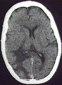 | Abscess, aspergillus in SLE patient | Abscess, aspergillus in SLE patient | Central Nervous System; Brain; Brain Abscess; Lupus Erythematosus, Systemic; Aspergillus; Infarction; Tomography, X-Ray, Computed | Slice of Life |
| 20 |
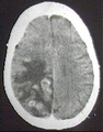 | Abscess, aspergillus in SLE patient | Infarct and granuloma in parietal and occipital. | Central Nervous System; Brain; Parietal Lobe; Occipital Lobe; Brain Abscess; Lupus Erythematosus, Systemic; Aspergillus; Infarction; Granuloma; Tomography, X-Ray, Computed | Slice of Life |
| 21 |
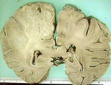 | Abscess, aspergillus in SLE patient | Abscess, aspergillus in SLE patient | Central Nervous System; Brain; Pareital Lobe; Brain Abscess; Lupus Erythematosus, Systemic; Aspergillus | Slice of Life |
| 22 |
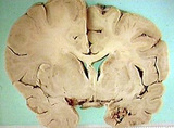 | Abscess, aspergillus in SLE patient, edema | Abscess, aspergillus in SLE patient, edema | Central Nervous System; Brain; Temporal Lobe; Brain Abscess; Brain Edema; Lupus Erythematosus, Systemic; Aspergillus | Slice of Life |
| 23 |
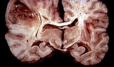
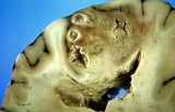 | Abscess, brain | Abscess, brain. A 27-83. Coronal plane. Photograph. Multimedia. | Brain Abscess; Central Nervous System; Anatomy | Slice of Life |
| 24 |
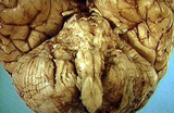
 | Abscess, brain in sepsis | Abscess, brain in sepsis. Brain abscess in sepsis. Photograph. Multimedia. | Brain; Abscess; Central Nervous System; Anatomy | Slice of Life |
| 25 |
 | Abscess, brain, basilar meninges involved | Abscess, brain, basilar meninges involved | Central Nervous System; Brain; Brain Abscess; Meninges | Slice of Life |