The Health Education Assets Library (HEAL) is a collection of over 22,000 freely available digital materials for health sciences education. The collection is now housed at the University of Utah J. Willard Marriott Digital Library.
TO
Filters: Collection: "ehsl_heal"
1 - 25 of 4
| Title | Description | Subject | Collection | ||
|---|---|---|---|---|---|
| 1 |
 |
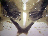
Optice chiasm thalamus iii ventricle | Optic chiasm thalamus iii ventricle. Close up, also massa intermedia. Coronal plane. Photograph. Multimedia. | Optic chiasm; Thalamus; Cerebral ventricle; Central nervous system; Anatomy | Slice of Life |
| 2 |
 |
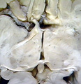
Thalamus close-up, optic nerve, chiasm and tract, posterior commissure, pineal gland, geniculates, third ventricle | Thalamus close-up, optic nerve, chiasm and tract, posterior commissure, pineal gland, genicula, third ventricle. Horizontal plane. Photograph. Multimedia. | Thalamus; Optic Nerve; Pineal Body; Cerebral Ventricles; Central Nervous System; Anatomy | Slice of Life |
| 3 |
 |
Cranial Nerve Exam: Abnormal Examples: Cranial Nerve 2 - Visual Fields | The patient's visual fields are being tested with gross confrontation. A right sided visual field deficit for both eyes is shown. This is a right hemianopia from a lesion behind the optic chiasm involving the left optic tract, radiation or striate cortex. NeuroLogic Exam has been supported by a gran... | Cranial Nerve Examination | NeuroLogic Exam: An Anatomical Approach |
| 4 |
 |
Cranial Nerve Exam: Abnormal Examples: Cranial Nerve 2 - Visual Fields (x2) | The patient's visual fields are being tested with gross confrontation. A right sided visual field deficit for both eyes is shown. This is a right hemianopia from a lesion behind the optic chiasm involving the left optic tract, radiation or striate cortex. NeuroLogic Exam has been supported by a gran... | Cranial Nerve Examination | NeuroLogic Exam: An Anatomical Approach |
1 - 25 of 4
