The Health Education Assets Library (HEAL) is a collection of over 22,000 freely available digital materials for health sciences education. The collection is now housed at the University of Utah J. Willard Marriott Digital Library.
TO
Filters: Collection: "ehsl_heal"
| Title | Description | Subject | Collection | ||
|---|---|---|---|---|---|
| 701 |
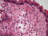 |
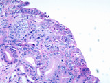
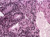
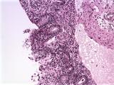
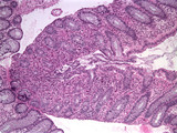
Duodenum amyloid deposition | Duodenum with amyloid deposition in lamina propria. Amyloid etiology unknown. Amyloid shows up as homogenous pink material in lamina propria and around blood vessels 40X. | Small Bowel | HEAL Reviewed Collection |
| 702 |
 |
Eosinophilic granuloma mastoid | High power view (40X) of a neoplastic proliferation of Langerhan's cells (Histiocytosis) in mastoid bone. | HEAL Reviewed Collection | |
| 703 |
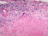 |
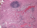
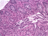
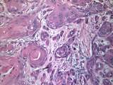
Adrenal gland with granulomatous reaction (gumma) to syphilis | This high power image (20X) shows a granuloma with a giant cell in the adrenal gland. The granuloma is a gumma formed during tertiary syphilis | gumma | HEAL Reviewed Collection |
| 704 |
 |
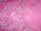
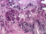
Adrenal gland with pheochromocytoma | Patient with clinical symptoms of hypertension. Image shows adjacent normal adrenal gland with paraganglioma (pheochromocytoma) involving adrenal medulla. | Adrenalin; Noradrenalin | HEAL Reviewed Collection |
| 705 |
 |
Adrenal gland with granulomatous reaction (gumma) to syphilis | This medium power image shows a granuloma in the adrenal gland. The granuloma is a gumma formed during tertiary syphilis. | Gumma | HEAL Reviewed Collection |
| 706 |
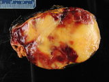 |
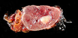
Adrenal myelolipoma | Gross photograph with ruler showing residual normal adrenal and mixture of yellow adipose tissue and red myeloid areas of myelolipoma. | benign | HEAL Reviewed Collection |
| 707 |
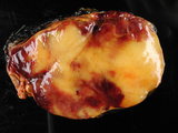 |
Adrenal myelolipoma | Gross photograph showing residual normal adrenal and mixture of yellow adipose tissue and red myeloid areas of myelolipoma. | benign | HEAL Reviewed Collection |
| 708 |
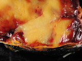 |
Adrenal myelolipoma | Close up showing residual normal adrenal and mixture of yellow adipose tissue and red myeloid areas of myelolipoma. | Benign | HEAL Reviewed Collection |
| 709 |
 |
Bladder carcinoma with lymphovascular invasion | Lymphovascular invasion by a bladder carcinoma away from the main tumor mass. | Urothelial Carcinoma | HEAL Reviewed Collection |
| 710 |
 |
Bladder carcinoma with lymphovascular invasion | Lymphovascular invasion by a bladder carcinoma away from the main tumor mass. | Urothelial Carcinoma | HEAL Reviewed Collection |
| 711 |
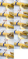 |
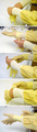
Procedure for venipuncture : changing tubes | Series of images showing changing of tubes during venipuncture | Venipuncture; Blood Collection Tubes | HEAL Reviewed Collection |
| 712 |
 |
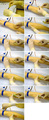
Minimize risks: procedure after venipuncture, 3 examples | A series of three images showing how to reduce the possibility of needle stick injuries after venipuncture by using a special safety engineered holder. | Venipuncture; Syringe Disposal Units, Sharps | HEAL Reviewed Collection |
| 713 |
 |
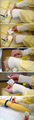
Procedure for venipuncture | Series of images showing the procedure of injection or venipuncture | Venipuncture | HEAL Reviewed Collection |
| 714 |
 |
Minimize risks: procedure before venipuncture | Series of three images showing how to reduce the possibility of needle stick injuries before venipuncture. | Venipuncture | HEAL Reviewed Collection |
| 715 |
 |
Procedure for sterilizing the puncture spot | Series of images showing the proceeding of sterilizing arm and puncture spot. | Venipuncture | HEAL Reviewed Collection |
| 716 |
 |
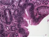
Barrett's Metaplasia with High Grade Dysplasia | Image of the esophagus showing intestinal or Barrett's metaplasia with associated high grade dysplasia at 20X. | High Grade Dysplasia | HEAL Reviewed Collection |
| 717 |
 |
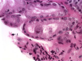
CMV Duodenitis | Patient with lung transplant developed CMV colitis, gastritis, and duodenitis. 40X showing viral inclusions on duodenum. | CMV; Cytomegalovirus Inclusions | HEAL Reviewed Collection |
| 718 |
 |
CMV Colitis | Patient with lung transplant developed CMV colitis, gastritis, and duodenitis. 20X showing viral inclusions on colon. | CMV; Cytomegalovirus Inclusions | HEAL Reviewed Collection |
| 719 |
 |
CMV Colitis | Patient with lung transplant developed CMV colitis, gastritis, and duodenitis. 40X showing viral inclusions on colon. | CMV; Cytomegalovirus Inclusions | HEAL Reviewed Collection |
| 720 |
 |
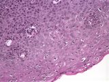
Vagina Condyloma | Image showing human papilloma virus affecting the vagina. | HPV; Human Papilloma Virus | HEAL Reviewed Collection |
| 721 |
 |
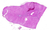
Larynx squamous cell carcinoma | Supraglottic carcinoma involving base of epiglottis. This whole mount slide of the larynx was taken in the coronal plane and photographed at 1X. Patient was a smoker and consumed alcohol in large quantities. | Cancer; Supraglottic | HEAL Reviewed Collection |
| 722 |
 |
Squamous cell carcinoma of the larynx | Tumor can be seen invading from the true vocal cord into anterior soft tissue passing between the thyroid and cricoid cartilage. | Cancer | HEAL Reviewed Collection |
| 723 |
 |
Barrett's Metaplasia with Low Grade Dysplasia | Image of the esophagus showing intestinal or Barrett's metaplasia with associated low grade dysplasia at 20X. | Low Grade Dysplasia | HEAL Reviewed Collection |
| 724 |
 |
CMV Duodenitis | Patient with lung transplant developed CMV colitis, gastritis, and duodenitis. 20X showing viral inclusions on duodenum. | CMV; Cytomegalovirus Inclusions | HEAL Reviewed Collection |
| 725 |
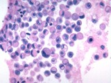 |
Breast carcinoma | Metastatic breast carcinoma in pleural fluid | Breast Cancer | HEAL Reviewed Collection |
| 726 |
 |
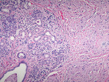
Adrenal cortical hyperplasia | Patient with ectopic ACTH production and diffuse adrenal cortical hyperplasia. | ACTH | HEAL Reviewed Collection |
| 727 |
 |
Preparation before needle stick | Series of images showing how to put on security gloves before venipuncture. | Venipuncture | HEAL Reviewed Collection |
| 728 |
 |
Procedure for removing cannula from vein | Series of images showing how to remove needle and cannula together with vacutainer holder | Venipuncture | HEAL Reviewed Collection |
| 729 |
 |
Invasive squamous cell carcinoma oral cavity | Image shows a moderately differentiated squamous cell carcinoma of the oral cavity with keratinization in patient who smokes and drinks. | Oral Cavity | HEAL Reviewed Collection |
| 730 |
 |
Granular Cell Tumor Sublingual Salivary Gland | Patient presented with nodule in roof of mouth as well as hoarseness with mass on vocal cord. Excision of sublingual gland showed a granular cell tumor as did the mass on the vocal cord. | HEAL Reviewed Collection | |
| 731 |
 |
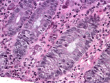
Barrett's Metaplasia with High Grade Dysplasia | Image of the esophagus showing intestinal or Barrett's metaplasia with associated high grade dysplasia at 40X. | High Grade Dysplasia | HEAL Reviewed Collection |
| 732 |
 |
CMV Gastritis | Patient with lung transplant developed CMV colitis, gastritis, and duodenitis. 60X showing viral inclusions on stomach. | CMV; Cytomegalovirus Inclusions | HEAL Reviewed Collection |
| 733 |
 |
CMV Colitis | Patient with lung transplant developed CMV colitis, gastritis, and duodenitis. 10X showing viral inclusions on colon. | CMV; Cytomegalovirus Inclusions | HEAL Reviewed Collection |
| 734 |
 |
Skin gout tophus | Low power microscope photograph showing a tophus in patient with gout. | Tophus; Urate Crystals | HEAL Reviewed Collection |
| 735 |
 |
Granular Cell Tumor Sublingual Salivary Gland | Patient presented with nodule in roof of mouth as well as hoarseness with mass on vocal cord. Excision of sublingual gland showed a granular cell tumor as did the mass on the vocal cord. | HEAL Reviewed Collection | |
| 736 |
 |
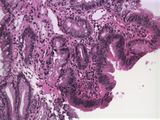
Barrett's Metaplasia with Low Grade Dysplasia | Image of the esophagus showing intestinal or Barrett's metaplasia with associated low grade dysplasia at 10X. | Low Grade Dysplasia | HEAL Reviewed Collection |
| 737 |
 |
Sebaceous Carcinoma Eyelid Orbital Exenteration | Surgical pathology specimen of skin with sebaceous carcinoma requiring orbital exenteration | Adnexal Tumor | HEAL Reviewed Collection |
| 738 |
 |
Skin gout tophus | high power microscope photograph showing a tophus in patient with gout. | Urate Crystals; Tophus | HEAL Reviewed Collection |
| 739 |
 |
Vagina Condyloma | Image showing human papilloma virus affecting the vagina. | HPV; Human Papilloma Virus | HEAL Reviewed Collection |
| 740 |
 |
Vagina Condyloma | Image showing human papilloma virus affecting the vagina. | HPV; Human Papilloma Virus | HEAL Reviewed Collection |
| 741 |
 |
CMV Colitis | Patient with lung transplant developed CMV colitis, gastritis, and duodenitis. High power (60X) showing CMV viral inclusions on colon. | CMV; Cytomegalovirus Inclusions | HEAL Reviewed Collection |
| 742 |
 |
Barrett's Metaplasia with Low Grade Dysplasia | Image of the esophagus showing intestinal or Barrett's metaplasia with associated low grade dysplasia at 40X. | Low Grade Dysplasia | HEAL Reviewed Collection |
| 743 |
 |
CMV Gastritis | Patient with lung transplant developed CMV colitis, gastritis, and duodenitis. 60X showing viral inclusions on stomach. | CMV; Cytomegalovirus Inclusions | HEAL Reviewed Collection |
| 744 |
 |
Eosinophilic granuloma mastoid | High power view (40X) of a neoplastic proliferation of Langerhan's cells (Histiocytosis) in mastoid bone | HEAL Reviewed Collection | |
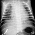
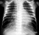
| 745 |
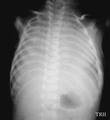 |
Anterior-posterior chest X-ray of meconium-stained severely asphyxiated infant | Anterior-posterior X-ray of meconium-stained severely asphyxiated infant with Type II Meconium Aspiration Syndrome (MAS) and shock lung (ARDS). | Anterior-Posterior Chest Diameter; Type II MAS; Shock Lung | Harris Pediatric Image Collection |
| 746 |
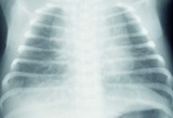 |
Anterior-posterior X-ray taken shortly after birth of infant with Type II MAS | Anterior-posterior chest X-ray taken shortly after birth of baby with Type II Meconium Aspiration Syndrome (MAS). | Anterior-Posterior Chest Diameter; Air Trapping; Type II MAS | Harris Pediatric Image Collection |
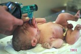
| 747 |
 |
Further suctioning of MAS infant before first breath taken | Further suctioning of Meconium Aspiration Syndrome (MAS) infant before baby takes his/her first breath. | Mouth Suctioning | Harris Pediatric Image Collection |
| 748 |
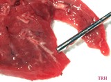 |
Wet hepatisized lung at autopsy | Wet hepatisized lung at autopsy of infant dying of Type II Meconium Aspiration Syndrome (MAS). | Type II MAS | Harris Pediatric Image Collection |
| 749 |
 |
Chest X-ray of (MAS) infant with severe Type I Meconium Aspiration Syndrome prior to respiratory failure | Chest X-ray of baby with severe Type I Meconium Aspiration Syndrome (MAS) just prior to respiratory failure (RF) requiring mechanical ventilation - noting patchy infiltrates or areas of atelectasis scattered throughout markedly over-expanded lungs (predominantly on the right) due to air-trapping. | Air Trapping; Respiratory Failure; Infiltrates | Harris Pediatric Image Collection |
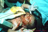
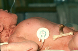
| 750 |
 |
Manual ventilation during successful delivery room resuscitation | Manual ventilation during successful delivery room resuscitation of a meconium-stained, initially depressed, postmature newborn infant male. | manual ventilation | Harris Pediatric Image Collection |
| 751 |
 |
Increased AP-diameter of chest in MAS infant due to air-trapping | Increased AP-diameter of chest in infant with Meconium Aspiration Syndrome (MAS) due to air-trapping. | Air Trapping; Anterior-Posterior Chest Diameter | Harris Pediatric Image Collection |
| 752 |
 |
Suctioning of the mouth in MAS infant before delivery of the body | Suctioning of the mouth of infant with Meconium Aspiration Syndome (MAS) before delivery of the body. | Mouth Suctioning | Harris Pediatric Image Collection |
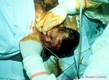
| 753 |
 |
Typical post-term meconium-stained infant receiving conventional mechanical ventilation | A typical post-term, meconium-stained infant receiving conventional ventilation for MAS complicated by right-sided peumothorax and requiring chest tube decompression and drainage. | mechanical ventilation; chest tube decompression | Harris Pediatric Image Collection |
| 754 |
 |
Post-term MAS infant | Post-term Meconium Aspiration Syndrome (MAS) infant with barrel chest and in need for oxygen; shows signs of postmaturity (peeling skin) and air-trapping (increased anterior-posterior diameter). | Air Trapping; Anterior-Posterior Chest Diameter; Barrel Chest | Harris Pediatric Image Collection |
| 755 |
 |
Suctioning of the perineum in a case of birth through meconium-stained amniotic fluid | Suctioning of the perineum in a case of birth through meconium-stained amniotic fluid; the nose is the first to appear. | perineum suctioning | Harris Pediatric Image Collection |
| 756 |
 |
Aspiration of particulate matter (meconium) producing ball-valve mechanism and air-trapping with subsequent air leak complication | Aspiration of particulate matter (meconium) producing ball-valve mechanism and air-trapping with subsequent air leak complication. If bronchus is only partially occluded during inspiration (A) when airways are dilated due to low intrathoracic pressure (represented by black arrows), gas may enter but... | Ball-Valve Mechanism; Air Trapping; Intrathoracic Pressure | Harris Pediatric Image Collection |
| 757 |
 |
Anterior-posterior chest X-ray of same MAS infant | Anterior-posterior chest X-ray of same Meconium Aspiration Syndrome (MAS) baby now on conventional mechanilcal ventilation - note worsening air-trapping. | Air Trapping; Anterior-Posterior Chest Diameter | Harris Pediatric Image Collection |
| 758 |
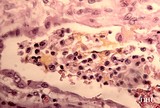 |
Meconium debris and inflammatory cells in occluded terminal bronchiole | Meconium debris and inflammatory cells in occluded terminal bronchiole in case of lethal prenatal aspiration of meconium-stained amniotic fluid. | Inflammatory Cells; Prenatal Aspiration | Harris Pediatric Image Collection |
| 759 |
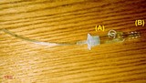 |
Meconium aspirator | Meconium aspirator attaches to standard 15mm ET tube adaptor at one end (A) and wall suction line on the other (B). | Meconium Aspirator | Harris Pediatric Image Collection |
| 760 |
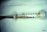 |
Meconium-stained mucus in DeLee Trap | Meconium-stained mucus in DeLee Trap. | DeLee Trap | Harris Pediatric Image Collection |
| 761 |
 |
Suturing | After placing four throws to create a square knot, the suture is then cut just above the knot. | Knowledge Weavers Dermatology | |
| 762 |
 |
Suturing | The suture is then tightened by crossing the non-dominant (left) hand over the dominant (right) hand. | Knowledge Weavers Dermatology | |
| 763 |
 |
Suturing | This demonstrates tightening of the double loop along the long axis of the wound using suture. | Knowledge Weavers Dermatology | |
| 764 |
 |
Shave technique | This demonstrates a shave technique on a pig's foot. Because there are no protruding lesions, I start by angling the blade at about 45 degrees from the skin surface, and while advancing the blade forward, I have a slight side to side sawing motion, and when I am about halfway through the target, I t... | Shave Biopsy | Knowledge Weavers Dermatology |
| 765 |
 |
Urticaria | This patient has developed rather severe urticaria, the cause of which was unknown. In urticaria, the skin swells and initially looks red and later can blanch as the amount of fluid increases within the skin. Internal organs can be involved in the process, and we are particularly concerned about the... | Knowledge Weavers Dermatology | |
| 766 |
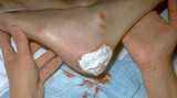 |
Silvadene | I placed Silvadene on the wound. Silvadene is an excellent antibacterial cream to apply to a wound after debridement. One must insure that an 1/8 inch to 1/4 inch coat is applied to help insure that the dressing doesn't absorb all the cream and allow the wound to subsequently dry out. | Knowledge Weavers Dermatology | |
| 767 |
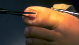 |
Ingrown nail | The nail plate is cut from the underlying nail bed with scissors. | Knowledge Weavers Dermatology | |
| 768 |
 |
Excision procedure | Demonstrates the same V. Summary) | Knowledge Weavers Dermatology | |
| 769 |
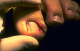 |
Ingrown nail | This shows the nail plate just barely attached. | Surgical Methods | Knowledge Weavers Dermatology |
| 770 |
 |
Excision: suturing | The double loop is tightened by pulling the hands into their natural position, but the knot cannot be adequately tightened by pulling in that direction; the knot must be tightened by pulling the suture ends along the length (long axis) of the wound. (This is shown in the following slide) | Knowledge Weavers Dermatology | |
| 771 |
 |
Scalpel | This is a cross-sectional view demonstrating the blade angled away from the center of the ellipse. | Knowledge Weavers Dermatology | |
| 772 |
 |
Dermatitis | Best control of the dermatitis is achieved by first soaking the skin for 10 minutes in lukewarm water. | Knowledge Weavers Dermatology | |
| 773 |
 |
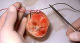
Ingrown nail | 90% phenol is one of the agents advocated for destroying the nail matrix that grows at the base of the cul-de-sac beneath the proximal nail fold. | Knowledge Weavers Dermatology | |
| 774 |
 |
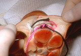
Basal cell carcinoma: excision removal | This person had a basal cell carcinoma, and the epidermis and dermis were excised. The danger area where the temporal branch of the facial nerve may lie and be quite close to the undersurface of the skin is shown by the straight lines drawn from ear to forehead. The surgeon should be careful to unde... | Surgical Methods | Knowledge Weavers Dermatology |
| 775 |
 |
Goodman | This is a picture of Dr. Goodman (of Goodman & Gillman fame). I once asked him how much you should know about a medication before using it, and he said,A lot! And you can quote me on that! | Knowledge Weavers Dermatology | |
| 776 |
 |
Punch biopsy | Punch biopsy is excellent for sampling a skin disease that has multiple essentially identical lesions, or sampling a lesion within the skin. It is designed for full thickness skin biopsy, and is not an effective tool for biopsying the fat. When using it, it should be held as shown, should be spun ve... | Knowledge Weavers Dermatology | |
| 777 |
 |
Keloid | Before injecting the keloid with the corticosteroid, I recommend injecting local anesthetic into the underlying fat, and waiting about five minutes for the anesthetic to take effect. The anesthetic should not be injected into the keloid as this reduces the amount of space into which one can inject t... | Knowledge Weavers Dermatology | |
| 778 |
 |
Suturing | This demonstrates the deep dermal suture properly placed with the loop, which will be just on the under-surface of the dermis, and the knot will be buried well within the fat. This will prevent the knot from extruding through the dermis and out through the wound. | Knowledge Weavers Dermatology | |
| 779 |
 |
Suturing | The loop is then tightened with the hands in their natural position. This is done four times and then the suture is cut leaving suture ends at .5 cm long. | Knowledge Weavers Dermatology | |
| 780 |
 |
Tearing action | They are then opened which creates a tearing action. | Knowledge Weavers Dermatology | |
| 781 |
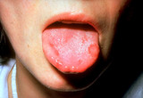 |
Aphthous stomatitis | Aphthous stomatitis (cankers) involving the tongue. Aphthous stomatitis ulcers are painful, and major aphthous stomatitis induces ulcers that last for many weeks, and sometimes induce scarring. | Canker Sore | Knowledge Weavers Dermatology |
| 782 |
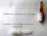 |
Epinephrine | Standard epinephrine (1:1,000) is shown at the top, and the crystalline form (Sus-Phrine) is a concentration of 1:200. Both are administered into the subcutaneous fat, and a dose of epinephrine 1:1,000 is 0.01 cc's per kg, and the dose of Sus-Phrine is half of that (0.005 cc's per kg), and the Sus-P... | Knowledge Weavers Dermatology | |
| 783 |
 |
Excision: suturing | AVis formed as the deep dermal suture exits from the wound, and the suture is pulled so that the short (non-needle bearing end) is about 2 cm long. Both strands of suture should come out on the same side of the loop, and the order must be: loop, short end (non-needle bearing), long end (needle bea... | Knowledge Weavers Dermatology | |
| 784 |
 |
Miliaria | Occlusion of the eccrine sweat glands produces a condition called miliaria; this appears as small red papules and/or vesicles, and associated inflammation. | Knowledge Weavers Dermatology | |
| 785 |
 |
Flea bites | Presumed flea bites. | Knowledge Weavers Dermatology | |
| 786 |
 |
Excision procedure | The following slides are a brief review of the excision procedure. The scalpel is angled outward away from the wound edge to ensure that the wound edges are beveled at about 45 o. | Surgical Methods | Knowledge Weavers Dermatology |
| 787 |
 |
Decubitus ulcer | A decubitus ulcer in an older woman in a nursing home. This was induced because the person was lying in one position for too long, and this compromised blood supply to the skin. | Knowledge Weavers Dermatology | |
| 788 |
 |
Forceps | The forceps are held with the non-dominant hand as shown here between the first two or three fingers. | Knowledge Weavers Dermatology | |
| 789 |
 |
Urticaria | Urticaria | Knowledge Weavers Dermatology | |
| 790 |
 |
Suturing | This demonstrates that a total of four throws should be made when tying the deep dermal suture, and if done properly, square knots are laid down. It is said that square knots provide optimum knot security. | Knowledge Weavers Dermatology | |
| 791 |
 |
Suturing | The short arm of the suture is then grasped with the needle holder, and then the non-dominant (left) hand crosses over the right hand as one tightens the knot pulling along the long axis of the wound. | Knowledge Weavers Dermatology | |
| 792 |
 |
Needle holder | The needle holder (sometimes called needle driver) can be held as shown, or | Knowledge Weavers Dermatology | |
| 793 |
 |
Excision procedure | This suture is tightened by crossing the non-dominant (left) and over the dominant (right) hand. This same sequence is continued until four loops have been thrown and then the suture is cut leaving ends approximately .5 cm long. | Knowledge Weavers Dermatology | |
| 794 |
 |
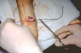
Skin tags | Skin tags. They are rather small, and have a pedunculated base giving them the appearance of a teardrop. | Skin Tags | Knowledge Weavers Dermatology |
| 795 |
 |
Suturing | I exit through the other side of the wound as superficially and closely as possible to the wound edge. Generally, I am about a mm away from the wound edge, and just barely entering the dermis. It is crucial to be as superficial within the dermis and as close to the wound edge on the second pass as i... | Knowledge Weavers Dermatology | |
| 796 |
 |
Necrosis | The purple/red area on the heel is an area of necrosis of the entire epidermis and dermis that is induced by the person lying in one position for too long. Again, the entire epidermis and dermis are necrotic, and need to be surgically removed. | Knowledge Weavers Dermatology | |
| 797 |
 |
Excision: suturing | The needle holder is then placed inside the long arm of the V, and a single loop is thrown around the needle holder. The short arm of the suture is then grasped. | Knowledge Weavers Dermatology | |
| 798 |
 |
Excision procedure | The central specimen is then removed ensuring that there is same thickness throughout the specimen. | Surgical Methods | Knowledge Weavers Dermatology |
| 799 |
 |
Shave technique | This demonstrates the shave technique. Local anesthetic can be injected into the fat beneath the target lesion, or within the lesion itself. One should be extremely careful to inject as little anesthetic as necessary to anesthetize the skin, because the anesthetic will artifactually enlarge and dist... | Knowledge Weavers Dermatology | |
| 800 |
 |
Suturing | This demonstrates placing the needle holder on the inside of the long arm of the V using actual suture. | Knowledge Weavers Dermatology |