The Health Education Assets Library (HEAL) is a collection of over 22,000 freely available digital materials for health sciences education. The collection is now housed at the University of Utah J. Willard Marriott Digital Library.
TO
Filters: Collection: "ehsl_heal"
| Title | Description | Subject | Collection | ||
|---|---|---|---|---|---|
| 2201 |
 |
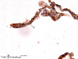
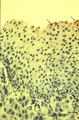
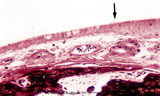
Keratin 7 staining in dysplastic epithelium of a bronchus (human, adult) | Stain: anti-keratin 7 antibody (Pan-Ck 7) immunoperoxidase staining (with aminoethylcarbazole (AEC) substrate). A red-brown staining with AEC indicates a positive reaction for cytokeratin 7. Disturbances in the growth and maturing might result in dysplasia of the epithelium with a changed reaction p... | Immunoperoxidase; Immuno-reaction | Poja Histology Collection - Respiratory System Subset |
| 2202 |
 |
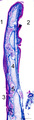
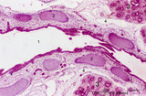
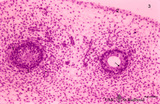
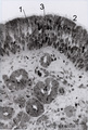
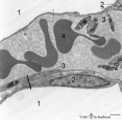
Pseudoglandular period of developing lung (human, embryo, low magnification) | Stain: Hematoxylin and eosin. Cross-sectioned future bronchial tubes (1), the surrounding mesenchyme becomes more condensed around the epithelium. The mesoderm of the future visceral pleura (2) as well as the future parietal pleura (3) and (4) indicates pleural cavity. The cartilagineous spinal colu... | Lung development; Visceral pleura; Parietal pleura; Pseudoglandular period; Bronchial tubes; Mesenchyme | Poja Histology Collection - Respiratory System Subset |
| 2203 |
 |
Pseudoglandular period of developing lung (human, embryo, low magnification) | Stain: Trichrome (Goldner). Cross-sectioned future bronchial tubes (1) of varying sizes. Note the apical position of the nuclei in these epithelial cells with light-stained basal parts (glycogen). The surrounding mesenchyme becomes more condensed (↓) around the epithelium and in between numerous ... | Lung development; Visceral pleura; Pseudoglandular period; Bronchial tubes; Mesenchyme | Poja Histology Collection - Respiratory System Subset |
| 2204 |
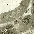 |
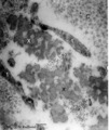
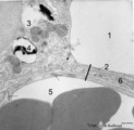
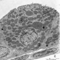
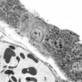
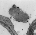
Nasal glands of the nasal vestibulum (rat) | Electron microscopy. At the top part of a striated draining duct with a wide lumen (*); these cuboidal ductal cells (1) contain many basolaterally located mitochondria. The ductal cells are enforced by interstitial fibroblast (2) and a capillary (4). Neighboring serous gland cells (5) contain dark s... | Nasal vestibulum; Seromucous glands; Nasal glands; Serous cells; Secretion granules; Intercalated duct; Striated duct | Poja Histology Collection - Respiratory System Subset |
| 2205 |
 |
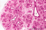
Small bronchus in the lung (human, adult) | Stain: Azan. Pseudostratified epithelium (1) with in between light-stained goblet cells and a blue stained basement membrane (↓) with the proper lamina. The thin smooth muscle layer (2) is purple stained and accompanies the mucosal layer. At (3) a small hyaline cartilage and seromucous bronchial g... | Small bronchus; Pseudostratified epithelium; Seromucous glands | Poja Histology Collection - Respiratory System Subset |
| 2206 |
 |
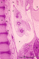
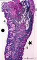
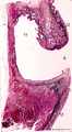
Survey of epiglottis (human) | Stain: Azan. Centrally light-stained elastic cartilage (4). Lingual side (2) is covered with squamous epithelium. At the laryngeal side (1) the squamous epithelium usually turns into respiratory epithelium with seromucous laryngeal glands (5). Note also lymphoid aggregations in this area (3 →). | Oral cavity; Laryngeal glands | Poja Histology Collection - Respiratory System Subset |
| 2207 |
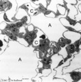 |
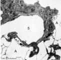
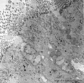
The terminal part of the airway: alveoli (dog) | Electron microscopy (low magnification). (A) indicate alveoli; (C) indicate capillaries. (1) type I alveolar cell (pneumocyte I, squamous alveolar cell); (2) type II alveolar cell (pneumocyte II, great alveolar cell). (3) alveolar macrophage. (4) endothelium of capillary. | Pneumocyte I; Pneumocyte II; Alveolar macrophage | Poja Histology Collection - Respiratory System Subset |
| 2208 |
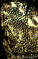 |
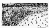
Capillary system of a lung alveolus (cat) | Stain: specimen injected with India ink via the pulmonary system. In an en face view of the alveolar wall the black- and granular-stained capillary plexus is well shown. The larger vessels represent branches of the pulmonary arteriole. | Poja Histology Collection - Respiratory System Subset | |
| 2209 |
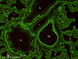 |
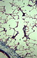
Heparan sulfates in the terminal sac period of the lung (mouse, fetus) | Stain: fluorescence microscopy with anti-heparan sulphate antibody (a phage-display antibody, HS4E4). Heparan sulfates are linear polysaccharides that belong to the group of the glycosaminoglycans (GAGs). These GAGs are found associated with basement membranes as shown here. The sulphated saccharide... | Bronchioli; Air spaces; Terminal sac period | Poja Histology Collection - Respiratory System Subset |
| 2210 |
 |
Epithelium of trachea (mammals, common respiratory epithelium) | Scheme electron microscopy. Columnar ciliated cells (1) with up to 200 motile cilia with an organelle-rich apex and in the middle a goblet cell (2) with accumulation of mucus secretion granules in the apex. Note that all epithelial cells (i.e., junctions) contact the basal lamina. Four basal cells (... | Respiratory epithelium ; Pseudostratified epithelium; Basal cells | Poja Histology Collection - Respiratory System Subset |
| 2211 |
 |
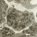
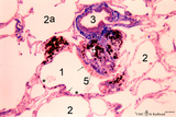
Epithelial lining of respiratory bronchiolus in the lung (rat) | Electron microscopy (low magnification). The epithelial lining of the respiratory bronchiolus contains ciliated cells (1) and Clara cells (2) with electron-dense secretory granules. At (4) interstitium with elastin, collagen and myofibroblasts; (5) shows a small pulmonary arteriole. At arrow (→) a... | Respiratory bronchiolus; Ciliated epithelium; Clara cells; Secretory granules; Pneumocytes; Alveolar cells; Interstitium | Poja Histology Collection - Respiratory System Subset |
| 2212 |
 |
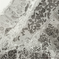
Detail of elastin in an alveolar tip in lung tissue (human, adult) | Electron microscopy. Note the lace-like pattern of lumps of amorph elastin (X) that appears more electron-dense than the collagen fibers (C). At this high magnification cross-sectioned microfibrils (that contain among others fibrillin) are present in close association (*) with different areas of ela... | Elastin-associated proteins; Elastoblast; Myofibroblast | Poja Histology Collection - Respiratory System Subset |
| 2213 |
 |
Type I alveolar cell in the lung (human, adult) | Electron microscopy. At the top the alveolar space is lined by type I alveolar cell (1, pneumocyte I). The cytoplasm is well provided with organelles and few electron-dense lysosomes, and pinocytotic vesicles. These pneumocyte I cells cover the interalveolar septa that contain fibroblasts and bundle... | Pneumocyte I; Interstitium | Poja Histology Collection - Respiratory System Subset |
| 2214 |
 |
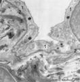
Nasal glands of the nasal vestibulum (rat) | Electron microscopy. A low-power magnification reveals serous acini of a seromucous gland; in the center and lower left corner lumina (*) of the acini are present and dense secretion granules of varying sizes represent the active cells. Between the acini the distended interstitium reveals a fibrobla... | Nasal vestibulum; Seromucous glands; Nasal glands; Serous cells; Secretion granules | Poja Histology Collection - Respiratory System Subset |
| 2215 |
 |
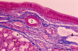
Survey of nose wing - human | Stain: Azan. At the top orofacial muscle (1, reddish) and hyaline cartilage (2, bluish) at the right. The external skin surface of the nose wing dot side (.) is covered with squamous epithelium, light-stained sebaceous glands (3) and hair structures (4). At the starred side (*) the nasal vestibulum... | Poja Histology Collection - Respiratory System Subset | |
| 2216 |
 |
Fibrillin in the alveoli of the lung (human, adult) | Stain: antifibrillin antibody immunoperoxidase staining with diaminobenzidin reaction. Fibrillin is one of the elastin-associated microfibrillar proteins. Here fibrillin (and elastin) is localized as brown threads in the alveolar septa and alveolar tips (1). The elastin is visible as white threads (... | Alveolar tips; Elastin-associated proteins; Fibrillin | Poja Histology Collection - Respiratory System Subset |
| 2217 |
 |
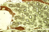
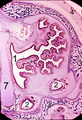
Pseudoglandular - canalicular period of developing lung (human, fetus) | Stain: Azan. Longitudinal section through a large bronchus (1) with cartilagineous rings (2). At (3) developing glandular structures in islets of bronchial tubes surrounded by condensed mesenchyme. At (4) lymph vessels. | Lung development; Pseudoglandular period; Canalicular period; Mesenchyme; Bronchial tubes | Poja Histology Collection - Respiratory System Subset |
| 2218 |
 |
Type II and type I alveolar cells in the lung (human, adult) | Electron microscopy. At the top the alveolar space (1) is lined by a thin cytoplasm (2) of type I alveolar cell (pneumocyte I). At the left side part of a type II alveolar cell (3, pneumocyte II) with electron-dense remnants of two characteristic multilamellar bodies (4) as well as many organelles. ... | Pneumocyte I; Pneumocyte II; Interstitial cell; Alveolar cell type I; Alveolar cell type II | Poja Histology Collection - Respiratory System Subset |
| 2219 |
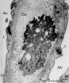 |
Elastin close near the alveolar tip in lung tissue (human, adult) | Electron microscopy. The alveolar tip in the alveolar spaces (Alv) is covered with flattened type I alveolar cells (2). The arrows at (↓1) indicate the junctions between these flattened cells. The amorph elastin (*) in the alveolar tip appears more electron-dense than the collagen fibers (Col). No... | Type I alveolar cells; Elastin-associated proteins | Poja Histology Collection - Respiratory System Subset |
| 2220 |
 |
Pseudoglandular period of developing lung (human, embryo) | Stain: Hematoxylin and eosin. Two cross-sectioned future bronchial tubes. Note the basal position of the nuclei with light-stained apical cytoplasm (↓, glycogen). The surrounding mesenchyme condenses (1) around the epithelium and developing capillaries and small blood vessels (*) are present. Futu... | Lung development; Visceral pleura; Pseudoglandular period; Bronchial tubes; Mesenchyme | Poja Histology Collection - Respiratory System Subset |
| 2221 |
 |
Clara cell in lung (gerbil) | Electron microscopy. In this species a non-ciliated Clara cell (1 = nucleus) contains numerous electron-gray secretion granules (2) of varying sizes, smaller more dense granules tend to localize in the apex of the cell. Between the granules agranular endoplasmic reticulum. Irregular blunt microvill... | Respiratory bronchiolus ; Clara cells; Secretory granules | Poja Histology Collection - Respiratory System Subset |
| 2222 |
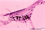 |
Alveolar duct in the lung (mouse) | Stain: PAS and hematoxylin. Part of an alveolar duct lumen (1) that shows bronchiolar characteristics such as cuboidal epithelium (2) covering a bundle of smooth muscle (3) and connective tissue containing macrophages (4) with black pigment deposits. Note PAS-positivity of these cuboidal cells (Clar... | Alveolar ducts; Clara cells | Poja Histology Collection - Respiratory System Subset |
| 2223 |
 |
Terminal sac period of developing lung (human, fetus) | Stain: Hematoxylin and eosin. The transition of a terminal bronchiolus (1) into two future respiratory bronchioli (2), present as dilated spaces (saccules derived from the primitive respiratory channels, hence the name terminal sac period). The surrounding cellular tissue is composed of developing p... | Lung development; Terminal sac period; Respiratory bronchioli; Mesenchyme | Poja Histology Collection - Respiratory System Subset |
| 2224 |
 |
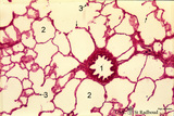
Peripheral alveolar area of the lung (human) | Stain: Azan. (1) Part of a pulmonary artery in a septum (2). (3) represents part of a bronchiolus respiratorius that continues into several alveolar ducts (4) and subsequently in alveolar sacs. Arrows (↓) indicate small foci of carbon deposits. | Respiratory bronchioli; Alveolar ducts; Alveolar sacs | Poja Histology Collection - Respiratory System Subset |
| 2225 |
 |
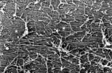
Surface of the nasal septum (rat) | Scanning electron microscopy of anterior part of the nasal septum. The superficial squamous cells of the stratified epithelium are large cells provided with small stubby microvilli. Cell borders are well indicated (↓). | Nasal vestibulum; Stratified epithelium | Poja Histology Collection - Respiratory System Subset |
| 2226 |
 |
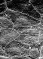
Surface of olfactory epithelium in the nose (rat) | Scanning electron microscopy. A carpet of fine long threads of olfactory cilia (*). On top of the carpet broken remnants of cilia clotted due to secretion products. | Olfactory epithelium | Poja Histology Collection - Respiratory System Subset |
| 2227 |
 |
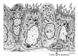
Olfactory mucosa in the nose (human) | Stain: Toluidine blue, one-micron Epon plastic section. Pseudostratified epithelium with light stained nuclei (1) (note distinct nucleoli) of olfactory cells. At the surface faintly stained cilia/microvilli (2). Nuclei of sustentacular (supporting) cells (3) are stained darker and predominantly in t... | Bowman glands; Olfactory glands; Olfactory mucosa; Pseudostratified epithelium | Poja Histology Collection - Respiratory System Subset |
| 2228 |
 |
The alveolus and air-blood barrier in the lung (rat) | Electron microscopy. The alveolus (1) is lined by a thin extension (2) of the alveolar epithelial cell type I (2), the pneumocyte I and the thin endothelium (3) of the capillary filled with erythrocytes (4) and blood platelets (5). The thin-walled air-blood barrier (↔) consists of the transition f... | Pneumocyte I; Alveolar cell type I | Poja Histology Collection - Respiratory System Subset |
| 2229 |
 |
Epithelial lining of respiratory bronchiolus in the lung (gerbil) | Electron microscopy (low magnification). Two major cell types line a bronchiolus: a cuboidal-shaped ciliated one that exhibits microvilli with many organelles (1); the non-ciliated cells (2, Clara cells) are taller and are well provided with numerous electron-grey and smaller electron-dense secretor... | Respiratory bronchiolus; Ciliated epithelium; Cuboidal epithelium; Clara cells; Secretory granules | Poja Histology Collection - Respiratory System Subset |
| 2230 |
 |
Pseudoglandular - canalicular period of developing lung (human, fetus) | Stain: Azan. Part of a future lung lobe with cross-sectioned larger bronchi (1) in close association with islets of future smaller bronchial tubes (*). Note the branching of the bronchial tubes within an islet. The mesenchyme within the islets of future tubes becomes more condensed (↓). Blood vess... | Lung development; Visceral pleura; Pseudoglandular period; Canalicular period; Mesenchyme | Poja Histology Collection - Respiratory System Subset |
| 2231 |
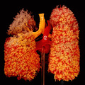 |
The bronchial tree and branches of the pulmonary artery (human, adult, posterior aspect) | Resin corrosion cast of left and right lung. Posterior aspect of lower trachea (1, yellow) with two principal bronchi (yellow) with red-stained pulmonary artery (2) and its branching. The cast of both lung lobes reveals especially the intricate divisions and branching of the bronchial tree (yellow) ... | Macroscopy | Poja Histology Collection - Respiratory System Subset |
| 2232 |
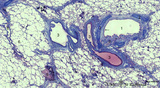 |
Survey of lung parenchym with bronchi and blood vessels (human, adult) | Stain: Azan. The bronchus (1) is surrounded with hyaline cartilage rings (2), seromucous glands (9) and muscle fibers (*), and lined with respiratory epithelium (←). Pulmonary arteries (3) in their neighborhood; a solitary pulmonary vein (8); (4) Carbon deposits of varying sizes (dust loaded macr... | Bronchiolus | Poja Histology Collection - Respiratory System Subset |
| 2233 |
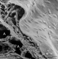 |
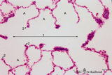
Surface of lung pleura (gerbil) | Scanning electron microscopy of visceral pleura. Alveoli (A) and capillaries (c) with scattered erythrocytes (artifactual due to preparation procedures). At (*) connective tissue and in between erythrocytes. The visceral surface is covered by a continuous sheet of flat squamous mesothelial cells (1,... | Visceral pleura; Mesothelium | Poja Histology Collection - Respiratory System Subset |
| 2234 |
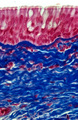 |
Respiratory epithelium of trachea (human, adult, high magnification) | Stain: Azan. The red-stained pseudostratified epithelium contains cilia and light goblet cells (white droplets) and a distinct basement membrane that continues in the intense blue-stained lamina propria and the submucosa consisting of fibrous tissue of elastic and collagenous fibers. | Tracheal glands; Excretory duct; Perichondrium | Poja Histology Collection - Respiratory System Subset |
| 2235 |
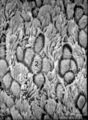 |
Respiratory epithelial cells in the nasal concha (rat) | Scanning electron microscopy. Bushes of long cilia (1) are present on the top of the epithelial cells. In between clusters of microvilli-studded goblet cells (2) are visible. | Respiratory epithelium; Pseudostratified epithelium | Poja Histology Collection - Respiratory System Subset |
| 2236 |
 |
Keratin 7 staining in squamous metaplasia and dysplasia of the epithelium of a bronchus (human, adult) | Stain: anti-keratin 7 antibody (Pan-Ck 7) immunoperoxidase staining (with aminoethylcarbazole (AEC) substrate). A red-brown staining with AEC indicates a positive reaction for cytokeratin 7. Note that the top layer only reacts weakly positive for keratin 7 (↓). Upon squamous metaplasia the bronch... | Squamous metaplasia; Immunoperoxidase; Immuno-reaction | Poja Histology Collection - Respiratory System Subset |
| 2237 |
 |
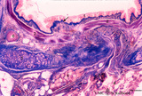
Frontal section of head (pig, fetus) | Stain: Azan. At the upper half the nasal septum (7) is a lightly stained plate (cartilago septi nasi). Developing conchae with supporting hyaline cartilage scaffolds (*) (light-stained) are present in the nasal chamber (1) and known as inferior (lowest), middle and superior turbinate bones. The whol... | Conchae nasales; Trabecular bone | Poja Histology Collection - Respiratory System Subset |
| 2238 |
 |
Transition of respiratory bronchiolus into alveolar duct in the lung (human) | Stain: Azan. The alveolar duct (1↔) exhibits an alveolated wall but at a few places (3) bronchiolar characteristics are evident such as cuboidal epithelium covering small areas of smooth muscle and connective tissue. They differ from the characteristic alveolar tips (2↓) of neighboring thin-wall... | Alveolar ducts; Alveolar tips | Poja Histology Collection - Respiratory System Subset |
| 2239 |
 |
Respiratory bronchiolus (human) | Stain: Azan. The lumen (1) shows an irregular lining. It is covered by low columnar-cuboidal cells (→). Alveolar pouches (*) within the same lumen are lined by thin alveolar epithelium as found in the surrounding alveoli (2). At (5) small patch of smooth muscle, (4) macrophages with phagocytized c... | Respiratory bronchiolus; Columnar epithelium; Cuboidal epithelium; Alveolar epithelium | Poja Histology Collection - Respiratory System Subset |
| 2240 |
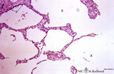 |
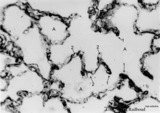
Progressing centrilobular lung emphysema (human, adult) | Stain: Hematoxylin and eosin. Emphysema is defined as enlargement of the airspaces (X) distal to the terminal bronchioles, with destruction of the alveolar walls. At (3) remnant of a respiratory bronchiolus. Alveolar walls are destroyed and other alveolar walls are thickened (1) and show an increas... | Alveolar tips | Poja Histology Collection - Respiratory System Subset |
| 2241 |
 |
Respiratory epithelium in the nasal septum (rat) | Electron microscopy. At the left pseudostratified epithelium (1) with ciliated columnar (light) cells and electron-dense basal cells (2). In the middle a wide venous capillary (3) with erythrocytes. At the lower part a serous area of a seromucous gland (4) with dark granules and well-developed rough... | Respiratory epithelium; Basal cells | Poja Histology Collection - Respiratory System Subset |
| 2242 |
 |
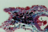
Bronchiolus and alveoli in lung (human, adult) | Stain: Hematoxylin and eosin. (1) indicates a bronchiolus with respiratory epithelium and muscle fibers in its wall. Alveolar spaces (2) are separated by alveolar cell types that lined thin septa with capillaries. The septa end in tips and knobs (3). | Bronchiolus; Alveolar septa; Pneumocyte I; Pneumocyte II; Alveolar cell types | Poja Histology Collection - Respiratory System Subset |
| 2243 |
 |
Ventricular fold (plica ventricularis) (human, higher magnification) | Stain: Azan. On top pseudostratified columnar, ciliated epithelium (1). Centrally below an excretory duct (2) of laryngeal seromucous gland (3) with some adipocytes (*). Blood vessels and two lymph vessels (4) are present. | Supraglottis; Ventricular fold; Laryngeal glands; Seromucous glands | Poja Histology Collection - Respiratory System Subset |
| 2244 |
 |
Scheme of trachea (human, adult) | On top pseudostratified ciliated epithelium (1) with goblet cells followed by a condensed layer of elastic fibres (2) and the transition (*) to seromucous tracheal glands (3) close to the edge of the perichondrium (4) of hyaline cartilage (5). | Pseudostratified epithelium ; Perichondrium | Poja Histology Collection - Respiratory System Subset |
| 2245 |
 |
Fibrillin in the alveoli of lung emphysema (human, adult) | Stain: antifibrillin antibody immunoperoxidase staining with diaminobenzidin reaction. Fibrillin is one of the elastin-associated microfibrillar proteins, and marks therefore the presence of elastin in lung tissue. In centrilobular emphysema (e.g., in lungs of smokers) the lesions are more common an... | Alveolar tips; Elastin-associated proteins; Fibrillin | Poja Histology Collection - Respiratory System Subset |
| 2246 |
 |
Detail of lamellar body (surfactant) and type I alveolar cell in lung (rat) | Electron microscopy. After fixation the extracellular lining of surfactant (phosphatidylcholine, phosphoglycerol, cholesterol and proteins) will often be present as free packed lamellae in the alveolar space (4). This so-called tubular myelin is observed as stacks (5) of lipid crystals and aqueous l... | Pneumocyte I ; Pneumocyte II; Alveolar cells; Tubular myelin | Poja Histology Collection - Respiratory System Subset |
| 2247 |
 |
Epithelial lining of bronchiolus in the lung (mammalia) | Scheme electron microscopy. Two major cell types line a bronchiolus, a more cuboidal shaped ciliated one (1) that also exhibits microvilli with many organelles (including electron-dense lysosomal structures). The non-ciliated cells (2, Clara cells) appeared taller and dome-shaped and protrude into t... | Bronchiolus; Ciliated epithelium; Clara cells | Poja Histology Collection - Respiratory System Subset |
| 2248 |
 |
Respiratory epithelial cells bordering the olfactory region in the nose (human) | Electron microscopy. The five respiratory cells (1) show many cross-sectioned cilia (2) in the lumen. Basal bodies (arrow heads) are regularly arranged at the apex of these cells, and in between, short, slender microvilli. Electron-light cytoplasmic protrusions (3) between the cilia are olfactory bu... | Respiratory epithelium | Poja Histology Collection - Respiratory System Subset |
| 2249 |
 |
Fibrillin in the alveoli in lung tissue (human, adult) | Stain: imunoperoxidase staining with anti-fibrillin antibodies and diaminobenzidin reaction (frozen section). Fibrillin is one of the elastin-associated microfibrillar proteins that wraps the elastin protein core and is localized in normal lung structures such as alveolar septa and tips. The immuno-... | Alveolar tips; Elastin-associated proteins; Fibrillin | Poja Histology Collection - Respiratory System Subset |
| 2250 |
 |
Survey frontal section of larynx (human) | Stain: Azan. A pseudostratified epithelium covers the mucosa of the laryngeal cavity and vestibulum. At the vocal fold edge (3) the epithelium appears to be nonkeratinizing squamous. The connective tissue is rigidly attached to the this edge and merges into the vocal ligament (dense elastic) and voc... | Vocal muscle; Vocal ligament; Laryngeal glands; Ventricular fold | Poja Histology Collection - Respiratory System Subset |
| 2251 |
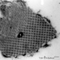 |
Free surfactant (tubular myelin) in alveolar space of the lung (rat) | Electron microscopy. After fixation the extracellular lining of surfactant (phosphatidylcholine, phosphoglycerol, cholesterol and proteins) will often be present as free packed lamellae in the alveolar space. This so-called tubular myelin (partly cross-sectioned), (1) is observed as stacks of lipid ... | Pneumocyte II; Tubular myelin | Poja Histology Collection - Respiratory System Subset |
| 2252 |
 |
Epithelium of respiratory bronchiolus (detail, human, high magnification) | Stain: Azan. An irregular lining of low columnar-cuboidal cells (1) and thin alveolar epithelium (2) are present. Note cuboidal Clara cells (↓). At (3) small patches of smooth muscles and at (4) macrophages with phagocytized carbon particles (black dots). Small pulmonary artery (5), lymph capillar... | Respiratory bronchiolus; Cuboidal epithelium; Alveolar epithelium; Lymph capillary | Poja Histology Collection - Respiratory System Subset |
| 2253 |
 |
Nasal concha with respiratory mucosa (dog, high magnification) | Stain: Hematoxylin and eosin. (↑) Indicates the transition of the olfactory epithelium (1) into the respiratory epithelium (2) with pseudostratified ciliated epithelium and goblet cells. The submucosa is richly vascularized (3). Lamellar bone of the turbinate (4) is stained reddish-black. | Conchae nasales; Olfactory epithelium; Respiratory epithelium | Poja Histology Collection - Respiratory System Subset |
| 2254 |
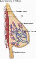 |
Breast - Cross Section - Ductal Carcinoma (Labeled) | Ductal carcinoma of breast. | Pectoralis Major; Breast Lobule; Ductule | Royal College of Surgeons in Ireland Illustrations |
| 2255 |
 |
Lymph Nodes of Head and Neck (Labeled) | Lymph nodes. Head. Neck. | Submental; Submandibular; Deep Cervical; Jugulodigastric; Mastoid; Occipital; Parotid | Royal College of Surgeons in Ireland Illustrations |
| 2256 |
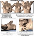 |
Monthly Breast Self-Exam (Labeled) | Monthly breast self-exam. | Cancer Screening | Royal College of Surgeons in Ireland Illustrations |
| 2257 |
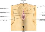 |
Vulva (Labeled) | External genitalia. Female. | Mons Pubis; Glans; External Urethral Orifice; Perineal Body; Opening of Vagina; Vestibule of Vagina; Labium Minus; Labium Majus | Royal College of Surgeons in Ireland Illustrations |
| 2258 |
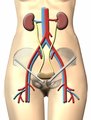 |
Urinary System | Urinary system. Kidneys. Ureter. Bladder. | Common Iliac Veins; Common Iliac Arteries | Royal College of Surgeons in Ireland Illustrations |
| 2259 |
 |
Aortocaval Fistula (Labeled) | Fistula between aorta and inferior vena cava. | Aortocaval Fistula | Royal College of Surgeons in Ireland Illustrations |
| 2260 |
|
Resection and Primary Anastomosis of Sigmoid Colon (Labeled) | Colon resection. Primary anastomosis. | Primary Anastomosis; Resection | Royal College of Surgeons in Ireland Illustrations |
| 2261 |
|
Mastectomy - Right Breast | Breast cancer. Incision site. Mastectomy. | Incision Site; Breast Cancer | Royal College of Surgeons in Ireland Illustrations |
| 2262 |
|
Resection and End Colostomy | Colon resection. End colostomy. Harmann's procedure. | End Colostomy; Colon Resection; Hartmann's Procedure | Royal College of Surgeons in Ireland Illustrations |
| 2263 |
|
Gastrointestinal Tract (Labeled) | Gastrointestinal tract showing upper GI, ligament of Treitz, and lower GI. | Upper GI; Upper Gastrointestinal Tract; Ligament of Treitz; Lower GI; Lower Gastrointestinal Tract | Royal College of Surgeons in Ireland Illustrations |
| 2264 |
|
Resection and Primary Anastomosis of Sigmoid Colon | Make private -- psd file. Colon resection. Primary anastomosis. | Primary Anastomosis; Resection | Royal College of Surgeons in Ireland Illustrations |
| 2265 |
|
Male Urogenital Diaphragm | Urogenital diaphragm. Male. | Ischiopubic Ramus; Perineal Membrane; Urogenital Diaphragm; Perineal Body; Penile Veins; Prostatic Venous Plexus | Royal College of Surgeons in Ireland Illustrations |
| 2266 |
|
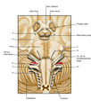
Exposed Facial Nerve with Retracted Parotid Gland and Stylohyoid Muscle (Labeled) | Exposed facial nerve with retracted parotid gland and stylohyoid muscle. | Stylohyoid Muscle | Royal College of Surgeons in Ireland Illustrations |
| 2267 |
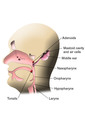 |
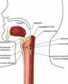
Pharynx (Labeled) | Pharyngeal structures. | Mastoid Cavity; Mastoid Air Cells | Royal College of Surgeons in Ireland Illustrations |
| 2268 |
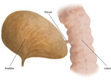 |
Bladder Fistula (Labeled) | Bladder Fistula. Colon. | Royal College of Surgeons in Ireland Illustrations | |
| 2269 |
|
Male Reproductive System | Anatomy of testicle. | Testicle; Glans Penis; Ductus Deferens | Royal College of Surgeons in Ireland Illustrations |
| 2270 |
|
Ruptured Thoracic Aorta (Labeled) | Ruptured thoracic aorta. | Oesophagus; Posterior Mediastinum; Anterior Mediastinum | Royal College of Surgeons in Ireland Illustrations |
| 2271 |
|
Male Urogenital System | Male urogenital system. | Ureteric Orifice; Sphincter Urethrae Muscle; Perineal Body; Levator Ani | Royal College of Surgeons in Ireland Illustrations |
| 2272 |
|
Brachial Plexus (Labeled) | Brachial plexus. | Lateral Cord; Posterior Cord; Axillary Nerve; C5-C8; T1 | Royal College of Surgeons in Ireland Illustrations |
| 2273 |
|
Gastric Lymph Nodes | Gastric lymph nodes. Lymphatic System. | Coeliac Nodes; Splenic Nodes; Pancreatic Nodes | Royal College of Surgeons in Ireland Illustrations |
| 2274 |
|
Mandible - Inner and Outer Surfaces (Labeled) | Mandible. | Coronoid Process; Ramus; Angle of Mandible; Mental Foramen; Mylohyoid Groove; Lingula; Mandibular Foramen; Mylohyoid Line; Condylar Process | Royal College of Surgeons in Ireland Illustrations |
| 2275 |
|
Prostate Gland and Ejaculatory Ducts | Prostate gland. Ejaculatory ducts. | Median Lobe of Prostate Gland; Right Ejaculatory Duct; Left Ejaculatory Duct | Royal College of Surgeons in Ireland Illustrations |
| 2276 |
|
Intestinal Obstructions | Mural tumor, luminal gallstone, and extramural hernia. | Intestinal Obstructions; Mural Tumor; Luminal Gallstone; Extramural Hernia | Royal College of Surgeons in Ireland Illustrations |
| 2277 |
|
Epididymal Cyst | Spermatocele. Epididymal cyst. | Epididymal Cyst | Royal College of Surgeons in Ireland Illustrations |
| 2278 |
|
Thoracic Lymph Nodes (Labeled) | Thoracic lymph nodes. Lymphatic System. | Right Lymph Duct; Internal Jugular Vein; Left Subclavian Vein; Left Brachiocephalic Vein; Cisterna Chyli; Bronchiomediastinal Trunk | Royal College of Surgeons in Ireland Illustrations |
| 2279 |
|
Inferior Epigastric Arteries and Veins | Inferior epigastric arteries. Inferior epigastric veins. | Inferior Epigastric Arteries; Inferior Epigastric Veins; Deep Inguinal Ring; Obturator Artery; Obturator Vein | Royal College of Surgeons in Ireland Illustrations |
| 2280 |
|
Brachial Plexus (Labeled) | Brachial plexus. | Lateral Cord; Posterior Cord; Axillary Nerve | Royal College of Surgeons in Ireland Illustrations |
| 2281 |
|
Digestive System | Digestive system. GI tract. Inferior vena cava. Mesenteric vessels. Aorta. | Royal College of Surgeons in Ireland Illustrations | |
| 2282 |
|
Vulva | External genitalia. Female. | Mons Pubis; Glans; External Urethral Orifice; Perineal Body; Opening of Vagina; Vestibule of Vagina; Labium Minus; Labium Majus | Royal College of Surgeons in Ireland Illustrations |
| 2283 |
|
Elbow Joint (Labeled) | Elbow. Bones. Ligaments. | Annular Ligament; Radial Collateral Ligament; Tendon of Bicep; Ulnar Collateral Ligament | Royal College of Surgeons in Ireland Illustrations |
| 2284 |
|
Testicle - Exposed | Testicle. Spermatic cord. Tunica vaginalis. | Tunica Vaginalis | Royal College of Surgeons in Ireland Illustrations |
| 2285 |
|
Male Urogenital System | Male urogenital system. | Levator Ani; Sphincter Ani Externus; Anococcygeal Ligament; Anal Canal; Perineal Body; Urogenital Diaphragm; Superficial Perineal Pouch; Navicular Fossa; Spongy Urethra; Urethra Bulb; Membranous Urethra; Prostatic Urethra; Colles' Fascia | Royal College of Surgeons in Ireland Illustrations |
| 2286 |
|
Mastectomy - Right Breast | Breast cancer. Incision site. Mastectomy. | Incision Site; Breast Cancer | Royal College of Surgeons in Ireland Illustrations |
| 2287 |
|
Esophagus, Heart and Thoracic Aorta (Labeled) | Esophagus. Heart. Thoracic Aorta. | Oesophagus | Royal College of Surgeons in Ireland Illustrations |
| 2288 |
|
Elbow Joint (Labeled) | Elbow. Bones. Ligaments. | Annular Ligament; Radial Collateral Ligament; Tendon of Bicep; Ulnar Collateral Ligament | Royal College of Surgeons in Ireland Illustrations |
| 2289 |
|
Cross-Section of Chest (Labeled) | Pericardium. Heart. Lungs. | Serous Pericardium, Parietal; Serous Pericardium, Visceral; Fibrous Pericardium; Pericardial Cavity; Right Atrium; Left Atrium; Right Ventricle; Left Ventricle; Oblique Sinus of Pericardial Cavity; Oesophagus; Pulmonary Veins | Royal College of Surgeons in Ireland Illustrations |
| 2290 |
|
Lymphatics | Lymphatic system. Thoracic structures and vessels. | T1; T10; Cisterna Chyli; Oesophagus; Descending Aorta | Royal College of Surgeons in Ireland Illustrations |
| 2291 |
|
Pelvic Girdle | Pelvic girdle. Os coxa. | Articular Surface; Fifth Lumbar Vertebra; ASIS; Obturator Foramen; Pubis; Pubic Tubercle; Ischial Spine; Promontory | Royal College of Surgeons in Ireland Illustrations |
| 2292 |
|
Puestow Procedure (Labeled) | Puestow procedure. Pancreas. | Puestow Procedure; Stricture; Jejunal Loop | Royal College of Surgeons in Ireland Illustrations |
| 2293 |
|
Male Reproductive System | Illustration of penis and male perineum. | Corpus Spongiosum; Corpus Cavernosum; Glans Penis; Corona; Bulb of Penis; Perineal Membrane; Crus Penis | Royal College of Surgeons in Ireland Illustrations |
| 2294 |
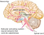 |
Neural Network Controlling Wakefulness (Labeled) | Brainstem reticular activating system. | Royal College of Surgeons in Ireland Illustrations | |
| 2295 |
 |
Ventral Surface of the Brain with Cranial Nerves - Basal (Labeled) | Ventral surface of brain. Cranial nerves. | Pituitary Stalk; Optic Tract; Nervus Intermedius; Choroid Plexus | Royal College of Surgeons in Ireland Illustrations |
| 2296 |
 |
Retropharyngeal Space (Labeled) | Retropharyngeal space. | Aryepiglottic Fold; Vestibular Fold; Ventricle; Posterior Lamina of Cricoid Cartilage; Cricothyroid Membrane; Thyrohyoid Membrane | Royal College of Surgeons in Ireland Illustrations |
| 2297 |
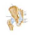 |
Ligaments of Hip - Anterior (Labeled) | Hip ligaments. | Posterior Sacroiliac Ligament; Sacrospinous Ligament; Sacrotuberous Ligament; Ischiofemoral Ligament | Royal College of Surgeons in Ireland Illustrations |
| 2298 |
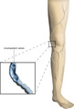 |
Varicose Veins - Incompetent Valves (Labeled) | Varicose veins. Incompetent valves. | Incompetent Valves | Royal College of Surgeons in Ireland Illustrations |
| 2299 |
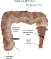 |
Closed Loop Obstruction of Colon (Labeled) | Closed loop obstruction of colon. Carcinoma of the descending colon. | Flaccid Distal Bowel; Distended Caecum; Competent Ileocaecal Valve; Dilated Proximal Bowel | Royal College of Surgeons in Ireland Illustrations |
| 2300 |
 |
Female Urogenital System (Labeled) | Urogenital system. Female. | Fimbria; Uterovesical Pouch; Anterior Abdominal Wall; Symphysis Pubis; Anal Canal | Royal College of Surgeons in Ireland Illustrations |