The Health Education Assets Library (HEAL) is a collection of over 22,000 freely available digital materials for health sciences education. The collection is now housed at the University of Utah J. Willard Marriott Digital Library.
TO
Filters: Collection: "ehsl_heal"
| Title | Description | Subject | Collection | ||
|---|---|---|---|---|---|
| 2101 |
 |
An introduction and literature references to the Human Placenta as part of the POJA collection on FEMALE REPRODUCTIVE ORGANS | An introduction and literature references to the Human Placenta as part of the POJA collection on FEMALE REPRODUCTIVE ORGANS (PDF File) | placenta; references; preface | Poja Histology Collection - Placenta |
| 2102 |
 |
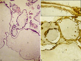
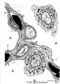
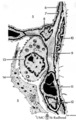
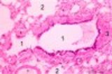
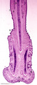
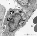
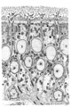
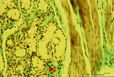
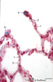
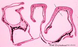
Anchoring villi (human placenta, early midpregnancy) | Stain: Hematoxylin -azophloxine. Survey (A) and detail (B). The maternal side at the bottom shows reddish matrix-type fibrinoid accumulations (1) (Rohr's fibrinoid layer) close to an anchoring villus (2) and the cytotrophoblastic cell columns (3). The anchoring villus is lined by cytotrophoblast... | placenta; chorionic villi; Hofbauer cell; anchoring villus; cytotrophoblast | Poja Histology Collection - Placenta |
| 2103 |
 |
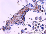
Anchoring villi (human placenta, early midpregnancy) | Stain: Hematoxylin -azophloxine. The maternal side at the bottom shows reddish matrix-type fibrinoid accumulations (1) (Rohr's fibrinoid layer) close to an anchoring villus (2) and a cytotrophoblastic cell column (3). The anchoring villus is lined by cytotrophoblasts (4) which are covered at th... | placenta; anchoring villus; fibrinoid; deciduas; cytotrophoblast | Poja Histology Collection - Placenta |
| 2104 |
 |
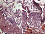
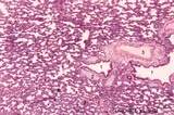
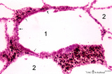
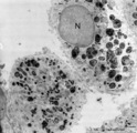
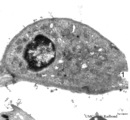
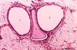
Complete hydatidiform mole (human) | Stain: (A) Hematoxylin-eosin. (B+C) Immunoperoxidase staining with diaminobenzidin (DAB) and hematoxylin counterstaining for keratin 7 (OVTL 12-30 antibody). (A)Trophoblast cells surround the dilated chorionic villi (1), with centrally large amounts of edematous mesenchymal stroma with almost no b... | trophoblast; complete hydatidiform mole; placenta; villus; immunohistochemistry; keratin-7 | Poja Histology Collection - Placenta |
| 2105 |
 |
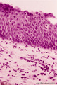
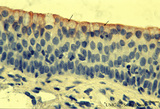
Epithelial lining of trachea and bronchiolus (mammals) | Left side trachea: Columnar ciliated cells (1) with up to 200 motile cilia with an organelle-rich apex and in the middle a goblet cell (2) with accumulation of mucus secretion granules in the apex. Note that all epithelial cells (i.e., junctions) contact the basal lamina. Four basal cells (3) do not... | Bronchiolus; Pseudostratified epithelium; Ciliated epithelium; Clara cells; Basal cells; Neuro-endocrine cells | Poja Histology Collection - Respiratory System Subset |
| 2106 |
 |
Air-blood barrier in the lung (mammals) | Scheme electron microscopy. (1, ↓) Represents type I pneumocytes lining alveolar spaces (A). Cell (2) represents a free alveolar macrophage. The type II pneumocyte (3) is adherent to type I pneumocyte extensions (note junctional connection), and contains multilamellar bodies (surfactant). A myofib... | Pneumocyte type I ; Pneumocyte type II | Poja Histology Collection - Respiratory System Subset |
| 2107 |
 |
Neuroepithelial body in terminal bronchiolus (golden hamster) | Electron microscopy. Three epithelial cells, as part of the N(euro) E(pithelial) B(ody), contribute to a cluster of neuroendocrine cells. They belong to the Amine Precursor Uptake and Decarboxylation cell system so-called APUD cells. The dense-core granules (↓) contain among others dopamine or ser... | Terminal bronchiolus ; Neuro-endocrine cells | Poja Histology Collection - Respiratory System Subset |
| 2108 |
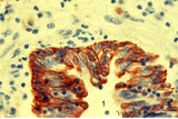 |
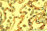
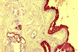
Keratin 7 in the bronchial epithelium of the lung (human, adult) | Stain: anti-keratin 7 antibody (Pan-Ck 7) immunoperoxidase staining (with aminoethylcarbazole (AEC) substrate). The epithelium of the bronchus (1) stains dark brown-red after the reaction with AEC indicating a positive reaction for cytokeratin 7. This antibody can be used in the study of development... | Bronchiolus; Immunoperoxidase; Immuno-reaction | Poja Histology Collection - Respiratory System Subset |
| 2109 |
 |
Surface of olfactory epithelium (rat) | Electron microscopy. Bottom left shows an olfactory bulb (1, vesicle) with cross-sectioned basal bodies. Right side part of the apex of a supporting cell (2) with microvilli (3). Parallel to the surface of the epithelium one long olfactory cilium (4) and several other cross-sectioned ones are detect... | Olfactory epithelium; Olfactory vesicle | Poja Histology Collection - Respiratory System Subset |
| 2110 |
 |
Alveolar cells in the lung (mammals) | Scheme electron microscopy. (5) alveolar space; (6) type I Pneumocyte; (7) basal lamina; (8) myofibroblast; (9) collagen and elastin fibers; (10) mesothelial cell of the visceral pleura; (11) capillary with erythrocyte; (12) endothelial cell lining the capillary; (13) type II pneum... | Pneumocyte type I ; Pneumocyte type I I | Poja Histology Collection - Respiratory System Subset |
| 2111 |
 |
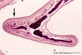
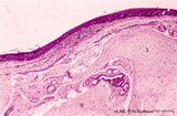
Scheme of the epiglottis (human, adult) | The laryngeal side of the epiglottis (1) is covered with respiratory epithelium, while the lingual side (2) is similar to the oral cavity epithelium (squamous type). The transitional zone to respiratory epithelium is marked by (3). The scaffold consists of elastic cartilage (4). Seromucous laryngea... | Laryngeal glands; Oral cavity | Poja Histology Collection - Respiratory System Subset |
| 2112 |
 |
Epithelial lining of bronchiolus in the lung (rat) | Scanning electron microscopy. Bushes of cilia indicate the presence of ciliated cells (1). Clustered non-ciliated cells (2, Clara cells) are dome-shaped with stubby microvilli at the surface, they protrude into the lumen to the tips of the cilia. | Bronchiolus ; Ciliated epithelium ; Clara cells | Poja Histology Collection - Respiratory System Subset |
| 2113 |
 |
Transitional region of epiglottis (human, high magnification) | Stain: Hematoxylin and eosin. Transition of squamous epithelium into pseudostratified epithelium. Note diapedesis of lymphocytes (darker stained rounded cells) through the epithelial barrier (→). The proper lamina presents some lymphocyte infiltration. | Diapedesis | Poja Histology Collection - Respiratory System Subset |
| 2114 |
 |
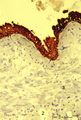
Keratin 7 in the lining alveolar epithelium of the lung (human, adult) | Stain: anti-keratin 7 antibody (Pan-Ck 7) immunoperoxidase staining (with aminoethylcarbazole (AEC) substrate). Both alveolar epithelial cell types I (1) and II (2) stain dark brown-red after the reaction with AEC indicating a positive reaction for cytokeratin 7. Note the white (non-stained) capilla... | Pneumocyte I; Pneumocyte II; Imunoperoxidase; Immuno-reaction | Poja Histology Collection - Respiratory System Subset |
| 2115 |
 |
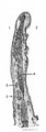
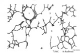
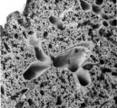
Arbor bronchialis, the bronchial tree (human, adult) | Resin corrosion cast of the lower trachea and bronchial tree (posterior aspect). The lobar and segmental bronchi and their main branches are coloured, different colours indicate areas supplied by different segmental bronchi. White-coloured lower trachea (Tr) divides into two principal bronchi (Bp)... | Segmental bronchi ; Macroscopy | Poja Histology Collection - Respiratory System Subset |
| 2116 |
 |
Respiratory bronchiolus (human) | Stain: Hematoxylin and eosin. A longitudinal section shows the lumen of a respiratory bronchiolus (1) with an irregular lining. It is covered by low columnar-cuboidal cells, a few pouches within the same lumen are lined by thin alveolar epithelium as found in the surrounding alveoli (2). Thin arrows... | Respiratory bronchiolus; Columnar epithelium; Cuboidal epithelium; Clara cells; Alveolar epithelium | Poja Histology Collection - Respiratory System Subset |
| 2117 |
 |
Nasal concha (dog, isolated turbinate bone) | Stain: Hematoxylin and eosin. This concha (turbinate with red-black-stained bone (1), is covered by a thin respiratory mucosa (3) and by a thick olfactory mucosa (2, arrow). Within the respiratory epithelium light stained goblet cells are visible between the low columnar/cuboidal epithelial cells (c... | Conchae nasales; Olfactory epithelium; Respiratory epithelium | Poja Histology Collection - Respiratory System Subset |
| 2118 |
 |
Small bronchus (golden hamster) | Electron microscopy. Two ciliated cells with nuclei (1). Supranuclearly electron-dense lysosomes (2, lipofucsin) are present, the apical parts contain mitochondria (3) and cross-sections of cilia (4) and microvilli (5) are visible in the lumen (*). Two mucoid goblet cells with packed secretion gran... | Ciliated epithelium | Poja Histology Collection - Respiratory System Subset |
| 2119 |
 |
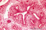
Striated duct of nasal gland (rat) | Electron microscopy. At (1) lumen of duct, (2) indicates nucleus of lining epithelial cell. Note the abundancy and palisade-like arrangement of mitochondria (3) in the basolateral regions, in the light microscope visible as a fine striation, hence the name striated duct or intralobular duct. (4) ind... | Nasal vestibulum; Nasal glands; Striated duct; Intralobular duct; Seromucous glands | Poja Histology Collection - Respiratory System Subset |
| 2120 |
 |
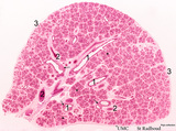
Late terminal sac period of developing lung (human, fetus, low magnification) | Stain: Hematoxylin and eosin. Cross-sections of a bronchus (1) with cartilage rings (2) closely associated with pulmonary arteries (3). The alveolar appearance is evident, and the open spaces represent future alveolar duct systems as well as alveolar sacs. The cellular septa are still thick due to n... | Lung development; Terminal sac period | Poja Histology Collection - Respiratory System Subset |
| 2121 |
 |
Elastin in alveolus of lung tissue (human, adult) | Immuno-electron microscopy (embedded in Lowicryl HM 20). Elastin (E) is labeled with electron-dense gold particles of 10 nm. Note that the amorph elastin appears pale after embedding in this type of resin. (A) indicates alveolar space, (F) unlabelled myofibrolast, (C) collagen fibers, and (1) the th... | Alveolar cells type I; Myofibroblasts; Immuno-electron microscopy; Immuno-staining | Poja Histology Collection - Respiratory System Subset |
| 2122 |
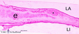 |
Survey of epiglottis (human) | Stain: Azan. Centrally light-stained elastic cartilage (e). Lingual side (LI) is covered with squamous epithelium. At the laryngeal side (LA) the squamous epithelium usually turns into respiratory epithelium with seromucous laryngeal glands (*). | Laryngeal gland; Oral cavity | Poja Histology Collection - Respiratory System Subset |
| 2123 |
 |
Lamellar body of a type II alveolar cell in the lung (dog) | Electron microscopy. Extrusion of surfactant material out of a type II alveolar cell (pneumocyte II, great alveolar cell). After fusion of a multilamellar body (1) (cytosome) with the apical cell membrane (note microvilli 2) the electron-dense lamellar content or surfactant (consisting of phosphatid... | Type II alveolar cell; Pneumocyte II; Multilamellar bodies; Cytosomes; Tubular myelin | Poja Histology Collection - Respiratory System Subset |
| 2124 |
 |
Pseudoglandular - canalicular period of developing lung (human, fetus) | Stain: Azan. Part of a bronchus (1) with young cartilage (2). The hyaline cartilage presents solitary chondrocytes embedded in the blue intercellular substance surrounded by a perichondrium (3). Note that the bronchus as well as the bronchial tubes (4) show an apical position of the epithelial nucle... | Lung development; Pseudoglandular period; Canalicular period; Mesenchyme | Poja Histology Collection - Respiratory System Subset |
| 2125 |
 |
Longitudinal section of trachea (human, adult) | Stain: Azan. On top: the epithelium (1) is pseudostratified with cilia and goblet cells. The dense lamina propria (2, ↓, deep blue) is demarcated from the submucosa with seromucous tracheal glands (3). C-shaped cartilage rings (C, light blue) maintain patency. The cartilage edges are connected by ... | Pseudostratified epithelium ; Seromucous glands; Tracheal glands; Fibroelastic membrane ; Adventitia | Poja Histology Collection - Respiratory System Subset |
| 2126 |
 |
Elastin in alveolus of lung tissue (human, adult) | Immuno-electron microscopy (embedded in Lowicryl HM 20). The amorph elastin (E) appears pale after embedding in this type of resin, but is antibody-labeled with electron-dense gold particles of 10 nm. (C) indicate bundles of collagen fibers (unlabeled) which are intermingled with swollen cytoplasmic... | Immuno-staining; Immuno-electron microscopy | Poja Histology Collection - Respiratory System Subset |
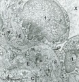
| 2127 |
 |
Nasal glands of the nasal vestibulum (rat, higher magnification) | Electron microscopy. Part of a serous acinus of a seromucous nasal gland demonstrates the central lumen (*) formed by four gland cells. Note junctional complexes (↑) and the close proximity of the dense secretion granules (2) ready to be released into the central lumen. These active cells contain ... | Nasal vestibulum; Seromucous glands; Nasal glands; Serous cells; Secretion granules | Poja Histology Collection - Respiratory System Subset |
| 2128 |
 |
Pseudoglandular period of developing lung (human, embryo) | Stain: Trichrome (Goldner). Cross-sectioned future bronchial tubes (1) of varying sizes, note the apical position of the nuclei with light-stained basal parts (glycogen). The surrounding mesenchyme condenses (↓) around the epithelium and in the neighbourhood small blood vessels (*) are present. At... | Lung development; Pseudoglandular period; Bronchial tubes; Mesenchyme | Poja Histology Collection - Respiratory System Subset |
| 2129 |
 |
Pseudoglandular period of developing lung (human, embryo) | Stain: Trichrome (Goldner). Cross-sectioned future bronchial tubes (1) of varying sizes, note the apical position of the nuclei with light-stained basal parts (glycogen). The surrounding mesenchyme becomes condensed (↓) around the epithelium and in between small blood vessels (*) are present. | Lung development; Pseudoglandular period; Bronchial tubes; Mesenchyme | Poja Histology Collection - Respiratory System Subset |
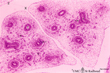
| 2130 |
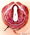 |
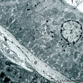
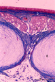
Survey of cross-section through larynx-esophagus (human) | Stain: Hematoxylin and bordeaux light. X = lumen of larynx (middle part); ↑ = esophagus (below) with an undulating mucosa covered with squamous epithelium. Elastic arythenoid cartilage (1); thyreoarythenoid muscle (2); thyroid cartilage (3); hyoid muscle (4). | Arythenoid cartilages | Poja Histology Collection - Respiratory System Subset |
| 2131 |
 |
Nostril area of nasal vestibulum - cutaneous region - human | Stain: Azan. Slightly keratinized squamous epithelium (1) with thin red cornified layer; (2) hair follicles; (3) vibrissae and (4) associated sebaceous glands. At the bottom skeletal muscle fibers; (5) transverse part of the musculous nasalis; (6) connective tissue. | Poja Histology Collection - Respiratory System Subset | |
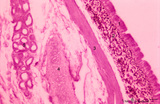
| 2132 |
 |
Survey nasal septum (dog) | Stain: Hematoxylin and eosin. The nasal septum consists of a cartilage skeleton present as one long (1, cartilago vomeronasalis) and two short plates (2, cartilago septi nasi) with centrally dark-stained areas of chondrocytes. Below, the distal tip is covered with keratinized stratified squamous epi... | Squamous stratified epithelium; Nasal vestibulum; Venous sinusoids; Venous plexus; Swell bodies | Poja Histology Collection - Respiratory System Subset |
| 2133 |
 |
Developing conchae in a frontal section of head (pig, fetus) | Stain: Azan. The fetal conchae develop like a branched gland (1) within the blue-stained trabecular bone (2) of the future skull. The fetal skin (3) is present at the top and bottom. Groups of cheek musculature (4) in middle left and right side. A broad sleeve of chin muscles below (5). The developi... | Conchae nasales; Trabecular bone | Poja Histology Collection - Respiratory System Subset |
| 2134 |
 |
Gross scheme of a lung lobule and its vascularization (human) | Scheme of branching of intrapulmonary bronchus. A small intrapulmonal bronchus with cartilage rings (1) divides into bronchioli (2), at (3) the terminal bronchioli that continues into respiratory bronchioli (4) followed by alveolar ducts (5) and at (6) the alveoli. A respiratory bronchiolus with alv... | Intrapulmonary bronchus; Bronchioli; Alveolar ducts; Lymphatic plexus; Visceral pleura | Poja Histology Collection - Respiratory System Subset |
| 2135 |
 |
Olfactory mucosa in the nose (dog) | Stain: Iron Hematoxylin and eosin (Heidenhain). The pseudostratified epithelium of the olfactory mucosa contains sensory cells, support cells and basal cells. The upper darker stained nuclei (1) belong to the supporting (sustentacular) cells showing a dark apical line (↑) at the top. Below lighte... | Bowman glands; Olfactory glands; Olfactory mucosa; Pseudostratified epithelium | Poja Histology Collection - Respiratory System Subset |
| 2136 |
 |
Olfactory gland (Bowman) in olfactory region of concha (human) | Electron microscopy. Beneath the olfactory epithelium tubuloalveolar glands of Bowman (1) are found containing cells that produce dense secretion granules arranged around a large lumen (*) with greyish secretion product. These cells produce a serous fluid in which odoriferous substances are dissolve... | Olfactory epithelium; Bowman glands; Olfactory glands; Secretion granules | Poja Histology Collection - Respiratory System Subset |
| 2137 |
 |
Olfactory epithelium in the nose (human) | Electron microscopy. At the top (lumen) cross-sections of olfactory bulbs (1) (vesicles) with cilia/basal bodies. Sustentacular (supporting) cells (2) exhibit numerous microvilli in the lumen, their cytoplasms contain many dispersed mitochondria and in the lower half aggregations of endoplasmic reti... | Olfactory epithelium; Olfactory vesicle | Poja Histology Collection - Respiratory System Subset |
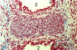
| 2138 |
 |
Alveolar sac in the lung (human) | Stain: Azan. Characteristic alveolar tips (arrows) of neighbouring thin-walled alveoli (1). The tips contain elastin masked by collagen (blue-stained). At (2) small pulmonary arteries. | Alveolar tips | Poja Histology Collection - Respiratory System Subset |
| 2139 |
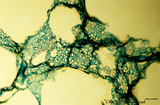 |
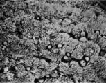
Capillary system of lung alveoli (cat) | Stain: specimen injected with trypan blue in gelatin via the pulmonary system. In a tangential view the blue-stained dense capillary plexus surrounding each alveolus is well depicted. | Poja Histology Collection - Respiratory System Subset | |
| 2140 |
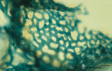 |
Capillary system of an alveolus (cat) | Stain: specimen injected with trypan blue in gelatin via the pulmonary system. In a tangential view the blue-stained dense capillary plexus of one alveolus is well shown. | Poja Histology Collection - Respiratory System Subset | |
| 2141 |
 |
Type II alveolar cell in the lung (rat) | Electron microscopy. At the top the alveolar space (1) is lined by a type II alveolar cell (pneumocyte II) (2) with blunt microvilli. In the cytoplasm the characteristic electron-dense multilamellar bodies (*) and light-stained swollen mitochondria are present. The lamellar bodies are responsible fo... | Pneumocyte I; Pneumocyte II; Alveolar cell type I; Alveolar cell type II; Multilamellar bodies; Interstitial cells | Poja Histology Collection - Respiratory System Subset |
| 2142 |
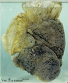 |
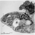
Bullous emphysema of the lung (honeycomb lung, human, adult) | Macroscopy of the left lung showing large apical bullae (3) due to total destruction of all alveoli leaving remnants of the covering pleura. (2) indicates small bullae of foci of destroyed lobules with permanent enlargement of the air spaces and (1) points to spots of anthracosis in less-affected lu... | Macroscopy; Honeycomb lung; Interstitial fibrosis; Hamman-Rich Syndrome | Poja Histology Collection - Respiratory System Subset |
| 2143 |
 |
Elastin in alveolar tip in lung tissue (human, adult) | Immuno-electron microscopy (embedded in Lowicryl HM 20). The amorph elastin (E) appears pale after embedding in this type of resin, but is antibody-labeled with electron-dense gold particles of 10 nm. (Col) indicate bundles of collagen fibers which are intermingled with swollen cytoplasmic extension... | Alveolar cell type I; Myofibroblast; Immuno-electron microscopy; Immuno-staining | Poja Histology Collection - Respiratory System Subset |
| 2144 |
 |
Scheme survey frontal section of larynx (human) | Stain: Azan. A pseudostratified epithelium covers the mucosa of the laryngeal cavity and vestibulum. At the vocal fold edge (3) the epithelium appears to be nonkeratinizing squamous. The connective tissue is rigidly attached to this edge and merges into the vocal ligament (dense elastic) and vocal m... | Vocal ligament; Laryngeal glands; Ventricular fold | Poja Histology Collection - Respiratory System Subset |
| 2145 |
 |
Pseudoglandular period of developing lung (human, embryo, low magnification) | Stain: Hematoxylin and eosin. The two future lung lobes contain many cross-sectioned future bronchial tubes (1). Note in these epithelial cells the apical position of the nuclei with light-stained basal parts (glycogen). The surrounding mesenchyme becomes more condensed (↓) around the epithelium a... | Lung development; Visceral pleura; Pseudoglandular period; Bronchial tubes; Mesenchyme | Poja Histology Collection - Respiratory System Subset |
| 2146 |
 |
Terminal sac period of developing lung (human, fetus, low magnification) | Stain: Hematoxylin and eosin. At the right cross-sections of a large bronchus (1) with cartilage rings (2). Close to them a pulmonary artery (3), and a bronchiolus (4) without cartilage. (5) indicates a respiratory bronchiolus. Most alveoli are not inflated, the numerous dilated spaces (6) are saccu... | Lung development ; Terminal sac period; Respiratory bronchioli | Poja Histology Collection - Respiratory System Subset |
| 2147 |
 |
Olfactory vesicle (bulb) in the nose (gerbil) | DUPLICATE RECORD - FOR DELETION Electron microscopy. A distinct olfactory bulb (1, vesicle) with horizontally extended cross-sectioned cilia (*) and basal bodies (↑) is surrounded by numerous microvilli and free olfactory cilia. Close to the first bulb a second one is about to protrude above th... | Olfactory epithelium; Olfactory vesicle | Poja Histology Collection - Respiratory System Subset |
| 2148 |
 |
Ventricular fold (plica ventricularis) (human) | Stain: Azan. On top pseudostratified epithelium (1). Lamina submucosa with predominantly mucous glands (3), fat cells (4) and blood vessels. The lamina propria also contains lymphoid follicles (2) with a germinal center (immune protection). | Laryngeal glands; Ventricular fold; Seromucous glands | Poja Histology Collection - Respiratory System Subset |
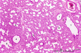
| 2149 |
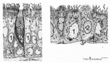
 |
Survey of a centrilobular lung emphysema: section of an lobule (human, adult) | Stain: Hematoxylin and eosin. The enlargement of large areas of the air spaces (X) is evident due to destruction of the walls of alveoli throughout the lobule. | Poja Histology Collection - Respiratory System Subset | |
| 2150 |
 |
Olfactory epithelium in the nasal cavity (mammals) | Scheme electron microscopy. Four olfactory bulbs (1, vesicles) with radially sprouting cilia are present at the surface of the epithelium. Their slender cell bodies (2, bipolar neurons) are flanked by sustentacular (supporting) broad cells (3) whose apices are filled with well developed organelles, ... | Pseudostratified epithelium; Olfactory epithelium; Basal cells; Olfactory vesicle; Bipolar neuron | Poja Histology Collection - Respiratory System Subset |
| 2151 |
 |
Keratin staining of bronchus epithelium (human, adult) | Stain: Anti-keratin-5-7-14-19 (OVTL 12/5 monoclonal antibody with immunoperoxidase staining on frozen sections). The pseudostratified epithelium (1) is stained keratin-positively (brown product) as well as the epithelial derivates such as the bronchial glands (2) and their excretory ducts (3). Other... | Bronchial epithelium; Keratin antibodies; Tumor diagnosis | Poja Histology Collection - Respiratory System Subset |
| 2152 |
 |
Keratin staining of bronchus epithelium (human, adult) | Stain: Anti-keratin-5-7-14-19 (OVTL 12/5 monoclonal antibody with immunoperoxidase staining on frozen sedctions). The pseudostratified epithelium (1) reacts positively (brown-red product), the nuclei of the basal cells are well visible (arrows). Other structures react negatively (blue stain) such as... | Tumor diagnosis; Bronchial epithelium; Lung parenchym; Keratin antibodies | Poja Histology Collection - Respiratory System Subset |
| 2153 |
 |
Longitudinal section of trachea (human, adult) | Stain: Azan. On top: the epithelium (1) is pseudostratified with cilia and goblet cells on a distinct basement membrane (↓). There is almost no demarcation (at this magnification) between lamina propria and submucosa due to the dense blue (2) staining of elastic fibers reinforced by collagen fiber... | Pseudostratified epithelium ; Tracheal glands; Fibroelastic membrane | Poja Histology Collection - Respiratory System Subset |
| 2154 |
 |
Respiratory bronchiolus (mouse) | Stain: Hematoxylin and eosin. The lumen (1) shows an irregular lining and is covered by low columnar-cuboidal cells (↓), locally disrupted by a few areas with thin alveolar epithelium as found in the surrounding alveoli (2). Thick arrows point to a row of light-stained Clara cells. At (*) discont... | Respiratory bronchiolus; Columnar epithelium; Cuboidal epithelium; Clara cells; Alveolar epithelium | Poja Histology Collection - Respiratory System Subset |
| 2155 |
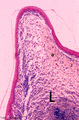 |
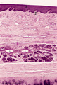
Vocal cord (plica vocalis) (human, glottis) | Stain: Azan. The true vocal cord consist of the vocal ligament (L) covered by stratified squamous epithelium and the striated vocal muscle (medial part of the thyreoarythenoid muscle, not depicted in this picture). At the tip of the vocal cord the stratified epithelium is distinct and the lamina pro... | Vocal ligament | Poja Histology Collection - Respiratory System Subset |
| 2156 |
 |
Pseudoglandular - canalicular period of developing lung (human, fetus) | Stain: Azan. Terminal budding and elongation of the future bronchial tree: branching of a bronchial tubes (1) within an islet where the mesenchyme condenses (4). A large blood vessel (2) as well as a lymph vessel (3) are recognizable. Note in the mesenchyme formation of blood capillaries in the vici... | Lung development; Pseudoglandular period ; Canalicular period; Mesenchyme | Poja Histology Collection - Respiratory System Subset |
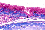
| 2157 |
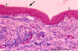 |
Transition surface of vocal cord into laryngeal surface (human, subglottis, higher magnification) | Stain:Stain: Azan. Transition (↓) from stratified squamous epithelium (1) into pseudostratified epithelium (2) in the subglottis region. Blood vessels and the cellularity of the proper lamina is evident. | Olfactory epithelium; Subglottis; Stratified epithelium; Pseudostratified epithelium | Poja Histology Collection - Respiratory System Subset |
| 2158 |
 |
Submucosa of trachea (human, adult) | Stain: Goldner trichrome. Mixed tracheal glands (1) of the submucosa (connective tissue light green) and bundles of smooth muscle fibers (2) close to the fibroelastic membrane between the cartilage edges. | Seromucous gland; Tracheal glands | Poja Histology Collection - Respiratory System Subset |
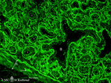
| 2159 |
 |
Heparan sulfates in the terminal sac-period of the lung (mouse, fetus) | Stain: fluorescence microscopy with anti-heparan sulphate antibody (a phage-display antibody, LKiv69). Heparan sulfates are linear polysaccharides that belongs to the group of the glycosaminoglycans (GAGs). The sulphated saccharide domains provide numerous docking sites for protein ligands. These GA... | Bronchioli; Air spaces; Terminal sac period | Poja Histology Collection - Respiratory System Subset |
| 2160 |
 |
Lung capillaries and air-blood barrier (rat) | Electron microscopy. Two neighboring capillaries (C) and the alveolar spaces (A). The endothelial cell (1) of the upper capillary contains an organelle-rich cytoplasm, a centriole (2), contractile filaments (↓) and electron-dense membrane-bound granules, the so-called Weibel-Palade bodies. These b... | Poja Histology Collection - Respiratory System Subset | |
| 2161 |
 |
Transition terminal bronchiolus - respiratory bronchiolus (human) | Stain: Azan. A longitudinal section shows the transition from the lumen of a terminal bronchiolus (1) into that of a respiratory bronchiolus (2). The epithelium of the terminal bronchiolus is regularly columnar and ciliated. The respiratory bronchiolus has an irregular lining and is covered by low c... | Terminal bronchiolus; Respiratory bronchiolus; Columnar epithelium; Cuboidal epithelium; Alveolar epithelium | Poja Histology Collection - Respiratory System Subset |
| 2162 |
 |
Pseudoglandular-canalicular period of developing lung (human, fetus, low magnification) | Stain: Azan. A future lung lobe with cross-sectioned larger bronchi (1) in close association with islets of future smaller bronchial tubes (*). Around the larger bronchi several cartilage structures (↓) are already present. Note that the mesenchyme around the tubes within the islets becomes more c... | Lung development; Visceral pleura; Pseudoglandular period; Canalicular period; Bronchial tubes; Mesenchyme | Poja Histology Collection - Respiratory System Subset |
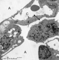
| 2163 |
 |
Alveolus in lung (dog) | Electron microscopy. (A) indicate alveolar space; (C) indicate capillary with erythrocyte. Drawing-pin represents endothelium. (1) type I alveolar cell; (↓) indicate thin cytoplasm of pneumocyte I. (2) type II alveolar cell. (3) alveolar macrophage. (*) are spots of elastin, intermingled with coll... | Pneumocyte I; Pneumocyte II; Alveolar cells; Alveolar macrophage | Poja Histology Collection - Respiratory System Subset |
| 2164 |
 |
Respiratory region of nasal vestibulum (human, higher magnification) | Stain: Azan. At the top pseudostratified, ciliated epithelium (1) with a distinct endoepithelial gland (2, pink-stained mucous cells). The proper lamina is cell-rich and draining ducts (3, enclosed by thicker blue-stained borders) and acini of the nasal glands (5) are present here as well as in the ... | Pseudostratified epithelium; Nasal glands; Seromucous glands; Endoepithelial glands | Poja Histology Collection - Respiratory System Subset |
| 2165 |
 |
Trachea (cat) | Stain: Orcein and eosin. At the top pseudostratified epithelium (1) followed by a thick layer filled with dark-stained elastic fibers (2). Bundle of smooth muscle fibers (3) (note: there is an artifactual space between the elastic layer and the smooth muscle), belonging to the tunica fibro-musculo-... | Tracheal glands; Seromucous glands | Poja Histology Collection - Respiratory System Subset |
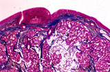
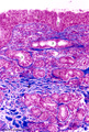
| 2166 |
 |
Epiglottis (human) | Stain: Hematoxylin and eosin. Stratified squamous epithelium (1) at the laryngeal side (top). Lymphocyte accumulation (2) in the lamina propria. Below elastic cartilage (3) and seromucous laryngeal glands (4) with draining ducts. | Squamous epithelium; Laryngeal glands | Poja Histology Collection - Respiratory System Subset |
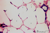
| 2167 |
 |
Lung alveoli (human) | Stain: Azan. Alveoli (diameter about 200 m) are formed by thin walls of epithelial cells (1) such as type I alveolar cells and (blue stained) fine collagen (3) and elastin intermingled with dilated capillaries (2). | Poja Histology Collection - Respiratory System Subset | |
| 2168 |
 |
Fibrillin in the alveoli of lung emphysema (human, adult) | Stain: antifibrillin antibody immunoperoxidase staining with diaminobenzidin reaction. Fibrillin is one of the elastin-associated microfibrillar proteins, and marks therefore also the presence of elastin in lung tissue. In centrilobular emphysema (e.g., in lungs of smokers) the lesions are more com... | Alveolar septa; Elastin-associated proteins; Fibrillin | Poja Histology Collection - Respiratory System Subset |
| 2169 |
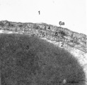 |
The air-blood barrier in the lung (rat) | Electron microscopy (high magnification). The alveolar space (1) and the capillary space (2) (with part of an erythrocyte, 3) are separated from each other respectively by cytoplasm of endothelial cell (5), the common basal lamina (6) with a lamina densa (6a) and the type I alveolar cell (4, pneumo... | Pneumocyte I; Alveolar cell type I | Poja Histology Collection - Respiratory System Subset |
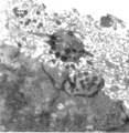
| 2170 |
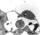 |
Brush cell in lung (rat) | Electron microscopy. In the proximal as well as in the terminal airways a special type of cell the so-called brush(-border) cell could be observed. In this picture this cell (X) is located in the alveolar space. At (*) the brush border, the dense cytoplasm contains many organelles. (C) is a capillar... | Brush cells | Poja Histology Collection - Respiratory System Subset |
| 2171 |
 |
Elastin in lung arteriole (human, adult) | Immuno-electron microscopy (embedded in Lowicryl HM 20). The alveolar septa contain a muscular pulmonary arteriole (lumen = X) with unlabelled endothelial cells (1). The elastic membranes in the arteriolar walls consist of more or less continuous bands of amorphous elastic lumps (E) antibody-labeled... | Alveolar septum; Immuno-electron microscopy; Immuno-staining | Poja Histology Collection - Respiratory System Subset |
| 2172 |
 |
Pseudoglandular - canalicular period of developing lung (human, fetus) | Stain: Azan. Location of a cartilagineous ring (1) between two cross-sectioned bronchi (2). Note in the bronchial epithelial cells the apical position of the epithelial nuclei and the light-stained basal part (↑) containing glycogen. The young hyaline cartilage presents solitary chondrocytes embed... | Lung development; Pseudoglandular period; Canalicular period; Mesenchyme | Poja Histology Collection - Respiratory System Subset |
| 2173 |
 |
Alveolar macrophage in lung (rat) | Electron microscopy. A wandering alveolar macrophage (1) migrates through an alveolar pore from an alveolar space (A1) into another (A2). Note the dark lysosomal structures and abundant organelles in its cytoplasm. The thin lining type I alveolar cell is hardly discernable (thin arrows →). Interst... | Pneumocyte I; Alveolar macrophages | Poja Histology Collection - Respiratory System Subset |
| 2174 |
 |
Epithelium of trachea (golden hamster) | Electron microscopy. Columnar ciliated cells (1) with cilia and organelle-rich apex; a goblet cell (2a) with accumulation of mucous secretion granules supranuclearly. Another goblet cell (2b) appears deprived; one basal cell (3) does not extend to the free surface. Light-grey aggregations of elastin... | Respiratory epithelium ; Pseudostratified epithelium; Basal cells | Poja Histology Collection - Respiratory System Subset |
| 2175 |
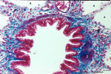 |
Bronchiolus in the lung (human, adult) | Stain: Azan. The lumen of the bronchiolus is lined with one layer of ciliated epithelium (1) and is folded due to the contraction of smooth muscle fibers (2) in the wall. Note the rich cellularity (3) in the stromal surrounding of the bronchiolus. Alveolar space (4). (5) pulmonary artery branches. | Ciliated epithelium; Bronchiolus | Poja Histology Collection - Respiratory System Subset |
| 2176 |
 |
Alveolar macrophages in lung (human, adult) | Electron microscopy. Two macrophages in the alveolar space (N = nucleus). Note the heterogeneity of many lysosomal structures containing among others carbon particles and the abundancy of organelles. | Alveolar macrophages | Poja Histology Collection - Respiratory System Subset |
| 2177 |
 |
Section of trachea (human, adult, high magnification) | Stain: Azan. On top: the epithelium (1) is pseudostratified with cilia and goblet cells (white droplets) on a distinct basement membrane (↓, undulating thick deep-red line) followed by a small proper lamina (2). The submucosa (3) starts where the fibrous tissue (elastic fibers reinforced with coll... | Pseudostratified epithelium ; Tracheal glands; Excretory duct; Perichondrium | Poja Histology Collection - Respiratory System Subset |
| 2178 |
 |
Laminin in the canalicular period of the lung (mouse, embryo) | Stain: Fluorescence microscopy with anti-laminin antibody. Laminin is a component of the basement membrane. Strong fluorescence visualizes the course of all basement membranes, especially around the bronchioles (B) and their branches in the developing lung tissue. In between the distal air spaces (t... | Bronchioli; Canalicular period | Poja Histology Collection - Respiratory System Subset |
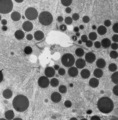
| 2179 |
 |
Multilamellar bodies of type II alveolar cell in the lung (mouse) | Electron microscopy. The cytoplasm of the type II alveolar cell (pneumocyte II) contains characteristic electron-dense multilamellar bodies (*) in different maturing stages. The lamellar bodies are responsible for the vacuolated appearance of these cells, and they give rise to surfactant (phospholip... | Pneumocyte II; Multilamellar bodies; Type II alveolar cell | Poja Histology Collection - Respiratory System Subset |
| 2180 |
 |
Upper part of nasal septum (dog, higher magnification) | Stain: Hematoxylin and eosin. Stratified squamous epithelium (1) with small papillae (2). The lamina propria contains many small and large thin-walled blood vessels also known as the venous plexus (sinusoids) of Auerbach (3, swell body). Nasal glands (4) with draining ducts (5, larger diameter) and ... | Squamous stratified epithelium; Nasal vestibulum; Venous sinusoids; Venous plexus; Swell bodies | Poja Histology Collection - Respiratory System Subset |
| 2181 |
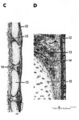 |
Scheme of trachea | C. Trachea (human, adult) magnification x 7.5 objective). D. Trachea (human, adult, detail of rectangle in C) magnification x 85 objective. (12) pseudostratified ciliated epithelium; (13) seromucous tracheal glands; (14) condensed layer of elastic fibers at the border of lamina propria and submu... | Pseudostratified epithelium ; Fibroelastic membrane; Adventitia | Poja Histology Collection - Respiratory System Subset |
| 2182 |
 |
Elastic fibers in the lung (human) | Stain: Orcein. At the left (1) several alveoli (A) are depicted with a faint staining of cellular elements. The elastic fibers within the alveoli are stained reddish-purple. At the right (2) a high magnification of a tangential-sectioned alveolus shows the thicker bundles as well as very thin branch... | Elastic fibers | Poja Histology Collection - Respiratory System Subset |
| 2183 |
 |
Alveolar septum in the lung (human, high magnification) | Stain: Azan. Two neighboring alveoli separated by a septum. (1) points to the tip containing bluish-pink elastin and collagen. The nucleus of a squamous alveolar cell (type I pneumocyte) is indicated by (2), and a free alveolar macrophage by (3). | Alveolar tip; Pneumocyte I | Poja Histology Collection - Respiratory System Subset |
| 2184 |
 |
Progressing centrilobular lung emphysema (human, adult) | Stain: Hematoxylin and eosin. Emphysema is defined as enlargement of the air spaces (X) distal to the terminal bronchioles, with destruction of the alveolar walls. The remaining alveolar walls are thickened (1). There is an increased cellularity and locally signs of chronic inflammation (↓) are pr... | Poja Histology Collection - Respiratory System Subset | |
| 2185 |
 |
Scheme of peripheral lung parenchym (human, adult) | (1) respiratory bronchiolus; (2) alveolar duct; (3) alveolar sac is a virtual sac formed by several alveoli, but continuous with the alveolar duct; (4) alveoli which end in small tips (↓) with elastin interwoven with collagen. | Alveolar duct | Poja Histology Collection - Respiratory System Subset |
| 2186 |
 |
Keratin 7 staining in squamous metaplasia of the epithelium of a bronchiolus (human, adult) | Stain: anti-keratin 7 antibody (Pan-Ck 7) immunoperoxidase staining (with aminoethylcarbazole (AEC) substrate). A red-brown staining with AEC indicates a positive reaction for cytokeratin 7. Note that the top layer only is stained positively for keratin 7 (↓). Upon squamous metaplasia the bronchio... | Bronchiolus; Squamous metaplasia; Immunoperoxidase; Immuno-reaction | Poja Histology Collection - Respiratory System Subset |
| 2187 |
 |
The alveolar tip in lung tissue (human, adult) | Electron microscopy. The lung tip is covered by a flattened alveolar cell type I (↓1) and a similar neighboring bulging one with nucleus (2). In the alveolar tip amorph elastin (5) appears more electron-dense than the collagen fibers (Col) and is interwoven with it. Small foci of calcifications a... | Alveolar tip; Elastoblast | Poja Histology Collection - Respiratory System Subset |
| 2188 |
 |
Alveolar cell type II (pneumocyte II) in an alveolus (dog) | Electron microscopy. At (X) the alveolar space, the bulging alveolar cell type II shows the characteristic multilamellar bodies (*, cytosomes) that contain precusor material of surfactant. The lamellar bodies are responsible for the vacuolated appearance of these cells, and they give rise to surfact... | Pneumocyte II; Alveolar cell type II | Poja Histology Collection - Respiratory System Subset |
| 2189 |
|
Olfactory vesicle (bulb) in the nose (gerbil) | Electron microscopy. A distinct olfactory bulb (1, vesicle) with horizontally extended cross-sectioned cilia (*) and basal bodies (↑) is surrounded by numerous microvilli and free olfactory cilia. Close to the first bulb a second one is about to protrude above the surface of the supporting cells b... | Olfactory epithelium; Olfactory vesicle | Poja Histology Collection - Respiratory System Subset |
| 2190 |
 |
Survey of bronchus and lung parenchym (human, adult) | Stain: Azan. (1) Lumen of small bronchus beside a lumen of pulmonary artery (2). Thin plates of hyaline cartilage (3) and connective tissue surrounding (1) and (2). Arrows (→) indicate lung alveoli with black-stained patches of carbon deposits. (4) Lung parenchym with alveoli. | Lung parenchym | Poja Histology Collection - Respiratory System Subset |
| 2191 |
 |
Bronchiolus in the lung (human, adult) | Stain: Azan. The lumen is lined with one layer of ciliated epithelium (1) without goblet cells. Patches of smooth muscle fibers (2) are present. Notice small bronchial arteries (*). Arrows (↓) indicate sites of carbon accumulations between the alveoli (3). | Bronchiolus; Ciliated epithelium | Poja Histology Collection - Respiratory System Subset |
| 2192 |
 |
Respiratory epithelium in the nasal vestibulum (rat) | Scanning electron microscopy. Note bushes of cilia (1) on the ciliary epithelium. In between areas of short microvilli-studded bulging or flattened goblet cells. Remnants of erythrocytes (2) are also found on top of ciliated cells. | Respiratory epithelium; Nasal vestibulum | Poja Histology Collection - Respiratory System Subset |
| 2193 |
 |
Survey of lung parenchym (gerbil) | Scanning electron microscopy. The photograph shows (1) bifurcation of several bronchi in the lung parenchym with alveoli (2). The long arrows indicate the route of alveolar ducts (4 ↕ length, and cross 4↑). Pulmonary vessels (3). | Lung parenchym; Pulmonary vessels | Poja Histology Collection - Respiratory System Subset |
| 2194 |
 |
Surface of trachea epithelium (rat) | Scanning electronmicroscopy. There is an abundancy of cilia with interspersed protrusions (↓) of goblet cells due to apical secretion. | Pseudostratified epithelium | Poja Histology Collection - Respiratory System Subset |
| 2195 |
 |
Epithelial lining of terminal bronchiolus (golden hamster) | Electron microscopy (low magnification). Two major epithelial cell types line the bronchiolus: (1) the ciliated cells with many organelles, and (2) the non-ciliated cells or Clara cells well provided with organelles, especially agranular endoplasmic reticulum and depending on their activity, numerou... | Terminal bronchiolus; Ciliated epithelium ; Clara cells; Neuro-endocrine cells; Pneumocytes; Alveolar cell types; Interstitium | Poja Histology Collection - Respiratory System Subset |
| 2196 |
 |
Brush cell in lung (rat) | Electron microscopy. In the proximal as well as in the terminal airways a special type of cell can be observed, the so-called brush (-border) cell. In this picture this cell is located in the alveolar space. At (1) the brush border of thick plump microvilli. In the cytoplasm a Gogi area, mitochondri... | Brush cell | Poja Histology Collection - Respiratory System Subset |
| 2197 |
 |
Survey of the nasal conchae (dog, isolated turbinate bones) | Stain: Hematoxylin and eosin. The conchae (turbinates) consist of three parts: the inferior, middle and superior turbinate bones (3, black-stained); they are covered by a respiratory mucosa (1) or by an olfactory mucosa (2). In humans only a small part of the superior concha exhibits olfactory epith... | Conchae nasales; Olfactory epithelium; Respiratory epithelium | Poja Histology Collection - Respiratory System Subset |
| 2198 |
 |
Transitional zone in the middle part of nasal vestibulum (comparable with red zone or vermilion border of lip, human) | Stain: Azan. At the right slightly cornified squamous epithelium (1) with small dermal papillae (2) and numerous blood vessels (3). To the left the transition to pseudostratified epithelium (4). In the submucosa branching seromucous nasal glands with draining ducts (5). | Stratified epithelium; Pseudostratified epithelium; Nasal glands; Seromucous glands | Poja Histology Collection - Respiratory System Subset |
| 2199 |
 |
Survey of bronchus and lung parenchym (human, adult) | Stain: Azan. Lumina of bifurcated bronchi (1, 2). Thin plates of hyaline cartilage (3) and connective tissue surround the bronchi. Arrows (→) indicate lung alveoli with black-stained patches of carbon deposits. (4) Lung parenchyma with alveoli. | Lung parenchym | Poja Histology Collection - Respiratory System Subset |
| 2200 |
 |
Olfactory epithelium in the nasal cavity (mammals) | Three-dimensional scheme electron microscopy. Six olfactory cells (1) surround one massive central supporting cell (2). A third, basal cell (3) is located at the basal plate aposing to the supporting cell. These olfactory cells are tall and slender bipolar nerve cells with chemoreceptive functions. ... | Pseudostratified epithelium; Olfactory epithelium; Basal cells; Olfactory vesicle; Bipolar neuron | Poja Histology Collection - Respiratory System Subset |