The Health Education Assets Library (HEAL) is a collection of over 22,000 freely available digital materials for health sciences education. The collection is now housed at the University of Utah J. Willard Marriott Digital Library.
TO
Filters: Collection: "ehsl_heal"
| Title | Description | Subject | Collection | ||
|---|---|---|---|---|---|
| 1901 |
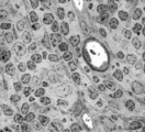 |
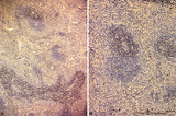
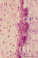
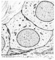
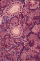
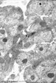
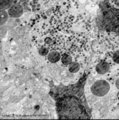
Survey of part of splenic lymphatic nodule (rat) | Electron microscopy. (1) shows an arteriolar branch of a central artery, and cross-sections of capillary branches (1a). Within the periarteriolar lymphatic sheath (PALS) mostly T lymphocytes (5) are present between concentric arranged reticular cells (2). Cells with larger lighter stained nuclei rep... | central artery ; electron microscopy; PALS; Antigen presenting cells (APC) | Poja Histology Collection - Lymphatic Tissues and Organs Subset |
| 1902 |
 |
Cystic corpuscle in thymus (mouse, young adult) | Electron microscopy. This cystic corpuscle is lined by specialized epithelioreticular cells and exhibits a lumen (1) which is filled with long microvilli however, curiously derived from a flattened epithelial cell (2, nucleus). The cell contains few electron-dense keratohyalin granules (3). (-->) po... | epithelioreticular cells; thymic corpuscle; thymic cyst; lymphoid tissue | Poja Histology Collection - Lymphatic Tissues and Organs Subset |
| 1903 |
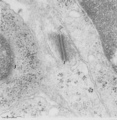 |
Thymus cortex (mouse, young adult) | Electron microscopy. (1) A well developed desmosome (1) of an epithelioreticular cells type II (TEC2). Apart from few free ribosomes, glycogen granules (2) are present in the electron-light cytoplasm. (3) part of an electron-grey thymocyte with many ribosomes. | epithelioreticular cell type II ; desmosome; lymphoid tissue | Poja Histology Collection - Lymphatic Tissues and Organs Subset |
| 1904 |
 |
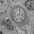
Dendritic cells in spleen (mouse) | Electron microscopy. The interdigitations (*) of the left dendritic cells (antigen-presenting cell or APC) (1 and 2a) are clearly shown. The right (2b) is a neighbour dendritic cell sectioned at the level of the Golgi area. (3) lymphocyte and (4) reticular cell. | electron microscopy; dendritic cell | Poja Histology Collection - Lymphatic Tissues and Organs Subset |
| 1905 |
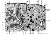 |
Scheme of the spleen (human) | Spleen: A. general diagram; B. adult; C. senium 1. capsule of dense irregular connective tissue with few elastic and smooth muscle fibers (it varies with the species); 2. trabecula (septum); 3. trabecular artery derived from the splenic artery; 4. trabecular vein; 5. when the pulpa artery ... | white pulp; red pulp | Poja Histology Collection - Lymphatic Tissues and Organs Subset |
| 1906 |
 |
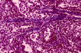
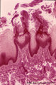
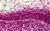
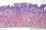
White pulp of spleen (mouse) | Stain: Hematoxylin & eosin in A and alkaline phosphatase in B (with substrate Naphtol Fast Blue RR). The general structure of the white pulp of the spleen and its specific microenvironment for T and B cells is well illustrated using alkaline phosphatase that strongly stains the capillaries around th... | alkaline phosphatase; PALS; T lymphocytes; white pulp | Poja Histology Collection - Lymphatic Tissues and Organs Subset |
| 1907 |
 |
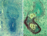
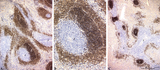
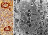
The effect of cyclophosphamide treatment on the B and T cells in spleen (rat) | Stain: Immunoperoxidase staining using diaminobenzidin (DAB)/ hematoxylin counterstained on frozen section with antibodies to B cells (Mark 1), CD3 and CD8 T cells. (A): B cells in the follicles, germinal centres (2) and corona are stained positive brown, while the PALS area (1) is negative (blue)... | CD3 lymphocytes; CD8 lymphocytes; B lymphocytes; cyclophosphamide | Poja Histology Collection - Lymphatic Tissues and Organs Subset |
| 1908 |
 |
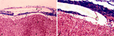
Age involution of thymus (human, postpuberal) | Stain: Azan. The size of the thymus is age-dependent, and undergoes a continuous process of involution, starting at postpuberal age. Due to depletion and reduced production of cortical thymocytes, as well as a gradual atrophy of the epithelial cells, the clear distinction between medulla (1) and cor... | thymus age; adipose cells; thymus involution; lymphoid tissue | Poja Histology Collection - Lymphatic Tissues and Organs Subset |
| 1909 |
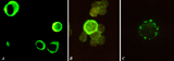 |
Immunohistochemical identification of splenic B cells (rat) | Stain: Immunofluorescence of FITC-labeled anti-Mark-1 antibody against rat B cells, carried out on spleen cells after fixation (A, B) or non-fixated spleen cells (C). (A) shows (young) plasma cells producing antibodies. (B) a green-stained B cell. The surrounding negative cells are likely T cells.... | immunofluorescence; B lymphocyte; capping | Poja Histology Collection - Lymphatic Tissues and Organs Subset |
| 1910 |
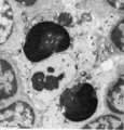 |
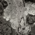
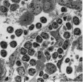
Thymus cortex (mouse, young adult) | Electron microscopy. Surrounded by thymocytes (2) a cortical macrophage (starry-sky macrophage) is seen and shows an electron-light nucleus (N) and a distinct nucleolus. The cell has engulfed two apoptotic thymocytes (1). The cytoplasm also contains small electron-dense lysosomes and myelin figures ... | cortical macrophage; epithelioreticular cell type II ; apoptotic thymocyte; lymphoid tissue | Poja Histology Collection - Lymphatic Tissues and Organs Subset |
| 1911 |
 |
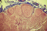
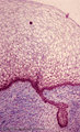
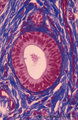
Survey of splenic trabecular artery (human) | Stain: Azan. The splenic artery (or lienal artery) is the blood vessel that provides oxygenated blood to the spleen. Branches of the splenic artery divide into trabecular arteries (1) which enter the white pulp as central arteries (4) that is surrounded with lymphocytes (5,periarteriolar lymphatic s... | trabecular artery; PALS; sinusoid | Poja Histology Collection - Lymphatic Tissues and Organs Subset |
| 1912 |
 |
Immunohistochemistry of B cells in splenic white pulp (rat) | Stain: Immunohistochemistry of Vector red for Mark-1-antibody-stained B cells. (1) indicates the PALS area populated with unstained T cells located around the central artery of the white pulp. (2) indicates the positively red stained B cells in the germinal centre of the follicle. (3) the B-cells i... | marginal zone; immunohistochemistry; follicle | Poja Histology Collection - Lymphatic Tissues and Organs Subset |
| 1913 |
 |
Penicillar arterioles in spleen (human) | Stain: Azan. The central or follicular artery in the follicle of the spleen splits into many arterioles. These arterioles spread as so-called penicillar arterioles (1) shown here. They are still surrounded by a very thin perilymphatic sheath (PALS) that disappears as the arteries in a brush-like pat... | penicillar arterioles; red pulp | Poja Histology Collection - Lymphatic Tissues and Organs Subset |
| 1914 |
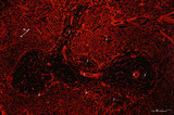 |
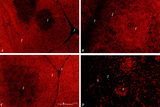
Localization of heparan sulfate (HS) in spleen (rat) | Stain: Immunofluorescence of Alexa 594 red labeled single chain antibody 3C3 for heparan sulfate (HS). The antibody stains HS epitopes of the meshwork of reticulum cells and the basal membrane of blood vessels. (1) central arteries in the T cell area of the PALS (2). The B cell area in the follic... | white pulp; heparan sulfate; PALS | Poja Histology Collection - Lymphatic Tissues and Organs Subset |
| 1915 |
 |
Thymic nurse cell (TNC) (mouse) | Electron microscopy. The thymic nurse cell (TNC) consists of an epithelial reticular cell (type II) enclosing thymocytes. The TNC exists as a sealed structure in situ, i.e. the lymphocytes within the TNC are isolated from the general thymic environment. TNC are located in the cortex, where mature ... | epithelioreticular cell type II; thymic nurse cell | Poja Histology Collection - Lymphatic Tissues and Organs Subset |
| 1916 |
 |
Scheme of lymph node (human) | Lymph node: A. survey; B. detail subcapsular (or marginal) sinus 1. adipose tissue; 2. capsule; 3. afferent lymph vessel with valves (B); 4. subcapsular (or marginal) sinus; 5. germinal centre of a secondary lymphatic nodule (or follicle); 6. crescent or mantle zone indicated by cap of per... | cortex; scheme; paracortex; germinal center | Poja Histology Collection - Lymphatic Tissues and Organs Subset |
| 1917 |
 |
Afferent lymph vessel in lymph node (human) | Stain: Azan. Left (A) and right (B): part of the cortex of a lymph node with the capsule (1) and subcapsular (or marginal) sinus (2) filled with lymphocytes. Left (A): (3) show perpendicularly localized reticular cells and fibres (blue) in the sinus. (4) indicate afferent lymph vessel with valves ... | lymph vessel; subcapsular sinus; follicle; cortex | Poja Histology Collection - Lymphatic Tissues and Organs Subset |
| 1918 |
 |
The effect of cyclophosphamide on splenic B cells (rat) | Stain: Immunoperoxidase staining using diaminobenzidin (DAB)/ hematoxylin counterstained on frozen section of B cells with the antibody Mark-1. The PALS area (1) contains T cells and remains unstained blue. The positively stained B cells (brown) are found in the germinal centres (2) and in the coron... | cyclophosphamide; immunosupression; B lymphocytes; Mark 1 antibody | Poja Histology Collection - Lymphatic Tissues and Organs Subset |
| 1919 |
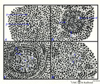 |
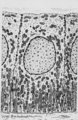
Scheme of development of germinal centre in lymphatic nodule (human) | A. primary nodule (or follicle) with mainly nave B lymphocytes and some memory cells, but no germinal centre; B. proliferation of lymphoblasts by mitosis (3,-->) and development of capillaries (*) in the centre of the follicle; C. so-called secondary nodule with lighter stained germinal centre wit... | follicle; antigen stimulation; scheme; germinal center | Poja Histology Collection - Lymphatic Tissues and Organs Subset |
| 1920 |
 |
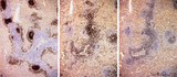
Age involution of thymus (human) | Stain: Azan. A: Although the adipose tissue in the thymus of a patient of 65 years is predominant it still contains areas of remnants (arrows 1+2) of cortical (2) and medullary portions (1) of the thymus. B+C: Degradation of thymic tissues is less progressed in adults and shows less replacement of... | thymus age; thymus involution; adipose cells; lymphoid tissue | Poja Histology Collection - Lymphatic Tissues and Organs Subset |
| 1921 |
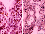 |
Splenic sinusoids (human) | Stain: A: Azan and B: Silver stain (Gomori). The splenic sinusoids are built in the form of a grid. The frame of the barrel consists of elongated endothelial cells (1) which are spirally wrapped around by reticular fibers (2). To this reticular fibers are attached many macrophages which control the... | sinusoid; reticular fibers | Poja Histology Collection - Lymphatic Tissues and Organs Subset |
| 1922 |
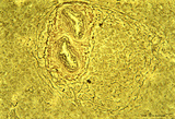 |
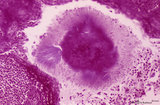
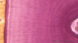
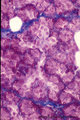
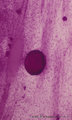
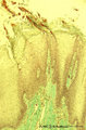
Circumscript reticular pattern of splenic lymphatic nodule (human) | Stain: Silver stain (Gomori). In this particular case the black-stained reticular network is beautiful organized in and around the nodule. Note also the reticulin staining between the myocytes in the wall of both cross-sectioned (1) central arteries. (2) follicle with lymphocytes. | sinusoid; reticular fibers; follicle | Poja Histology Collection - Lymphatic Tissues and Organs Subset |
| 1923 |
 |
High endothelial venules (HEV) (rat lymph node, human palatine tonsil) | Stain: (A) Anti-laminin-antibody and immunoperoxidase staining using diaminobenzidin (DAB)/ hematoxylin counterstained on frozen section of lymph node; (B) electron microscopy (palatine tonsil). (A): Using an antibody against laminin brown-stained basement membranes are outlined demonstrating postc... | palatine tonsil; high endothelial venule (HEV); laminin; immunohistochemistry | Poja Histology Collection - Lymphatic Tissues and Organs Subset |
| 1924 |
 |
Thymus cortex (rat, young adult) | Electron microscopy. Type I epithelioreticular cells separate connective tissue compartment from the thymic parenchyma. With occludens junctions and desmosomes as barriers they form wide-mesh networks creating specific microenvironments for developing T cells. The extensions of type I cells (5) are... | thymus cortex; epithelioreticular cell type I; epithelioreticular cell type II; lymphoid tissue | Poja Histology Collection - Lymphatic Tissues and Organs Subset |
| 1925 |
 |
Immunohistochemistry with cellular markers in thymus (rat) | Stain: Alexa 594 red immunofluorescence. (1) medulla; (2) cortex; (3) septa. (A): Strong CD8 staining of the thymic cortical lymphocytes (single CD8+ and double positive CD4+CD8+ thymocytes). (B): OX19 staining for almost all T cells, equivalent to T1 human marker and Ly-1 mouse marker. Note that... | cyclophosphamide; CD8 monoclonal antibody; OX19 monoclonal antibody; ER1 monoclonal antibody | Poja Histology Collection - Lymphatic Tissues and Organs Subset |
| 1926 |
 |
Hassall's corpuscle in thymus (mouse) | Electron microscopy. The centre of a small Hassall's corpuscle consists of darker-stained cells (1) which are keratinizing and represent degenerating cytoplasmic remnants (2). (3) is an infiltrating monocyte and (*) indicate the presence of free keratohyalin granules from disintegrated epithelial ce... | thymic corpuscle (Hassalls); lymphoid tissue ; epithelioreticular cell (ERC) | Poja Histology Collection - Lymphatic Tissues and Organs Subset |
| 1927 |
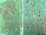 |
MHC-class II dendritic cells in spleen (rat) | Stain: Immunohistochemistry of Vector red staining of MHC-class II with ER13 antibody. The survey in (A) shows that MHC-class II expressing cells are conspicuously concentrated in dendritic cell types (B) in the transition zone between red pulp (2) and white pulp (1). These dendritic or antigen-pres... | MHC class II; dendritic cells; immunofluorescence | Poja Histology Collection - Lymphatic Tissues and Organs Subset |
| 1928 |
 |
Thymus medulla (rat, young adult) | Electron microscopy. Epithelioreticular cell of the medulla with an electron-light cytoplasm contains cross-sections of vacuoles with small finger-like cytoplasmic extrusions in the lumen (1). Electron-dense lysosomal structures (2) are also present as well as bundles of intermediate filaments (kera... | medullar epithelioreticular cell ; keratin filaments; lymphoid tissue | Poja Histology Collection - Lymphatic Tissues and Organs Subset |
| 1929 |
 |
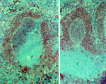
Immunoperoxidase stained CD3 positive T cells in spleen (rat) | Stain: Immunoperoxidase staining using diaminobenzidin (DAB)/ hematoxylin counterstained on frozen section with anti CD3 antibodies. Survey (A) and detail (B) show that CD3 marker for mature T cells stains positively (brown) in the PALS area (1) around the central arteries, while the adjacent follic... | CD3 lymphocytes; immunohistochemistry; PALS | Poja Histology Collection - Lymphatic Tissues and Organs Subset |
| 1930 |
 |
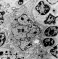
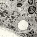
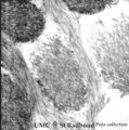
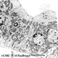
: Lymph node (rat) | Electron microscopy. A low magnification of a part of the medulla showing medullary cords surrounded by labyrinthine medullary sinus (*). In this picture the medullar cord runs from left bottom corner to right top corner, and is lined by flat reticular cell types. Within the cord one finds a star-sh... | medulla; electron microscopy; sinusoid | Poja Histology Collection - Lymphatic Tissues and Organs Subset |
| 1931 |
 |
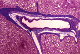
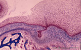
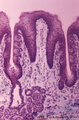
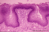
Calcified bacterial plaque in palatine tonsil ('gut-associated lymphatic tissue' or GALT) (human) | Stain: Azan. A tonsillar crypt lined by squamous epithelium (2) that is infiltrated with lymphocytes (1). It is a normal finding that within the crypts free cells, plugs of lymphocytes (1) and calcified epithelial debris as well as colonies of oral commensally bacteria are present. (3) shows a calc... | bacterial plaque | Poja Histology Collection - Lymphatic Tissues and Organs Subset |
| 1932 |
 |
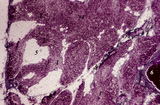
Cortex of lymph node (human) | Stain: Azan. The cortex of the lymph node comprises the (secondary) follicles with germinal centres (1) and corona or mantle zone (2), all filled with B lymphocytes. The paracortical area (3) below the follicles largely comprises T cells. Afferent lymph vessels enter via the capsule (6) into the m... | cortex; paracortex | Poja Histology Collection - Lymphatic Tissues and Organs Subset |
| 1933 |
 |
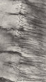
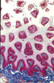
Cross-section of tooth in alveolar bone (cat, adult) | Stain: Picric acid and hematoxylin. From left to right: Periodontal ligament with blood vessels. Acellular cementum (dark purple rim). Dentin with radiair arrangement of dentinal tubules; fine incremental (imbrication) lines of von Ebner run at right angles to these tubules. These lines represent di... | oral cavity; incremental lines; von Ebner | Poja Histology Collection - Oral Cavity Subset |
| 1934 |
 |
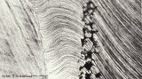
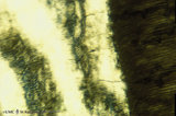
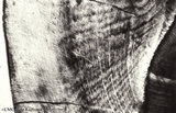
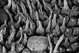
Dentinal tubules in longitudinal section of tooth - human, adult. Thin ground section, polarizing microscopy - optical axes of the polarizing plates are crossed at 60. | Slightly oblique section shows dentinal tubules depicted by brown-stained hollow fiber-like structures containing odontoblastic processes (Tomes' fibers) in a semi-three dimensional way. Numerous fine secondary branches of these processes are anastomosing with those of neighboring tubules. | oral cavity; Tomes' Fibers; dentinal tubules | Poja Histology Collection - Oral Cavity Subset |
| 1935 |
 |
Cross section of tooth in alveolar bone - cat; low magnification | Stain: Picric acid and hematoxylin. From left to right: alveolar bone tissue with osteons; periodontal ligament with blood vessels; acellular cementum (dark purple rim); dentin with (purple) radiair arrangement of dentinal tubules; fine incremental (imbrication) lines of von Ebner run at right angl... | oral cavity; incremental lines; alveolar bone | Poja Histology Collection - Oral Cavity Subset |
| 1936 |
 |
Dentinal tubules in longitudinal section of tooth - human, adult. Thin ground section, polarizing microscopy - optical axes of the polarizing plates are crossed at 60. | Slightly oblique section shows dentinal tubules depicted by brown-stained hollow fiber-like containing odontoblastic processes (Tomes' fibers) in a semi-three dimensional way ('stubble-field' aspect). Darker stained clusters represent anastomosing secondary branches. | oral cavity; Tomes' Fibers; dentinal tubules | Poja Histology Collection - Oral Cavity Subset |
| 1937 |
 |
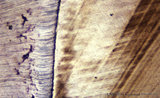
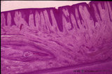
Dentinoenamel junction in longitudinal section of tooth (human, adult). Thin ground section. | From left to right: Enamel with fine striation (course of enamel rods or prisms). Few enamel tufts (left, dark) consisting of hypocalcified enamel rods and interprismatic substance arise from the junction. Scallop-like course of dentinoenamel junction. Dentin with dentinal tubules to the dentinoenam... | oral cavity; enamel tufts; dentinoenamel junction; dentinal tubules | Poja Histology Collection - Oral Cavity Subset |
| 1938 |
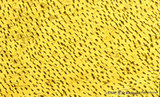 |
Dentinal tubules in longitudinal section of tooth - human, adult. Thin ground section, polarizing microscopy - optical axes of the polarizing plates are crossed at 60. | Slightly oblique section shows dentinal tubules depicted by brown-stained stubble structures containing odontoblastic processes (Tomes' fibers) in a semi-three dimensional way. Numerous fine branches of these processes are obvious in dentin. | oral cavity; Tomes' Fibers; dentinal tubules | Poja Histology Collection - Oral Cavity Subset |
| 1939 |
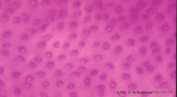 |
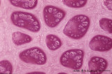
Dentin in cross section of tooth - human, adult | Stain: Hematoxylin and eosin. Cross-sectioned dentinal tubules demonstrate: a light stained center with the odontoblastic process (Tomes' fiber); darker stained peritubular dentin (highly mineralized), also called Neuman's sheath. Intertubular dentin (less mineralized) is present between the tubule... | oral cavity; dentinal tubules; secondary dentin | Poja Histology Collection - Oral Cavity Subset |
| 1940 |
 |
Dentin in cross section of tooth (human, adult) | Stain: Hematoxylin and eosin. Longitudinally sectioned dentinal tubules are parallely arranged, and numerous side branches are visible. | oral cavity; dentinal tubules | Poja Histology Collection - Oral Cavity Subset |
| 1941 |
 |
Dentinoenamel junction in the tooth (human, adult). Thin ground section of crown. | From left to right: Enamel with fine striation (composition of enamel rods or prisms); darker zones almost perpendicular to the striation are the incremental lines (Retzius) due to successive apposition of layers of enamel as the crown is formed. Dentinoenamel junction is shown as a narrow fissure f... | oral cavity; dentinal tubules; dentinoenamel junction; interglobular dentin; lines of Retzius | Poja Histology Collection - Oral Cavity Subset |
| 1942 |
 |
Dentinoenamel junction in the tooth - human, adult. Thin ground section of crown. | From left to right: Superficial dentin (bluish) in the crown with S-shaped course of dentinal tubules; They pass uninterrupted through the irregular black structures (due to filling with air) representing hypocalcified areas (interglobular dentin); Mineralization of dentin starts in small globular ... | oral cavity; dentinal tubules; dentinoenamel junction; interglobular dentin; lines of Retzius | Poja Histology Collection - Oral Cavity Subset |
| 1943 |
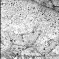 |
Dentinogenesis in tooth development - bell stage, gerbil, postnatal | Electronmicroscopy. At the top right corner side the distal cytoplasmic parts of presecretory ameloblasts resting on a thin grey basal lamina. In the central area predentin with collagen fibers (grey patches) and cross-sectioned small odontoblastic branches. In between them dispersed numerous dark-s... | oral cavity; predentin; matrix vesicles | Poja Histology Collection - Oral Cavity Subset |
| 1944 |
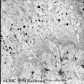 |
Dentinogenesis in tooth development - bell stage, gerbil, postnatal | Electronmicroscopy. At the bottom side of the predentin partly cross-sectioned odontoblasts with some organelles and many vesicular structures, the dark ones containing hydroxyapatite. Close to the odontoblasts a high concentration of secreted collagen fibers. Further away numerous matrix vesicles (... | oral cavity; predentin; matrix vesicles | Poja Histology Collection - Oral Cavity Subset |
| 1945 |
 |
Dentinoenamel junction in longitudinal section of tooth - human, adult. Thin ground section. | From left to right: enamel with fine striation (composed of stalks of enamel rods or prisms); enamel tufts (dark) arise at the dentinoenamel junction, and these tufts consist of hypocalcified enamel prisms and interprismatic substance; dentin with dentinal tubules up to the dentinoenamel. | oral cavity; enamel tufts; dentinoenamel junction; dentinal tubules | Poja Histology Collection - Oral Cavity Subset |
| 1946 |
 |
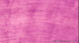
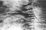
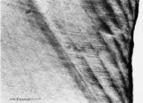
Enamel in longitudinal section of tooth - human, adult. Thin ground section. | From left to right: dentin with dentinal tubules; dentinoenamel junction; enamel with arrows pointing to bands (lines) of Hunter-Schreger; these alternating light and dark strips originate at the dentinoenamel junction and do not reach the enamel surface. This optical phenomenon is the result of the... | oral cavity; Hunter-Schreger bands; Retzius | Poja Histology Collection - Oral Cavity Subset |
| 1947 |
 |
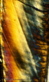
Enamel in longitudinal section of tooth - human, adult. Thin ground section, polarizing microscopy - optical axes of the polarizing plates are crossed at 60. | Using polarizing microscopy the birefringence of the crystalline structure of enamel is colorful demonstrated. Incremental lines (striae) of Retzius are well shown as straight oblique zones. Note the parallel lines of enamel stacks at the left corner of the picture. At the right side the lightly col... | oral cavity; Retzius; dentinoenamel junction | Poja Histology Collection - Oral Cavity Subset |
| 1948 |
 |
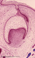
Early cap stage in tooth development - human, embryo; low magnification | Stain: Azan. From top to bottom: top left vestibular groove with gland formation; stratified ectoderm with dark red rim of basal cells from which a dental lamina sprouts downwards into the dental crypt (bony cavity) (bottom right corner); bone stains dark blue; connective tissue/mesenchym stains lig... | oral cavity; tooth development; dental lamina | Poja Histology Collection - Oral Cavity Subset |
| 1949 |
 |
Enamel in longitudinal section of tooth - human, adult. Thin ground section, polarizing microscopy - optical axes of the polarizing plates are crossed at 60. | Using polarizing microscopy the birefringence of the crystalline structure of enamel is colorful demonstrated. Incremental lines (striae) of Retzius are well shown as straight oblique zones. Top right a long dark fissure-like structure representing a crack. | oral cavity; Retzius; dentinoenamel junction | Poja Histology Collection - Oral Cavity Subset |
| 1950 |
 |
Enamel in longitudinal section of tooth - human, adult. Thin ground section, polarizing microscopy - optical axes of the polarizing plates are crossed at 60. | Left side enamel and at right side dark-stained striation of dentin. Wavy course of enamel prisms from dentinoenamel junction (right side, scalloped appearance) to the left. The transparent areas represent enamel lanes at a different polarizing angle. | oral cavity; dentinoenamel junction | Poja Histology Collection - Oral Cavity Subset |
| 1951 |
 |
Enamel in longitudinal section of tooth - human, adult. Thin ground section. | From left to right: incremental lines (striae) of Retzius are distinctly shown as oblique broad zones (at the left) to the enamel surface; during formation of the crown successive apposition of layers of enamel is deposited and results in these so-called incremental grow lines; surface of enamel in ... | oral cavity; incremental lines; Retzius | Poja Histology Collection - Oral Cavity Subset |
| 1952 |
 |
Early cap stage in tooth development - human, embryo; low magnification | Stain: Azan. From top to bottom: top left vestibular groove with gland formation; stratified ectoderm with ingrowth of the dental lamina (in the middle); bulbous growing end of dental lamina; bottom left alveolar bone formation (dark blue); and connective tissue/mesenchym stains light blue. | oral cavity; tooth development; dental lamina | Poja Histology Collection - Oral Cavity Subset |
| 1953 |
 |
Enamel (odontogenic) organ in tooth development - bell stage, human, embryo | Stain: Azan. Outer surface of bell; from left to right: (avascular) Stellate reticulum; capillaries in this stage proliferate and invaginate between the outer dental epithelial cells; Part of fibrous tooth follicle. | oral cavity | Poja Histology Collection - Oral Cavity Subset |
| 1954 |
 |
Early cap stage in tooth development - human, embryo | Stain: Azan. From top to bottom: stratified ectoderm with ingrowth of the dental lamina; knob-like end of the dental lamina; and collagen fibers of lamina propria are blue. | oral cavity; tooth development; dental lamina | Poja Histology Collection - Oral Cavity Subset |
| 1955 |
 |
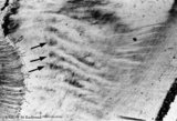
Enamel in longitudinal section of tooth - human, adult. Thin ground section. | At the left side surface of enamel in cuspal region; the parallel horizontal arrangement of stacks of rods (prisms) is evident. At the right side area of dentin (dark area). Incremental lines (striae) of Retzius run as curved lines (from bottom to middle top) and presented successive apposition of l... | oral cavity; incremental lines; Retzius; Hunter-Schreger bands | Poja Histology Collection - Oral Cavity Subset |
| 1956 |
 |
Enamel in longitudinal section of tooth - human, adult. Thin ground section. | Enamel is compact and acellular, and consists of vertical stacks of rods (prisms) as well as interrod (interprismatic) regions with less calcifying substance parallel to each other. Each prism is surrounded by an enamel sheath (a non-mineralized organic substance). From left to right: surface of ... | oral cavity; enamel rods; enamel prisms | Poja Histology Collection - Oral Cavity Subset |
| 1957 |
 |
Enamel rods (prisms) in tooth development - gerbil, postnatal | Electronmicroscopy. Cross-section of rods (prisms) demonstrate round aggregates with hydroxyapatite crystals. Between the round rods the interprismatic substance with crystals orientated in a different course. Note that twisting of the crystallites can be seen in the longitudinal bundles where grey ... | oral cavity; enamel prisms; hydroxyapatite crystals | Poja Histology Collection - Oral Cavity Subset |
| 1958 |
 |
Epithelial tooth bud in tooth development - tooth germ, human, embryo | Stain: hematoxylin. At the top stratified ectoderm. The proliferating basal cell layers are palisade-arranged with a light cytoplasm close to the basement membrane (right side). Below the basement membrane subepithelially an accumulation of inductive neural crest-derived mesenchymal cells is locally... | oral cavity; tooth bud; tooth development | Poja Histology Collection - Oral Cavity Subset |
| 1959 |
 |
Exocrine gland (salivary gland) | Scheme electronmicroscopy. Part of an acinus of mucous cells with different amount of mucous secretion. Supranuclearly the Golgi areas with maturing mucous secretion granules, basolateral the endoplasmic reticulum. At the top the lumen. The left cell shows an accumulation of the secretory droplets, ... | oral cavity; mucous gland | Poja Histology Collection - Oral Cavity Subset |
| 1960 |
 |
Exocrine gland (salivary gland) | Scheme electronmicroscopy. Part of an acinus of serous cells, basolaterally a well developed rough endoplasmic reticulum and apically secretion granules with different maturity towards the small lumen. On top stages of early formed secretion granule (left) and more matured ones (right). Note small j... | oral cavity; serous gland | Poja Histology Collection - Oral Cavity Subset |
| 1961 |
 |
Exocrine gland - intercalated duct - salivary glands, pancreas | Scheme electronmicroscopy. Part of an intercalated duct, the low cuboidal epithelial cells contain sparsely organelles and some desmosomal structures. Between the lining cells and the basal lamina is squeezed part of a filament-rich myoepithelial cell. Lumen side top left quadrant. | oral cavity; intercalated duct | Poja Histology Collection - Oral Cavity Subset |
| 1962 |
 |
Exocrine gland - intercalated duct - submandibular gland, human | Stain: Azan. Branching intercalated duct draining several serous acini, the lining duct cells are lighter stained due to the content of organelles. Serous cells are darker stained (many organelles) with round nuclei. Note few fat cells, the septa are blue stained. | oral cavity; intercalated duct; serous gland | Poja Histology Collection - Oral Cavity Subset |
| 1963 |
 |
Exocrine gland - striated (intralobular) duct - salivary glands, pancreas | Scheme electronmicroscopy. Part of a striated duct with basally membrane infoldings and mitochondria accumulations in the tall columnar cells. Small junctional complexes as well as apically mitochondria and numerous vesicles are present. | oral cavity; striated duct | Poja Histology Collection - Oral Cavity Subset |
| 1964 |
 |
Exocrine gland - parotid gland, rat | Electronmicroscopy. Part of a striated duct with basally membrane infoldings and numerous mitochondria perpendicularly orientated to the basal membrane. Upper side is luminal side. Apically junctional complexes as well as mitochondria and many vesicles are present. | oral cavity; serous gland | Poja Histology Collection - Oral Cavity Subset |
| 1965 |
 |
Exocrine gland - striated (intralobular) ducts - parotid gland, human | Stain: Azan. Cross-sections of striated ducts in upper part between serous acini. Note at the right bottom part of (lighter stained) lining cells of an intercalated duct. Connective tissues between the structures are blue stained. | oral cavity; striated duct | Poja Histology Collection - Oral Cavity Subset |
| 1966 |
 |
Exocrine gland - interlobular duct - submandibular gland, human | Stain: Azan. An interlobular duct with partly columnar as well as pseudostratified columnar epithelium. Note the dense connective tissue of the interlobular septum with small blood vessels. | oral cavity; seromucous glands | Poja Histology Collection - Oral Cavity Subset |
| 1967 |
 |
Exocrine gland - intercalated duct - submandibular gland, rat | Electronmicroscopy. The dark cells form an intercalated duct from the left lower corner to the right upper corner, and end in the acinus with lightly stained cells (serous). | oral cavity; intercalated duct; serous gland | Poja Histology Collection - Oral Cavity Subset |
| 1968 |
 |
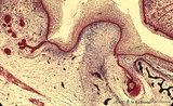
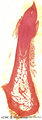
Foliate papillae of the tongue (dorsal side, rabbit) | Stain: Goldner trichrome. Foliate papil with primary and secondary connective tissue projections. Taste buds are localized in the lining, non-keratinized epithelium of the grooves. The serous gland of von Ebner (minor salivary gland) drains via an enlarged duct into the left groove of the papil. | oral cavity; foliate papillae; von Ebner | Poja Histology Collection - Oral Cavity Subset |
| 1969 |
 |
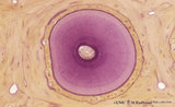
Free denticulus (pulp stone) in the tooth - longitudinal section of root pulp; human, adult | Stain: Hematoxylin and eosin. In the center of pulp connective tissue a free pulp stone (false denticulus). Dispersed through the pulp several small stones. Left of the central stone a longitudinal sectioned thin-walled blood vessel; right of the stone bundles of nerve fibers. | oral cavity; denticulus; pulp stone | Poja Histology Collection - Oral Cavity Subset |
| 1970 |
 |
Fungiform papillae of the tongue (dorsal side, human) | Stain: Hematoxylin and eosin. A broad prominent papil of connective tissue (without secondary papils) covered by non-keratinized stratified epithelium. This specimen does not show any taste bud. Well vascularized lamina propria. | oral cavity; fungiform papillae | Poja Histology Collection - Oral Cavity Subset |
| 1971 |
 |
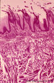
Filiform papillae of the tongue - dorsal side, human | Stain: Goldner trichrome. Detail of the threadlike keratinized extensions (red) of the stratified epithelium. Primary connective tissue papillae (green) divide in several small secondary ones. Note the absence of taste buds in filiform papillae. | oral cavity; filiform papillae | Poja Histology Collection - Oral Cavity Subset |
| 1972 |
 |
Filiform papillae of the tongue (dorsal side, human) | Stain: Heidenhain light bordeaux. Detail of the threadlike keratinized extensions of the stratified epithelium. Primary connective tissue papillae with 2 to 3 secondary papillae. Note the absence of taste buds in filiform papillae. | oral cavity; filiform papillae | Poja Histology Collection - Oral Cavity Subset |
| 1973 |
 |
Exocrine gland - submandibular gland, gerbil | Electronmicroscopy. Part of a serous acinus of this mixed gland. Note characteristic electron-dense secretion granules, the mature ones are larger. At the lower bottom is shown part of a lining cell of the intercalated duct. | oral cavity; serous gland | Poja Histology Collection - Oral Cavity Subset |
| 1974 |
 |
Filiform papillae of the tongue (dorsal side, human) | Stain: Azan. Oblique cross-section through the top of the filiform papillae. At the top of the picture keratinized extensions. Primary connective tissue papillae (blue) divide in several small secondary ones. | oral cavity; filiform papillae | Poja Histology Collection - Oral Cavity Subset |
| 1975 |
 |
Filiform papillae of the tongue - dorsal side, human | Stain: Heidenhain light bordeaux. Transverse section through the middle of the filiform papillae showing secondary connective tissue papillae (lightly stained). | oral cavity; filiform papillae | Poja Histology Collection - Oral Cavity Subset |
| 1976 |
 |
Filiform papillae of the tongue (dorsal side, human, neonate) | Scanning electronmicroscopy. Slender, thin threadlike extensions of the filiform papillae. In between a broad fungiform papilla. | oral cavity; filiform papillae; fungiform papilla | Poja Histology Collection - Oral Cavity Subset |
| 1977 |
 |
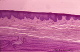
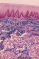
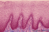
Gingiva - 'attached' gingiva of decalcified alveolar bone, human | Stain: Hematoxylin and eosin. Stratified squamous epithelium with parakeratosis (reddish) and deep papillae of the lamina propria. At the bottom, lamellar bone tissue (alveolar bone) with thickened periosteum (part of the periodontal ligament). At the bottom dentin. | oral cavity | Poja Histology Collection - Oral Cavity Subset |
| 1978 |
 |
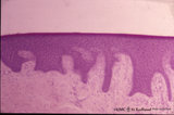
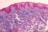
Gingiva ('attached' gingiva of decalcified alveolar bone, human, adult) | Stain: Hematoxylin and eosin. Thick layer of stratified squamous epithelium with parakeratosis (reddish) and shallow papillae of the lamina propria. At the bottom lamellar bone tissue (alveolar bone) with thickened periosteum. | oral cavity; alveolar bone | Poja Histology Collection - Oral Cavity Subset |
| 1979 |
 |
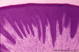
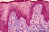
Gingiva - 'free' gingiva of decalcified alveolar bone, human | Stain: Hematoxylin and eosin. Thick layer of stratified squamous epithelium with parakeratosis (reddish) and broad papillae of the lamina propria. At the left, dense collagen tissue (projections of the the periodontal ligament). | oral cavity | Poja Histology Collection - Oral Cavity Subset |
| 1980 |
 |
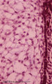
Gingiva ('free' gingiva of decalcified tooth, human, adult) | Stain: Hematoxylin and eosin. Thick layer of stratified squamous epithelium with parakeratosis (reddish) and many narrow papillae of the lamina propria. | oral cavity | Poja Histology Collection - Oral Cavity Subset |
| 1981 |
 |
Late cap stage in tooth development - human, embryo | Stain: Azan. From top to bottom: Top side stellate reticulum (enamel pulp) consisting of a network of ectoderm-derived cells; Right side outer dental epithelium with part of the fibrous tooth follicle. This epithelium will further develop downwards as the outer layer of the Hertwig's epithelial root... | oral cavity | Poja Histology Collection - Oral Cavity Subset |
| 1982 |
 |
Late cap stage in tooth development - human, embryo | Stain: Azan. The stellate reticulum (enamel pulp) consists of a network of ectoderm-derived branched cells and fluid-filled spaces (a.o. proteoglycans). It is a specialized avascular layer as a support and protection for the inner dental epithelial cells. At the right side one layer of cuboidal oute... | oral cavity; tooth development; outer dental epithelium; stellate reticulum | Poja Histology Collection - Oral Cavity Subset |
| 1983 |
 |
Late cap stage in tooth development - human, embryo | Stain: Azan. From top to bottom: At the top stellate reticulum (enamel pulp) consisting of a loose network of ectoderm-derived cells; Darker stained cell layers of the stratum intermedium; Columnar inner dental epithelium (presecreetory ameloblasts) at the distal side (secretion area) oriented towar... | oral cavity | Poja Histology Collection - Oral Cavity Subset |
| 1984 |
 |
Late cap stage of tooth development - human, embryo; low magnification | Stain: Azan. From top to bottom: Stratified ectoderm with a distinct basal layer (red line) of cuboid cells; Dental lamina giving rise to the cap stage (center) and to the primordium of permanent tooth (right); Odontogenic organ or enamel organ (future deciduous tooth surrounded by fibrous tooth fol... | oral cavity; dental lamina | Poja Histology Collection - Oral Cavity Subset |
| 1985 |
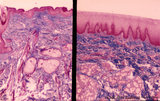 |
Lip (human), outer (left) and inner side (right) | Stain: Azan. Left image: keratinized squamous epithelium with thin red cornified layer, hair follicle, sebaceous glands and skeletal muscle fibers (orbicularis oris). Right image: non-keratinized epithelium with high, narrow dermal papillae and more capillaries. Mixed labial glands (seromucous), f... | oral cavity; lining mucosa | Poja Histology Collection - Oral Cavity Subset |
| 1986 |
 |
Lip (human), region between red zone (vermilion border) and mucosa inner surface | Stain: Azan. Bundles of skeletal muscle fibers (musculus orbicularis oris), highly vascularized lamina propria. Note the narrow dermal papillae on the left side (inner lip) and the broad irregular papillae on the right side (red zone of the lip). | oral cavity; lining mucosa; red zone; vermilion border | Poja Histology Collection - Oral Cavity Subset |
| 1987 |
 |
Lip (human), mucous inner surface | Stain: Azan. Non-keratinized epithelium with high, narrow dermal papillae and many capillaries. Mixed labial glands (seromucous) and few skeletal muscle fibers (orbicularis oris) in the submucosa. | oral cavity; lining mucosa; labial glands | Poja Histology Collection - Oral Cavity Subset |
| 1988 |
 |
Lip (human), mucoserous labial glands in mucous inner surface | Stain: Azan. Mixed labial glands with serous demilunes (von Ebner-Giannuzzi), a few myoepithelial cells, and a striated duct (right upper corner). | oral cavity; lining mucosa; labial glands | Poja Histology Collection - Oral Cavity Subset |
| 1989 |
 |
Lip (human), mucous inner surface | Stain: Azan. Non-keratinized epithelium with high, narrow dermal papillae and many capillaries. Note lymphocytic infiltrates and the capillaries in the dermal papillae. | oral cavity; lining mucosa | Poja Histology Collection - Oral Cavity Subset |
| 1990 |
 |
Lip (human), transitional zone (red zone or vermilion border) | Stain: Azan. Slightly cornified epithelium with high irregular dermal papillae and many capillaries. Note the epithelium is thicker, but less cornified than the epidermis. The red color of the lips is due to the rich vascularity of the lamina propria and the lucidity of the epithelium. | oral cavity; lining mucosa; red zone; vermilion border | Poja Histology Collection - Oral Cavity Subset |
| 1991 |
 |
Lip (human), transitional zone (red zone or vermilion border) | Stain: Azan. Slightly cornified epithelium with high irregular dermal papillae and many capillaries. Fat cells and skeletal muscle cells are located in the submucosa. | oral cavity; lining mucosa; red zone; vermilion border | Poja Histology Collection - Oral Cavity Subset |
| 1992 |
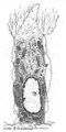 |
Odontoblast in tooth development - mammalian embryo | Scheme electronmicroscopy. A columnar and irregular shaped cell (mesenchymal origin) with a taperwise odontoblastic process within the predentin. The bottom side is adjacent to the future pulp cells. The cell body contains many organelles and especially a well-developed Golgi area with prosecretoy g... | oral cavity | Poja Histology Collection - Oral Cavity Subset |
| 1993 |
 |
Parotid gland (human) | Stain: Mallory trichrome. Survey: at the left bottom a large interlobular duct (in lumen remnants of secretion products) within a septum of dense connective tissue. At the top thinner (blue) septum, a thick (red-bluish) septum at the left. In the center three (intralobular) striated ducts between th... | oral cavity; serous gland | Poja Histology Collection - Oral Cavity Subset |
| 1994 |
 |
Papillae circumvallatae of the tongue (dorsal side, human) | Stain: Azan. Three broad papillae with taste buds facing the grooves in which the serous von Ebner glands drain. Striated skeletal muscles (musculus verticalis linguae and musculus longitudinalis superior) and lightly stained mucous glands (posterior lingual glands). | oral cavity; von Ebner; lingual muscles; lingual glands | Poja Histology Collection - Oral Cavity Subset |
| 1995 |
 |
Papillae filliformes of the tongue (dorsal side, human) | Stain: Heidenhain light bordeaux. Threadlike keratinized extensions of the stratified epithelium. Primary connective tissue papillae with 2 to 3 secondary papillae. The skeletal muscle fibers are arranged in three directions. | oral cavity; filiform papillae | Poja Histology Collection - Oral Cavity Subset |
| 1996 |
 |
Parotid gland (human) | Stain: Azan. The parotid gland: in most species the gland is composed entirely of serous acini. At the right a small (intralobular) striated duct; centrally one large interlobular duct with blood vessels. Scattered a few (white) fat cells. | oral cavity; serous gland | Poja Histology Collection - Oral Cavity Subset |
| 1997 |
 |
Papillae circumvallatae of the tongue (dorsal side, human) | Stain: Azan. A broad papilla with taste buds (lightly stained spots) facing the grooves in which the serous glands (von Ebner) drain. | oral cavity; von Ebner; circumvallate papillae | Poja Histology Collection - Oral Cavity Subset |
| 1998 |
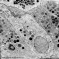 |
Parotid gland (rat) | Electronmicroscopy. Part of a serous acinus with characteristic secretion granules supranuclearly. Note different densities of the granules without any signs of fusion. | oral cavity; serous gland | Poja Histology Collection - Oral Cavity Subset |
| 1999 |
 |
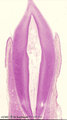
Permanent tooth - canine, human, adult; low magnification of labiolingual section | Stain: Hematoxylin and scarlet red. From top to bottom: Crown region with dentin but without enamel (decalcified specimens); Neck region at the attachment of the gingiva to dentin (left and right); Cementum is visible as a dark small rim from the neck region to the bottom of the tooth; Periodontal l... | oral cavity; alveolar process | Poja Histology Collection - Oral Cavity Subset |
| 2000 |
 |
Premolar permanent tooth (human, adult; low magnification of labiolingual section) | Stain: Hematoxylin and eosin. From top to bottom: Crown region with enamel-dentin boundary (there is no enamel present in decalcified specimens); dentin (blue-pink) enwraps the whole pulp chamber (light) and here the lines of dentinal tubules appear S-shaped. Neck region at the attachment of the gin... | oral cavity; pulp canal | Poja Histology Collection - Oral Cavity Subset |