The Health Education Assets Library (HEAL) is a collection of over 22,000 freely available digital materials for health sciences education. The collection is now housed at the University of Utah J. Willard Marriott Digital Library.
Filters: Collection: "ehsl_heal"
| Title | Description | Subject | Collection | ||
|---|---|---|---|---|---|
| 26 |
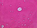 |
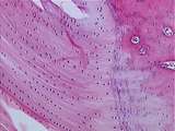
Bone - Haversian System | Concentric layers of collagen (the remnant of decalcified bone) surround the Haversian canal. Osteocyte nuclei can be seen within lacunae. UCLA Histology Collection. | Haversian canal | UCLA Histology |
| 27 |
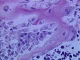 |
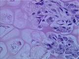
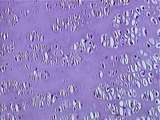
Bone - Osteoclasts and Osteocytes | Osteoclasts are cells with multiple nuclei which result from the fusion of many monocytes. Osteocytes are cells which have differentiated from osteoblasts and have surrounded themselves with bone. UCLA Histology Collection. | UCLA Histology | |
| 28 |
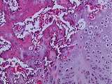 |
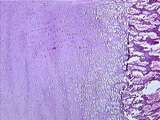
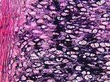
Bone / Cartilage | At a higher magnification of this fracture, we can identify cartilage cells undergoing hypertrophy, calcified cartilage, and woven bone. UCLA Histology Collection. | bone; cartilage | UCLA Histology |
| 29 |
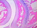 |
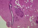
Bone / Cartilage | There is articular cartilage on each side of the joint space. Bone is stained pink. Find the synovial fold that generates the synovial fluid which lubricates the joint. Also find bone marrow spaces and the epiphyseal growth plate. UCLA Histology Collection. | articular cartilage; bone; epiphyseal growth plate | UCLA Histology |
| 30 |
 |
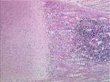
Bone / Cartilage | Between the bone spicules can be seen various bone cell types, including osteoblasts and osteoprogenitor cells. The left side of this image shows hypertrophied chondrocyte lacunae. UCLA Histology Collection. | Bone; Cartilage; osteoblasts, osteoprogenitor | UCLA Histology |
| 31 |
 |
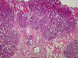
Bone / Cartilage | A healing fracture recapitulates the events of fetal endochondral ossification. A hyaline cartilage fracture callus forms to hold together the two ends of bone. Woven bone develops around (and eventually replaces) the cartilage, connecting the broken bone. Fracture healing occurs within the perioste... | bone; cartilage; periosteum | UCLA Histology |
| 32 |
 |
Bone / Cartilage - Cartilage Hypertrophy | This is an area of cartilage hypertrophy. To the right is the beginning of bone formation; mostly what can be seen are spicules of calcified cartilage. UCLA Histology Collection. | cartilage hypertrophy | UCLA Histology |
| 33 |
 |
Bone / Cartilage - Endochondral Ossification | The long bones develop by endochondral ossification, in which a cartilage model is laid down and then transformed into bone. UCLA Histology Collection. | UCLA Histology | |
| 34 |
|
Bone / Cartilage - Endochondral Ossification | The long bones develop by endochondral ossification, in which a cartilage model is laid down and then transformed into bone. UCLA Histology Collection. | UCLA Histology | |
| 35 |
 |
Bone / Cartilage - Epiphysial Growth Plate | In this low power view of the epiphysial growth plate, identify hyaline cartilage, spongy bone, and bone marrow filled with developing blood cells. UCLA Histology Collection. | hyaline cartilage | UCLA Histology |
| 36 |
 |
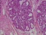
Breast | This active, lactating breast contains a large number of lobules of tightly packed alveoli. The connective tissue of the interlobular elements is highly compressed. Note the lactating glands, the excretory duct and some fat. UCLA Histology Collection. | Breast; lactating breast | UCLA Histology |
| 37 |
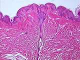 |
Breast | The male nipple has a rudimentary duct system. The epidermis is thin with enlarged dermal papillae. The bundles of smooth muscle found within the dermis allow the nipple to become erect. In female this response facilitates feeding. The lactiferous duct lined by a cubiodal epithelium, arise from the ... | Breast; male nipple | UCLA Histology |
| 38 |
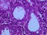 |
Breast | Alveoli of a lactating breast. The apical surface of the simple cuboidal cells is filled with lipid droplets which may be secreted from the cell. Other cells release the protein component of the breast milk. Each alveolus is invested within a basal lamina which contains a number of slender myoepithe... | Breast; lactating breast | UCLA Histology |
| 39 |
 |
Breast | Note the numerous glands of the nipple located within the dermis. Be sure to identify the epidermis marked with numerous dermal papillae. UCLA Histology Collection. | Breast; female nipple | UCLA Histology |
| 40 |
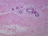 |
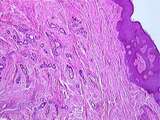
Breast | The female prepubertal breast does not differ from the nulliparous breast with respect to its attenuated glandular elements.The remaining lobules are composed primarily of ducts with no glandular or secretory alveoli. Identify the connective tissue and note the adipose tissue. UCLA Histology Collect... | Breast; prepubertal breast | UCLA Histology |
| 41 |
 |
Breast | This section is from the breast of a nulliparous women. Note the rudimentary duct system and secretory portion of the gland. Even during lactation the ducts basically serve an excretory function. Note the proliferation of interlobular connective tissue, the lactiferous sinus and the deposits of adip... | Breast; nulliparous breast | UCLA Histology |
| 42 |
 |
Breast | This magnification of a mammary alveolus clearly depicts the packed nature of the alveoli within the lactating breast. Notice the connective tissue of the interlobular septum. Compare this image with 243-40-1. UCLA Histology Collection. | Breast; lactating breast | UCLA Histology |
| 43 |
 |
Breast | The lactiferous ducts arise in the depths of the glandular tissue and are lined with a cubiodal epithelium. As a lactiferous ducts approaches the nipple it expands markedly to form the sometimes accordion-shaped lactiferous sinus which are lined with stratified cuboidal or even more complexed epithe... | Breast; female nipple; lactiferous ducts | UCLA Histology |
| 44 |
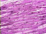 |
Cardiac Muscle | Locate and identify a number of anastamosing branches, central nuclei, and intercalated disks in this section of cardiocytes. Also note the abundant connective tissue spaces between cardiocytes. These areas contain many capillaries, providing the cardiocytes with an excellent vascular supply. UCLA H... | Cardiac Muscle; Heart | UCLA Histology |
| 45 |
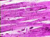 |
Cardiac Muscle | Note the special features of cardiac cells: including their anastomosing branches and the intercalated disks which link cells together. Note their centrally located nucleus and the short sarcomeres which give cardiac muscle its striated appearance. UCLA Histology Collection. | Cardiac Muscle; Heart; intercalated disks | UCLA Histology |
| 46 |
 |
Cardiac Muscle | Note the special features of cardiac cells: including their anastomosing branches and the intercalated disks which link cells together. Note their centrally located nucleus and the short sarcomeres which give cardiac muscle its striated appearance. UCLA Histology Collection. | Cardiac Muscle; Heart | UCLA Histology |
| 47 |
 |
Cartilage | This is a higher power view of the Achilles tendon insertion. Fibrocartilage is considered to be a cross between hyaline cartilage and tendon (we see little tendon here). Fibrocartilage consists of coarse bundles of collagen, between which the chondrocytes usually line up in rows. UCLA Histology Col... | fibrocartilage; tendon | UCLA Histology |
| 48 |
 |
Cartilage | On the left is cartilage proliferation, followed by cartilage maturation on the right. UCLA Histology Collection. | cartilage | UCLA Histology |
| 49 |
 |
Cartilage | At a higher power, the black elastic fiber deposits are clearly seen. Pinkish collagen is also a component of the matrix. Also note chondrocytes and perichondrium. UCLA Histology Collection. | chondrocytes; Elastic Cartilage; perichondrium | UCLA Histology |
| 50 |
 |
Cartilage | This is the zone of cartilage proliferation, with many of the chondrocytes in clusters. This demonstrates that these cells have recently divided but they have not had the time to produce enough matrix to separate the cells. UCLA Histology Collection. | UCLA Histology |