The Health Education Assets Library (HEAL) is a collection of over 22,000 freely available digital materials for health sciences education. The collection is now housed at the University of Utah J. Willard Marriott Digital Library.
TO
Filters: Collection: "ehsl_heal"
| Title | Description | Subject | Collection | ||
|---|---|---|---|---|---|
| 26 |
 |
Recessive epidermolysis bullosa | A baby with recessive epidermolysis bullosa. This patient links adequate numbers of anchoring fibrils (collagen type 7) to attach epidermis to the underlying dermis. | Knowledge Weavers Dermatology | |
| 27 |
 |
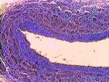
Spleen | The simple squamous cells of the mesothelium covering the spleen can be clearly seen. Elastin appears as reddish streaks among the blue-stained collagen fibers in the capsule. UCLA Histology Collection. | mesothelium; spleen; trichrome | UCLA Histology |
| 28 |
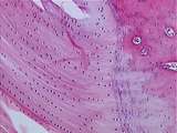 |
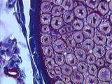
Cartilage | This is a higher power view of the Achilles tendon insertion. Fibrocartilage is considered to be a cross between hyaline cartilage and tendon (we see little tendon here). Fibrocartilage consists of coarse bundles of collagen, between which the chondrocytes usually line up in rows. UCLA Histology Col... | fibrocartilage; tendon | UCLA Histology |
| 29 |
 |
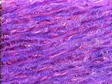
Circulatory System | In this aorta stained with Trichrome, identify pink elastin, blue collagen, and reddish muscle cells. Here the boundary between the tunica intima and tunica media is less distinct. However, the lumen is quite obvious. UCLA Histology Collection. | Aorta; Circulatory System; elastic artery; trichrome; tunica intima; tunica media | UCLA Histology |
| 30 |
 |
Circulatory System | Due to a special stain, the elastin of the tunica media is visible as dark, squiggly lines. The pink-stained areas between elastic fibers are occupied by collagen fibers and smooth muscle cells. UCLA Histology Collection. | aorta; Circulatory System; elastic artery; tunica media | UCLA Histology |
| 31 |
 |
Connective Tissue | The lining of the gallbladder is a tall simple columnar epithelium. The underlying loose connective tissue is characterized by a large number of cells relative to collagen fibers. Look for plasma cells here. UCLA Histology Collection. | Connective Tissue; Gall bladder; Liver | UCLA Histology |
| 32 |
 |
Epithelium - Cornea | The cornea of the eye is lined by simple squamous epithelium on its inner surface and stratified squamous epithelium on its outer surface. The stroma of the cornea consists of regularly arranged collagen fibers. UCLA Histology Collection. | simple squamous epithelium; stratified squamous epithelium | UCLA Histology |
| 33 |
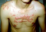 |
Keloids | Keloids. These are composed of excessive amounts of collagen, elastin, and ground substance. They occur generally on the upper chest and back, and also over areas of stretch such as the elbows and knees. | Knowledge Weavers Dermatology | |
| 34 |
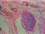 |
Lymph Nodes | This image shows a cross section of an efferent lymphatic vessel in the hilar region. Like veins, lymphatic vessels are thin-walled. Unlike veins, lymphatic vessels are filled with lymphocytes. The hilum contains large amounts of collagen fibers. UCLA Histology Collection. | Lymph Node; lymphatic vessel | UCLA Histology |
| 35 |
 |
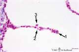
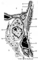
Scheme of peripheral lung parenchym (human, adult) | (1) respiratory bronchiolus; (2) alveolar duct; (3) alveolar sac is a virtual sac formed by several alveoli, but continuous with the alveolar duct; (4) alveoli which end in small tips (↓) with elastin interwoven with collagen. | Alveolar duct | Poja Histology Collection - Respiratory System Subset |
| 36 |
 |
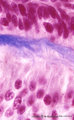
Skin | Observe the basal layer of the epidermis. In addition to the keratinocytes one can observe melanocytes. Note the difference between the cells of the basal layer and the spiny layer. Note the collagen and fibroblasts of the dermis. UCLA Histology Collection. | epidermis; keratinocytes; melanocytes; Skin | UCLA Histology |
| 37 |
 |
Alveolar septum in the lung (human, high magnification) | Stain: Azan. Two neighboring alveoli separated by a septum. (1) points to the tip containing bluish-pink elastin and collagen. The nucleus of a squamous alveolar cell (type I pneumocyte) is indicated by (2), and a free alveolar macrophage by (3). | Alveolar tip; Pneumocyte I | Poja Histology Collection - Respiratory System Subset |
| 38 |
 |
Circulatory System | In this high power view of the tunica media, identify pink elastic fibers, which appear thin and glassy. Smooth muscle appears thick and purple, and collagen fibers appear blue. Note the laminar arrangement of fibers and muscle cells. UCLA Histology Collection. | Aorta; Circulatory System; elastic artery; trichrome; tunica media | UCLA Histology |
| 39 |
 |
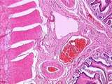
Circulatory System - Coronary Artery | A higher magnification of a coronary artery, focusing on the tunica adventitia, which contains collagen fibers and fibroblasts. Adipocytes, vasa vasorum (containing blood cells), and nerve bundles are also present. The muscular tunica media is visible to the lower right. UCLA Histology Collection. | Circulatory System; Coronary artery; tunica adventitia | UCLA Histology |
| 40 |
 |
Circulatory System - Muscular Artery | The tunica adventitia of this muscular artery is thicker than that of elastic arteries; it contains collagen, adipose tissue, vasa vasorum, and nerve bundles. The tunica media contains abundant smooth muscle cells, and less elastin than an elastic artery. UCLA Histology Collection. | Circulatory System; muscular artery; tunica adventitia | UCLA Histology |
| 41 |
 |
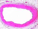
Circulatory System - Vein | In this Trichrome stained large vein, identify the tunica intima, tunica media, and the thick tunica advetitia. The tunica adventitia primarily contains collagen (blue) and dark-red smooth muscle cells. Note the collapsed lumen, caused by the lack of elasticity in venous walls. UCLA Histology Collec... | Circulatory System; Trichrome; tunica adventitia | UCLA Histology |
| 42 |
 |
Dendritic cells in spleen (rat) | Electron microscopy. The interdigitating dendritic cells (1) (so-called antigen-presenting cell, APC) exhibit numerous slender cell projections (1). (2) shows a macrophage with large lysosomes with heterogeneous contents. Small elongated fibroblastic reticular cells (3) form a structural framework ... | electron microscopy; dendritic cell | Poja Histology Collection - Lymphatic Tissues and Organs Subset |
| 43 |
 |
Peripheral Nervous System | This cross sectioned peripheral nerve demonstrates the relatively thick myelin covering of axons, as well as the endoneurium which is located between the myelin sheaths. Also note the blue-stained collagen of the epineurium and a small blood vessel. UCLA Histology Collection. | axons; endoneurium; myelin; Peripheral Nervous System | UCLA Histology |
| 44 |
 |
Presecretory ameloblasts in tooth development - bell stage, gerbil, postnatal | Electronmicroscopy. Well-arranged epithelial formation of presecretory ameloblasts (active nuclei) with their distal secretion sides towards the thin grey basal lamina. Predentin at the bottom close to the basal lamina and comprises collagen fibers, odontoblastic extensions and dispersed calcified m... | oral cavity; predentin; matrix vesicles | Poja Histology Collection - Oral Cavity Subset |
| 45 |
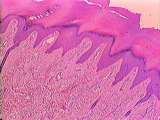 |
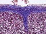
Skin - Sweat Gland | This example of thick skin has a sweat gland duct that travels through the dermis and epidermis to reach the bodys surface. Identify the dermal papillae, basal layer, spiny layer, granular layer, and thick cornified layer of the epidermis. Note the abundant collagen fibers of the reticular dermis. U... | UCLA Histology | |
| 46 |
 |
Spleen | Note the mesothelium of the visceral peritoneum covering the splenic capsule. Some of the pink streaks among the blue-stained collagen fibers in the capsule and trabeculae are elastin fibers and a few smooth muscle fibers may be present. Sinusoids are present in the red pulp. UCLA Histology Collecti... | red pulp; spleen; trichrome; visceral peritoneum | UCLA Histology |
| 47 |
 |
Alveolar cells in the lung (mammals) | Scheme electron microscopy. (5) alveolar space; (6) type I Pneumocyte; (7) basal lamina; (8) myofibroblast; (9) collagen and elastin fibers; (10) mesothelial cell of the visceral pleura; (11) capillary with erythrocyte; (12) endothelial cell lining the capillary; (13) type II pneum... | Pneumocyte type I ; Pneumocyte type I I | Poja Histology Collection - Respiratory System Subset |
| 48 |
 |
Ameloblasts and odontoblasts in tooth development - early bell stage, human, embryo; high magnification | Stain: Azan. From top to bottom: Presecretory ameloblasts with the distal side (secretion area) oriented towards the basal lamina intertwined with the tangential cut blue stained Korff's fibers (collagen) in predentin. Columnar odontoblasts in a epithelioid arrangement with their secretion area clos... | oral cavity; predentin; Korff's fibers | Poja Histology Collection - Oral Cavity Subset |
| 49 |
 |
Circulatory System - Veins | Walls of large veins have the same three tunics described for arteries, but the tunics are less distinct, and the elastic and muscular components are less developed. Note the lumen, tunica intima, tunica media, and tunica adventitia. The tunica adventitia is the thickest of the three coats, containi... | Circulatory System; tunica adventitia | UCLA Histology |
| 50 |
 |
Gastrointestinal Tract | This image of jejunum is taken from the base of a plica circularis. Observe the submucosa which contains collagen fibers, thick-walled arteries, thin-walled veins, and even thinner-walled lymphatic vessels. Also identify the inner circular muscle and outer longitudinal muscle layers, between which i... | gastrointestinal tract; small intestine | UCLA Histology |