The Health Education Assets Library (HEAL) is a collection of over 22,000 freely available digital materials for health sciences education. The collection is now housed at the University of Utah J. Willard Marriott Digital Library.
TO
Filters: Collection: "ehsl_heal"
| Title | Description | Subject | Collection | ||
|---|---|---|---|---|---|
| 1701 |
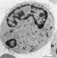 |
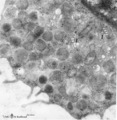
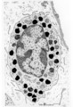
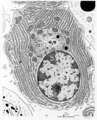
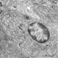
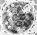
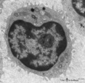
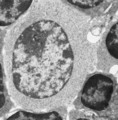
Monocyte (peripheral blood, human) | Electron microscopy. In this picture the large nucleus of this cell (diameter of 12-20 μm) is twice sectioned (1). Golgi areas (2), a few profiles of rough endoplasmic reticulum (3) and many free ribosomes are present. There are many mitochondria (4) as well as small-sized light vesicles (5). The c... | Poja Histology Collection - Blood & Bone Marrow Subset | |
| 1702 |
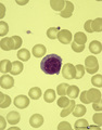 |
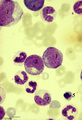
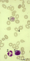
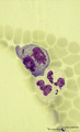
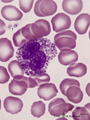
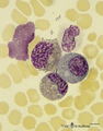
A plasmacytoid lymphocyte in peripheral blood smear (human) | Stain: May-Grnwald-Giemsa (MGG). The plasmacytoid lymphocyte is an activated B lymphocyte to be transformed into a plasma cell. The cytoplasm is more basophilic and the chromatin pattern is more clumped than in a virgin small lymphocyte. When the cell contains numerous immunoglobulin inclusions (glo... | Poja Histology Collection - Blood & Bone Marrow Subset | |
| 1703 |
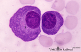 |
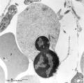
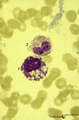
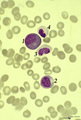
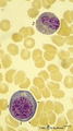
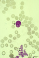
Plasma cells in bone marrow smear (human) | Stain: May-Grnwald-Giemsa (MGG). The two plasma cells are mature antibody-producing cells. The basophilic cytoplasm is filled up with rough endoplasmic reticulum (RER). The lighter zone (1) represents the Golgi areas. The nucleus is situated eccentrically and contains strands of condensed chromatin ... | Poja Histology Collection - Blood & Bone Marrow Subset | |
| 1704 |
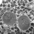 |
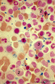
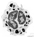
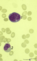
Survey of megakaryocytes (peripheral blood, human) | Electron microscopy. In a section of a so-called buffy coat collected from peripheral blood two huge polyploid megakaryocytes (1) (diameter up to 160 μm) are present. Note the dark granules in the future platelets. (2) erythroblasts; (3) reticulocytes and erythrocytes; (4, 5) monocytes. The arrows ... | Poja Histology Collection - Blood & Bone Marrow Subset | |
| 1705 |
 |
Orthochromatic erythroblast (bone marrow, rabbit) | Electron microscopy. The orthochromatic erythroblast (1) or late normoblast shows the early stage of extrusion of the nucleus though the latter appears not yet fully pyknotic. The cytoplasm contains few slender mitochondria (2) as well as scattered clumped polysomes. (3) Part of a reticulocyte. | Poja Histology Collection - Blood & Bone Marrow Subset | |
| 1706 |
 |
Survey of bone marrow section (human) | Stain: modified hematoxylin and eosin. In this survey the white spots (1) are fat cells. The marrow is well filled with cells of erythropoietic and myelopoietic series. The very large cells are usually megakaryocytes. Cells with very dark condensed nuclei belong mostly to the erythropoietic series. | Poja Histology Collection - Blood & Bone Marrow Subset | |
| 1707 |
 |
Megakaryoblast in bone marrow smear (human) | Stain: May-Grnwald-Giemsa (MGG). The megakaryoblast (1) is not yet producing platelets and has a relative small amount of basophilic cytoplasm. The nucleus is large with fine disperse chromatin and nucleoli. Most other nucleated cells are of the myeloid cell line. | Poja Histology Collection - Blood & Bone Marrow Subset | |
| 1708 |
 |
Erythroblast and neutrophilic myelocytes in bone marrow smear (human) | Stain: May-Grnwald-Giemsa (MGG). (1) an older basophilic erythroblast with slightly condensed nuclear chromatin. (2) two neutrophilic myelocytes with azurophilic primary granules. | Poja Histology Collection - Blood & Bone Marrow Subset | |
| 1709 |
 |
Band form of neutrophilic granulocyte | Scheme electron microscopy. From CFU-S (colony forming units-spleen) stem cells arise CFU-GM (colony forming unit-granulocyte/monocyte) stem cells. The latter divide by mitoses and differentiate via promyeloblasts and myeloblasts into neutrophilic myelocytes (the last proliferative stage). The next ... | Poja Histology Collection - Blood & Bone Marrow Subset | |
| 1710 |
 |
Mast cell (lung, human) | Electron microscopy. Mast cells (mastocytes) are frequently found perivascularly or perineurally. A part of this mast cell shows thin microvilli at the surface and the cytoplasm is provided with a moderate amount of organelles. At (*) the Golgi area (1) is seen in close association with granules tha... | Poja Histology Collection - Blood & Bone Marrow Subset | |
| 1711 |
 |
Iron (Perls) staining of sideroblasts in bone marrow smear (human) | Stain: Perl's stain for iron (Prussian blue). The intense blue stained hemosiderin in the erythroblasts (1) is visible as siderotic granules (pathologic siderosomes). These pathologic siderosomes are sometimes distributed in a circle around the nucleus (ringed sideroblasts). The myeloid cell types (... | Poja Histology Collection - Blood & Bone Marrow Subset | |
| 1712 |
 |
Neutrophilic granulocyte | Scheme electron microscopy. The neutrophil is a phagocytic cell (12-15 m) with a segmented lobular nucleus (3-5 lobes) and many cytoplasmic granules filled with degradative enzymes. These PMN cells (polymorph nuclear leukocytes) are the major cell types mediating acute inflammatory response to bacte... | Poja Histology Collection - Blood & Bone Marrow Subset | |
| 1713 |
 |
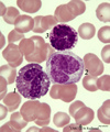
Differentiation stages of myeloid cells in bone marrow smear (human) | Stain: May-Grnwald-Giemsa (MGG). (1) Promyelocytes with coarse primary azurophilic granules and nucleoli. (2) Myelocyte with starting indentation of the nucleus (primary plus secondary granules). (3) Neutrophilic metamyelocytes with kidney shaped nuclei. (4) Band form of neutrophilic granulocytes (b... | Poja Histology Collection - Blood & Bone Marrow Subset | |
| 1714 |
 |
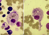
Contamination with squamous cell cells in peripheral blood smear (A) and in a bone marrow smear (B) (human) | Stain: May-Grnwald-Giemsa (MGG). In blood and bone marrow smears incidentally squamous epithelial cells from the skin tissues are found. (A-1) is an epithelial cell from the keratinized stratified (upper) epidermis; (A-2) a young neutrophil with a few vacuoles in the cytoplasm (probably degeneration... | Poja Histology Collection - Blood & Bone Marrow Subset | |
| 1715 |
 |
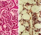
Granuloma in bone marrow smear (human) | Stain: (A) Hematoxylin and eosin. (B) Silver stain. Granuloma composed of giant cells, epithelioid cells, lymphocytes and reticular fibers (B). Granulomas in the bone marrow may be found in various inflammatory reactions as well as hematological and non-hematological malignancies and as a reaction t... | Poja Histology Collection - Blood & Bone Marrow Subset | |
| 1716 |
|
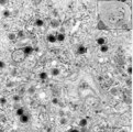
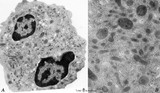
Circulating phagocytic cells | Electron microscopy, a set of the ultrastuctural features of different types of phagocytic cells. Top left side (A): Circulating monocyte with phagocytized latex particles (peripheral blood, human). The nucleus is large and with a long small indentation. Close to the nucleus several individually in... | Poja Histology Collection - Blood & Bone Marrow Subset | |
| 1717 |
|
Neutrophilic metamyelocyte in peripheral blood smear (human) | Stain: May-Grnwald-Giemsa (MGG). The neutrophilic metamyelocyte is differentiating nearly to a juvenile unsegmented (band form) neutrophil, but the nuclear chromatin is only partly condensed. Hardly visible dust-like granules can be observed in the still pale bluish cytoplasm. | Poja Histology Collection - Blood & Bone Marrow Subset | |
| 1718 |
 |
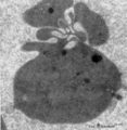
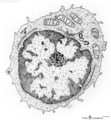
Monocyte | Scheme electron microscopy. The diameter of monocytes ranges from 12-20 m. Characteristic is the large (kidney-shaped) nucleus with one or more nucleoli (1) and several indentations. The cytoplasm contains: (2) Golgi areas, (3) electron-dense lysosomal granules or primary or azurophilic granules wit... | Poja Histology Collection - Blood & Bone Marrow Subset | |
| 1719 |
 |
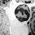
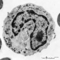
Monocyte (peripheral blood, human) | Electron microscopy. A large cell with an indented nucleus (1), few Golgi areas (2) and numerous organelles. The cytoplasm contains scattered homogeneous electron-dense lysosomal granules (3) (azurophilic granules with acid phosphatase, arylsulfatase) in variable amounts and few vacuoles. Small pseu... | Poja Histology Collection - Blood & Bone Marrow Subset | |
| 1720 |
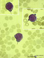 |
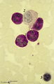
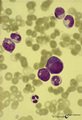
Normal proerythroblasts in bone marrow smear (human) | Stain: May-Grnwald-Giemsa (MGG). The three mounted proerythroblasts are easily recognized by their so called ears i.e., the blue cytoplasmic projections or extensions (arrows). The basophilic cytoplasm as well as the nucleoli point to the blast character of the cells. | Poja Histology Collection - Blood & Bone Marrow Subset | |
| 1721 |
 |
Neutrophilic granulocyte (peripheral blood, human) | Electron microscopy. Two nuclear lobes of the segmented nucleus are visible in a cytoplasm with a moderate amount of organelles but with abundant granules of varying sizes. The motile human neutrophil (9-14 μm) contains at least four types of granules. However, by routine electron microscopy primar... | Poja Histology Collection - Blood & Bone Marrow Subset | |
| 1722 |
 |
Dhle bodies in mature neutrophils in peripheral blood smear (human) | : May-Grnwald-Giemsa (MGG). In adults the usual response to a bacterial infection is a neutrophil leukocytosis with a shift to the left, toxic granulation, Dhle bodies and, when the infection is severe, cytoplasmic vacuolation and swelling occur. Dhle bodies (1, arrow) are small, pale-blue-grey cyto... | Poja Histology Collection - Blood & Bone Marrow Subset | |
| 1723 |
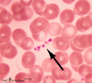 |
Platelets in peripheral blood smear (human) | Stain: May-Grnwald-Giemsa (MGG). The platelets or thrombocytes are small cell fragments released from megakaryocytes. The platelets (→) measure 1-3 μm in diameter. They contain fine azurophilic granules which may be dispersed throughout the cytoplasm or concentrated in the centre; in the latter c... | Poja Histology Collection - Blood & Bone Marrow Subset | |
| 1724 |
 |
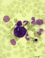
Monoblast, myeloid and erythropoietic cells in bone marrow smear (human) | Stain: May-Grnwald-Giemsa (MGG). (1) late promyelocyte or early myelocyte with nucleoli in the nucleus and ample azurophilic granules. (2) monoblast with indented nucleus and nucleoli (3) polychromatic erythroblast that will turn into an orthochromatic erythroblast (4), with progressing chromatin co... | Poja Histology Collection - Blood & Bone Marrow Subset | |
| 1725 |
 |
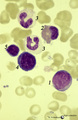
Shift to the left response of granulocytes in blood smear (human) | Stain: May-Grnwald-Giemsa (MGG). During infection a shift towards a relative increase of the number of neutrophilic granulocytes (3) with reduced lobulation of the nuclei and immature white cells, occurs in blood. (1) promonocyte. (2) (pro)myelocyte. | Poja Histology Collection - Blood & Bone Marrow Subset | |
| 1726 |
 |
Large lymphocyte in peripheral blood smear (human) | Stain: May-Grnwald-Giemsa (MGG). The large lymphocyte has a dense chromatin structure and a large transparent cytoplasm with some granules (→). (On the contrary a monocyte exposes a more transparent nucleus and a slightly greyish cytoplasm). | Poja Histology Collection - Blood & Bone Marrow Subset | |
| 1727 |
 |
Orthochromatic erythroblasts in bone marrow smear (human) | Stain: May-Grnwald-Giemsa (MGG). Two orthochromatic erythroblasts (1) with condensed nuclear chromatin and a transition of cytoplasmic stain towards the color of normal erythrocytes. (2) a segmented neutrophilic granulocyte. | Poja Histology Collection - Blood & Bone Marrow Subset | |
| 1728 |
 |
Orthochromatic erythroblast and lymphocyte in bone marrow smear (human) | Stain: May-Grnwald-Giemsa (MGG). The cytoplasm of this orthochromatic erythroblast (2) shows a faint polychromatic tint. The chromatin is arranged in clumps and stains deeply. (2) activated lymphocyte. | Poja Histology Collection - Blood & Bone Marrow Subset | |
| 1729 |
 |
Normal mature neutrophilic granulocyte in peripheral blood smear (human) | Stain: May-Grnwald-Giemsa (MGG). Neutrophilic granulocyte with five nuclear lobes or segments, (not hypersegmented). The arrow indicates a drumstick. Note the fine dust-like granules. | Poja Histology Collection - Blood & Bone Marrow Subset | |
| 1730 |
 |
Mast cell | Mast cells (mastocytes) are oval (12 m) or spindle-shaped and are frequently found perivascularly or perineurally. The cytoplasm is provided with a moderate amount of organelles and small thin or blunt microvilli at the surface. Most obvious is the presence of large granules varying in shape and siz... | Poja Histology Collection - Blood & Bone Marrow Subset | |
| 1731 |
 |
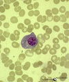
Toxic granulation and vacuolization in neutrophilic myelocytes in peripheral blood smear (human) | Stain: May-Grnwald-Giemsa (MGG). The myelocyte (1) and the metamyelocyte (2) show toxic granulation, vacuolisation and cytoplasmic swelling. Toxic granulation is characterized by violet-purple granules of varying size in the cytoplasm; generally admixed with normal pink granules. The phenomenon occu... | Poja Histology Collection - Blood & Bone Marrow Subset | |
| 1732 |
 |
Howell-Jolly bodies in reticulocytes in peripheral blood smear (human) | Stain: May-Grnwald-Giemsa (MGG). The Howell-Jolly-bodies (1) in the reticulocyte are remnants of the nucleus (nuclear chromatin granules) of an erythroblast during anomalous development due to vitamin B12 deficiency, folic acid deficiency or in megaloblastic and hemolytic anemias. (2) Neutrophil wit... | Poja Histology Collection - Blood & Bone Marrow Subset | |
| 1733 |
 |
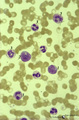
Neutrophilic and basophilic granulocytes in peripheral blood smear (human) | Stain: May-Grnwald-Giemsa (MGG). (1) A hypersegmented (>5 segments) neutrophilic granulocyte with clear fine granules. (2) Represents a mature basophilic granulocyte with clear distinguishable, coarse purple granules and a few vacuoles (because the granules dissolve in water during the staining proc... | Poja Histology Collection - Blood & Bone Marrow Subset | |
| 1734 |
 |
Flaming plasma cell in peripheral blood smear (human) | Stain: May-Grnwald-Giemsa (MGG). The so-called flaming plasma cell (1) is characterized by fiery fringes, which are formed by pseudopodic cytoplasmic projections (arrows) that stain with carmin red. These peripheral cytoplasmic spots contain numerous dilated endoplasmic reticulum cisterns, which are... | Poja Histology Collection - Blood & Bone Marrow Subset | |
| 1735 |
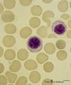 |
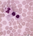
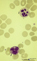
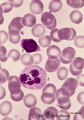
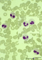
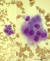
Plasmacytoid lymphocytes (activated B cells) in peripheral blood smear (human) | Stain: May-Grnwald-Giemsa (MGG). Two different plasmacytoid lymphocytes or activated young B cells (up to 15 μm) contain a dark-stained nucleus and a slightly basophilic cytoplasm with a kind of a 'nuclear hof' indicating the Golgi area. These cells will develop into plasma cells. | Poja Histology Collection - Blood & Bone Marrow Subset | |
| 1736 |
 |
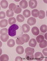
Monoblast in bone marrow smear (human) | Stain: May-Grnwald-Giemsa (MGG). The monoblast (1) has a large nucleus with an irregular border, nucleoli and fine disperse chromatin, and a basophilic cytoplasm with no or a limited number of granules. (2) Two neutrophilic band forms. | Poja Histology Collection - Blood & Bone Marrow Subset | |
| 1737 |
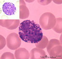 |
Basophilic granulocyte in peripheral blood smear (human) | Stain: May-Grnwald-Giemsa (MGG). The basophilic granulocyte is characterized by large, coarse, aggregated dark purple granules. The nuclear lobes are usually not very well visible and masked by the granules (see also inset). | Poja Histology Collection - Blood & Bone Marrow Subset | |
| 1738 |
 |
Dividing cells (mitosis) in bone marrow smear (human) | Stain: May-Grnwald-Giemsa (MGG). (1) Shows a dividing cell (mitotic figure) possibly an erythroblast cell type. (2) Shows two segmented neutrophils. | Poja Histology Collection - Blood & Bone Marrow Subset | |
| 1739 |
 |
Plasma cell | Scheme electron microscopy. The mature plasma cell or plasmacyte (12-15 m) shows an eccentric nucleus (1) with a characteristic chunky distribution of heterochromatin along the inner nuclear membrane ('spoke-wheel' effect in light microscopy). Juxta-nuclearly an elaborate Golgi area (2) with secreti... | Poja Histology Collection - Blood & Bone Marrow Subset | |
| 1740 |
 |
Basophilic granulocyte | Scheme electron microscopy. Basophils are non-phagocytic granulocytes that account for 0.5% to 1.0% of the circulating white blood cells. Their granulated cytoplasm stains with basic dyes, hence the name basophil. Electron microscopy reveals a multilobed nucleus (1); few mitochondria (2); numerous ... | Poja Histology Collection - Blood & Bone Marrow Subset | |
| 1741 |
 |
Eosinophilic myelocyte and neutrophilic myelocyte in bone marrow smear (human) | Stain: May-Grnwald-Giemsa (MGG). The eosinophilic myelocyte (1) contains brown-like granules (not orange) in the bluish basophilic cytoplasm. Nucleoli are still visible. Maturation of eosinophils parallels that of neutrophils except for the production of the secondary, specific granules in myelocyte... | Poja Histology Collection - Blood & Bone Marrow Subset | |
| 1742 |
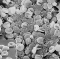 |
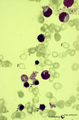
Mature erythrocytes (spleen, gerbil) | Scanning electron microscopy. Normal mature erythrocytes (1) are mostly biconcave and discoid. They might change their forms due to mechanical pressures. (2) a mature lymphocyte recognizable by many short stubby microvilli at the surface. (3) granulocytes are larger with fewer but longer microvilli.... | Poja Histology Collection - Blood & Bone Marrow Subset | |
| 1743 |
 |
Neutrophilic granulocyte with drumstick in peripheral blood smear (human) | Stain: May-Grnwald-Giemsa (MGG). This neutrophil has a segmented lobulated nucleus with one drumstick (→) and one non-specific appendage (small club). The cytoplasm is filled with very fine granules. (2) small (smudged) lymphocyte with a dark condensed, indented nucleus and a small rim of cytopla... | Poja Histology Collection - Blood & Bone Marrow Subset | |
| 1744 |
 |
Reticulocyte (peripheral blood, human) | Electron microscopy. An anucleate reticulocyte with fimbriated processes (asymmetrical folding of the cell membrane) after nuclear extrusion. The cytoplasm contains a heavy amount of hemoglobin, electron-dense lysosomes, vesicular remnants as well as scattered clumped polysomes. | Poja Histology Collection - Blood & Bone Marrow Subset | |
| 1745 |
 |
Megakaryocyte | Scheme electron microscopy. The megakaryocyte is derived from a pro-megakaryocyte which originates from splenic stem cells (CFU-S, colony forming units-spleen). The megakaryocyte is a giant polyploid cell (35-160 m) and contains a large multilobate nucleus (1). The perinuclear area shows Golgi areas... | Poja Histology Collection - Blood & Bone Marrow Subset | |
| 1746 |
 |
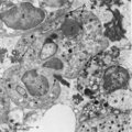
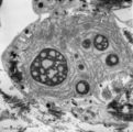
Megakaryocyte (bone marrow, mouse) | Electron microscopy. A detail of the cytoplasm of a megakaryocyte (see inset) demonstrates distinctly a part of the intermediate zone subdivided by an interconnected tubular system (so-called demarcation membrane system or open canalicular system: OCS) that is in continuity with the cell surface. Th... | Poja Histology Collection - Blood & Bone Marrow Subset | |
| 1747 |
 |
Reticulocytes in peripheral blood smear (human) | Stain: Brilliant cresyl blue. The blue stained reticulum strands and aggregates (1) are ribosomal residues in the young red blood cells that just have expelled their nuclei. The reticulocytes are not to be confused with large Heinz bodies. Heinz bodies (↓, arrows) are red cell inclusions composed ... | Poja Histology Collection - Blood & Bone Marrow Subset | |
| 1748 |
 |
Eosinophilic granulocyte (peripheral blood, human) | Electron microscopy. (A) Overview of the cell. Note two nuclear lobes (1) due to the section. There are numerous specific eosinophilic granules (2) and a Golgi area (3). Few thin filopodia (↓, arrows) are present. (B) Detail: some vesicles and many specific eosinophilic granules of varying sizes. ... | Poja Histology Collection - Blood & Bone Marrow Subset | |
| 1749 |
 |
Homozygotic Pelger-Huet's nuclear anomaly of granulocytes in peripheral blood smear (human) | Stain: May-Grnwald-Giemsa (MGG). In this case of homozygotic Pelger-Hut anomaly the neutrophil (1) as well as the eosinophil (2) have non-lobed nuclei with condensed chromatin. Characteristic fine dust-like granules are present in the neutrophil, while the eosinophil has separate recognizable coarse... | Poja Histology Collection - Blood & Bone Marrow Subset | |
| 1750 |
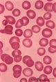 |
Erythrocytes in peripheral blood smear (human) | Stain: May-Grnwald-Giemsa (MGG). Normal erythrocytes in a blood smear of peripheral blood. The mature human erythrocytes do not have nuclei. Their normal diameter is about 7.5 μm. Macrocytes have a diameter >9 μm, while microcytes are smaller than 6 μm. The thickness of an erythrocyte in the cent... | Poja Histology Collection - Blood & Bone Marrow Subset | |
| 1751 |
 |
Reticulocytes in peripheral blood smear (human) | Stain: Brilliant cresyl blue. After expulsion of the erythroblastic nucleus the remaining cell is called reticulocyte. This staining visualizes the remained aggregated strands of polysomes as a fine network. The expulsed nucleus will be phagocytosed by macrophages and reticular cells in the bone mar... | Poja Histology Collection - Blood & Bone Marrow Subset | |
| 1752 |
 |
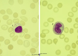
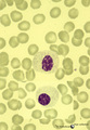
Large lymphocyte and small plasmacytoid lymphocyte in peripheral blood smear (human) | Stain: May-Grnwald-Giemsa (MGG). (1) large lymphocyte with a transparent light cytoplasm in which few fine granules are visible. (2) (inset) smaller plasmacytoid lymphocyte (activated young B cell) with light basophilic cytoplasm. | Poja Histology Collection - Blood & Bone Marrow Subset | |
| 1753 |
 |
Giant neutrophilic metamyelocyte in bone marrow smear (human) | Stain: May-Grnwald-Giemsa (MGG). (1) Giant neutrophilic metamyelocyte. (2) Normal neutrophilic metamyelocytes. (3) Smudged eosinophilic granulocyte. (4) Orthochromatic erythroblast. | Poja Histology Collection - Blood & Bone Marrow Subset | |
| 1754 |
 |
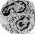
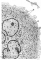
Plasma cells (spleen, mouse) | Electron microscopy. The mature plasma cell shows an eccentric nucleus with a characteristic chunky distribution of heterochromatin along the inner nuclear membrane ('spoke-wheel' effect in light microscopy). Juxta-nuclearly an elaborate Golgi area (2) with secretion vacuoles is localized (cytocentr... | Poja Histology Collection - Blood & Bone Marrow Subset | |
| 1755 |
 |
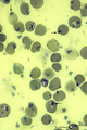
Phagocytosis of latex by neutrophil (peripheral blood, human) | Electron microscopy. The primary function of neutrophilic granulocytes is phagocytosis, ingestion of and destroying microbes or, as shown here, of latex particles. Several electron-light latex particles (1) (with a dense core) are internalized after incubation of free circulating granulocytes with a... | Poja Histology Collection - Blood & Bone Marrow Subset | |
| 1756 |
 |
Heterozygotic Pelger-Hut's nuclear anomaly of neutrophilic granulocytes in peripheral blood smear (human) | Stain: May-Grnwald-Giemsa (MGG). The majority of neutrophils has bilobed nuclei, the lobes being rounder than normal and the chromatin is more condensed. About 30% of the cells appear as band forms (imitating shift to the left). A characteristic 'pince-nez' shape is common. Note the fine dust-like g... | Poja Histology Collection - Blood & Bone Marrow Subset | |
| 1757 |
 |
Proerythroblast and eosinophilic metamyelocyte in bone marrow smear (human) | Stain: May-Grnwald Giemsa (MGG). The late proerythroblast (1) has a deep blue-stained basophilic cytoplasm and so called ears (→, slight bulging) of sidewards accumulated ribosomes (in electron microscopy) in the cytoplasm. The eosinophilic metamyelocyte (2) has characteristic large, solitary brow... | Poja Histology Collection - Blood & Bone Marrow Subset | |
| 1758 |
 |
Polycythemia in bone marrow smear (human) | Stain: Hematoxylin and eosin. Low power view. Polycythemic phase. Hypercellular marrow as a result of excessive proliferation of erythroid cells (numerous dark nuclei) and of the megakaryocytic lineage (tendency to cluster). (1) fat cells in bone marrow. (2) megakaryocytes. (3) erythropoietic cell t... | Poja Histology Collection - Blood & Bone Marrow Subset | |
| 1759 |
 |
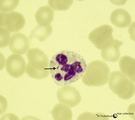
Eosinophilic granulocyte in peripheral blood smear (human) | Stain: May-Grnwald-Giemsa (MGG). The eosinophil (11-15 μm) contains a bilobed nucleus and numerous large solitary brown-orange granules. The eosinophils are the first line of defense against parasites but also take part in allergic reactions (bronchial asthma). Arrow (↓) points to two platelets. | Poja Histology Collection - Blood & Bone Marrow Subset | |
| 1760 |
 |
Proerythroblasts in bone marrow smear (human) | Stain: May-Grnwald-Giemsa (MGG). The two proerythroblasts (1) are large cells with condensed nuclear chromatin and a deep blue-stained basophilic cytoplasm with so-called ears (→, bulging) where ribosomes (in electron microscopy) are aggregated or shifted towards the periphery of the cell. (2) smu... | Poja Histology Collection - Blood & Bone Marrow Subset | |
| 1761 |
 |
A bare megakaryocyte nucleus in bone marrow smear (human) | Stain: May-Grnwald-Giemsa (MGG). (1) indicates a naked, large-sized polyploid nucleus of a megakaryocyte with visible lobes. No cytoplasm nor platelets are left around the nucleus. Most surrounding cells are of myeloid origin, except one orthochromatic erythroblast (2). | Poja Histology Collection - Blood & Bone Marrow Subset | |
| 1762 |
 |
Anemic bone marrow smear in iron deficiency (human) | Stain: May-Grnwald-Giemsa (MGG). Due to iron deficiency the erythrocytes (1) look empty, while also the erythroblasts (2) are relatively small and show a ruffled cell border. | Poja Histology Collection - Blood & Bone Marrow Subset | |
| 1763 |
 |
Fibrin phagocytosis by neutrophils (peripheral blood, mouse) | Electron microscopy. At site of tissue damage motile neutrophils are among the first to be involved actively in phagocytosis. In between a cluster of neutrophilic granulocytes fibrin depositions (1) are present. At (1→) fibrin accumulations are close attached to the surface of a neutrophil indicat... | Poja Histology Collection - Blood & Bone Marrow Subset | |
| 1764 |
 |
Megakaryocyte in bone marrow smear (human) | Stain: May-Grnwald-Giemsa (MGG), (black and white print). The giant polyploid cell (35-160 μm) contains a large multilobate nucleus. Note the ring-like pattern of the nuclei (1). The light-stained perinuclear zone (2) contains Golgi areas, within the encompassing greyish (intermediate) zone (3) gra... | Poja Histology Collection - Blood & Bone Marrow Subset | |
| 1765 |
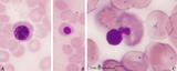 |
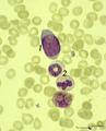
Nucleus expulsion in polychromatic-orthochromatic erythroblasts in bone marrow smear (human) | Stain: May-Grnwald-Giemsa (MGG). The composed pictures show three sequential stages in the final maturation stage of red blood cells. (A) polychromatic erythroblast with a condensed nucleus and an almost acidophilic cytoplasm. (B) the nucleus in the orthochromatic erythroblast is completely condense... | Poja Histology Collection - Blood & Bone Marrow Subset | |
| 1766 |
 |
Lymphocyte (mature) (peripheral blood, human) | Electron microscopy. Mature B and T lymphocytes are ameboid cells (5-8 m) with a high nucleus-cytoplasmic ratio. The cytoplasm contains a juxta-nuclear Golgi area with centrioles (6), few large mitochondria (4). Profiles of rough endoplasmic reticulum (RER) (5) are sparsely present. The surface exhi... | Poja Histology Collection - Blood & Bone Marrow Subset | |
| 1767 |
 |
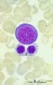
Large lymphocyte and monocyte in peripheral blood smear (human) | Stain: May-Grnwald-Giemsa (MGG). The large lymphocyte (1) has a slightly condensed nucleus and the cytoplasm contains some fine azurophilic granules, and is therefore sometimes called large granular lymphocyte (LGL) or killer cell. The monocyte (2) is generally larger than the lymphocyte with a roun... | Poja Histology Collection - Blood & Bone Marrow Subset | |
| 1768 |
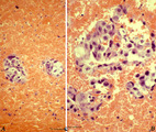 |
Prostatic carcinoma metastasis in bone marrow of sternum (human) | Stain: Hematoxylin and eosin. Survey (A) and detail (B) of a prostate carcinoma metastasized to the bone marrow, (sternum aspiration). Clusters of tumour cells; clearly foreign to the marrow. Common primary tumours that metastasize to the bone marrow include breast, prostate, lung, thyroid, kidney, ... | Poja Histology Collection - Blood & Bone Marrow Subset | |
| 1769 |
 |
Survey and detail of a peripheral blood smear contaminated with endothelial cells (human) | Stain: May-Grnwald-Giemsa (MGG). Survey (A) and details (B) show aggregates of elongated cells with a large nucleus, mostly situated at the end of a smear between the erythrocytes and granulocytes. These endothelial cells are contaminants derived from blood vessels during puncture. | Poja Histology Collection - Blood & Bone Marrow Subset | |
| 1770 |
 |
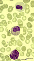
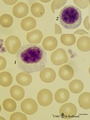
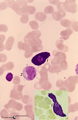
Eosinophil, monocyte and basophil in blood smear (human) | Stain: May-Grnwald-Giemsa (MGG). (1) eosinophilic granulocyte with two nuclear lobes and large eosinophilic granules in the cytoplasm. (2) monocyte with a large indented nucleus that is much more transparent than the nuclei of the two other cells. (3) basophilic granulocyte with aggregated dark purp... | Poja Histology Collection - Blood & Bone Marrow Subset | |
| 1771 |
 |
Homozygotic Pelger-Hut's nuclear anomaly of neutrophilic granulocytes in peripheral blood smear (human) | Stain: May-Grnwald-Giemsa (MGG). The neutrophils in this patient with homozygotic Pelger-Hut's anomaly have non-lobed nuclei. The chromatin is condensed and fine dust-like granules are present. | Poja Histology Collection - Blood & Bone Marrow Subset | |
| 1772 |
 |
Monoblast, erythroblasts and granulopoietic cells in bone marrow smear (human) | Stain: May-Grnwald-Giemsa (MGG). The monoblast (1) has a large nucleus with an irregular border, nucleoli and fine disperse chromatin, and a basophilic cytoplasm (greyish blue) with no or hardly any granules. (2) Eosinophilic metamyelocyte with large, brown granules. (3) Juvenile unsegmented (band f... | Poja Histology Collection - Blood & Bone Marrow Subset | |
| 1773 |
 |
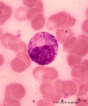
Normal plasma cell in peripheral blood smear (human) | Stain: May-Grnwald-Giemsa (MGG). The mature plasma cell has a large basophilic cytoplasma for IgG production, a clearly visible unstained (white) Golgi area, and a nucleus with well-condensed heterochromatin. Plasma cells can be found in the peripheral blood as a result of antigenic stimulation or c... | Poja Histology Collection - Blood & Bone Marrow Subset | |
| 1774 |
 |
Contamination with osteoblasts of a bone marrow puncture (human) | Stain: May-Grnwald-Giemsa (MGG). In bone marrow smears from a sternum puncture sometimes osteoblasts are found. Osteoblasts may occur in groups, are oval with a usually eccentric, relatively small nucleus. The chromatin is often coarse with 1-3 nucleoli. The cytoplasm is mostly light blue and may co... | Poja Histology Collection - Blood & Bone Marrow Subset | |
| 1775 |
 |
Contamination with osteoclasts of a bone marrow puncture (human) | Stain: May-Grnwald-Giemsa (MGG). In the procedure for preparing a bone marrow smear from a sternum puncture osteoclasts are frequently found in the bone marrow smears. The osteoclasts are multinucleated giant cells for the destruction of bone in the process of remodelling and growth. The cytoplasm m... | Poja Histology Collection - Blood & Bone Marrow Subset | |
| 1776 |
 |
Survey and detail of bone marrow section (human) | Stain: modified hematoxylin and eosin (A, B). The bone marrow consists of bone trabecles or crests (1) covered with osteoclasts and osteoblasts. Between the trabecles bone marrow parenchym (2) consisting of progenitor cells, reticular cells and fat cells (3). Numerous capillaries (B, 4) and enlarged... | Poja Histology Collection - Blood & Bone Marrow Subset | |
| 1777 |
 |
Orthochromatic erythroblast (bone marrow, rabbit) | Electron microscopy. The orthochromatic erythroblast or late normoblast is surrounded by young granulocytes (3) and part of a megakaryocyte (2). The cell shows the early stage of extrusion of the nucleus (1) that is already slightly pyknotic. The cytoplasm contains few small mitochondria (arrows) an... | Poja Histology Collection - Blood & Bone Marrow Subset | |
| 1778 |
 |
Orthochromatic erythroblast in bone marrow smear (human) | Stain: May-Grnwald-Giemsa (MGG). The orthochromatic erythroblast (1) has a dark condensed (pyknotic) nucleus located at one site of the cell, ready for being expulsed from the cell. (2) Erythrocyte. (3) Represents a platelet or thrombocyte. | Poja Histology Collection - Blood & Bone Marrow Subset | |
| 1779 |
 |
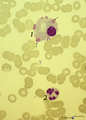
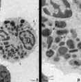
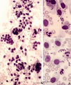
Hemophagocytosis by reticular cells in bone marrow smear (human) | Stain: May-Grnwald-Giemsa (MGG). (A) shows a reticular cell that has phagocytosed several erythrocytes (*). The cell is surrounded by darkly stained expulsed nuclei. In (B) a similar cell has phagocyized blood platelets (arrows). | Poja Histology Collection - Blood & Bone Marrow Subset | |
| 1780 |
 |
Neutrophilic myelocyte in bone marrow smear (human) | Stain: May-Grnwald-Giemsa (MGG). In the neutrophilic myelocyte the nucleus is located eccentrically at one side of the cell. Nucleoli are visible as well as the primary azurophilic granules in the cytoplasm. | Poja Histology Collection - Blood & Bone Marrow Subset | |
| 1781 |
 |
Erythron or erythropoietic island in bone marrow smear (human) | Stain: May-Grnwald-Giemsa. The picture shows a reticular cell (1) surrounded by almost exclusively erythroblasts at different stages of development and maturation (2). The cells marked with (*) are myeloid cell types. The reticular cell has phagocytized nuclear debris of expulsed nuclei (→). The r... | Poja Histology Collection - Blood & Bone Marrow Subset | |
| 1782 |
 |
Monoblast | Scheme electron microscopy. The precursor of this cell is the promonoblast derived from the common stem cell CFU-GM (colony forming unit-granulocyte/monocyte). After proliferation of the promonoblasts they transform into monoblasts (up to 10 μm). The nucleus of the monoblast shows an indentation as... | Poja Histology Collection - Blood & Bone Marrow Subset | |
| 1783 |
 |
Neutrophilic myelocyte and basophilic erythroblast in reactive bone marrow smear (human) | Stain: May-Grnwald-Giemsa (MGG). The neutrophilic myelocyte (1) has a nucleus (with nucleoli) located at one side of the cell. Cytoplasm contains many azurophilic granules. The early basophilic erythroblast (2) has a strong basophilic cytoplasm without granules, and a nucleus with condensed chromati... | Poja Histology Collection - Blood & Bone Marrow Subset | |
| 1784 |
 |
Lymphocyte (mature) | Scheme electron microscopy. Mature B and T lymphocytes are ameboid cells (5-8 m) with a high nucleus-cytoplasmic ratio. The cytoplasm contains a juxta-nuclear Golgi area (1) with centrioles (2), few large mitochondria (3) and sparsely profiles of rough endoplasmic reticulum (4) are present. The surf... | Poja Histology Collection - Blood & Bone Marrow Subset | |
| 1785 |
 |
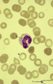
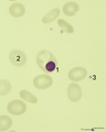
Monocyte in peripheral blood smear (human) | Stain: May-Grnwald-Giemsa (MGG). The nucleus of the mature monocyte has a kidney bean- or horseshoe-shape, is rather transparent compared to lymphocytes and granulocytes, and the cell is much larger (12-20 μm). The fine granular cytoplasm contains lysosomes, phagocytic vacuoles. Note platelets at (... | Poja Histology Collection - Blood & Bone Marrow Subset | |
| 1786 |
 |
Lymphoblast (spleen, rat) | Electron microscopy. Interspersed between small lymphocytes the lymphoblast with a high nucleus-cytoplasm ratio is a larger cell with a distinct nucleolus. The cytoplasm is studded with numerous polysomes and ribosomes, only few mitochondria and a single strand of rough endoplasmic reticulum are pre... | Poja Histology Collection - Blood & Bone Marrow Subset | |
| 1787 |
 |
Eosinophilic (meta)myelocytes in bone marrow smear (human) | Stain: May-Grnwald-Giemsa (MGG). Three eosinophilic (meta)myelocytes (1) at slightly different maturation stages. Notice the brown blue large solitary granules. (2) neutrophilic metamyelocyte. (3) smudged cell. | Poja Histology Collection - Blood & Bone Marrow Subset | |
| 1788 |
 |
Basophilic myelocyte in bone marrow smear (human) | Stain: May-Grnwald-Giemsa (MGG). The basophilic myelocyte (1) contains dark bluish-purple (metachromatic), coarse granules in the cytoplasm. (2) Platelets. | Poja Histology Collection - Blood & Bone Marrow Subset | |
| 1789 |
 |
Promonocyte in bone marrow smear (human) | Stain: May-Grnwald-Giemsa (MGG). The young promonocyte (1) shows clear nucleoli and a distinct indentation of the nucleus (arrow). (2) Neutrophilic myelocyte. | Poja Histology Collection - Blood & Bone Marrow Subset | |
| 1790 |
 |
Neutrophilic granulocyte (peripheral blood, human) | Electron microscopy. Survey (A) and detail (B) of a neutrophilic granulocyte. Two nuclear lobes (1) of the segmented nucleus are visible in a cytoplasm with a moderate amount of organelles but with abundant granules (2, 3) of varying sizes. The detail shows in the cytoplasm large amounts of granules... | Poja Histology Collection - Blood & Bone Marrow Subset | |
| 1791 |
 |
Late band-form of neutrophilic granulocyte (peripheral blood, human) | Electron microscopy. Band forms (9-12 μm) are the earliest stages of the motile two-lobed granulocytes. The horseshoe shaped nucleus (3) is irregularly and indicates progressing lobulation. The neutrophil contains many granules of varying sizes and densities. On base of routine electron microscopy ... | Poja Histology Collection - Blood & Bone Marrow Subset | |
| 1792 |
 |
Leukocyte Alkaline Phosphatase (LAP/NAP score) in peripheral blood smear (human) | Stain: Alkaline phosphatase staining (Ackerman). In mature neutrophils granules contain alkaline phosphatase. In eosinophils no alkaline phosphatase activity is present. A considerable number of brown stained neutrophils can be indicative for a patient with infection (leukemoid reaction, A). Image i... | Poja Histology Collection - Blood & Bone Marrow Subset | |
| 1793 |
 |
PAS-positively stained neutrophilic granulocytes in bone marrow smear (human) | Stain: Periodic acid-Schiff reaction (PAS). Mature neutrophils (1) have fine positive granules within negatively (un)stained cytoplasm, whereas eosinophils (2) and basophils show a positive cytoplasmic reaction contrasting with the negative granules. In lymphocytes (1) PAS-positive granules are rare... | Poja Histology Collection - Blood & Bone Marrow Subset | |
| 1794 |
 |
Plasma cell with Russell bodies (nose septum, rat) | Electron microscopy. This mature plasma cell exhibits in the cytoplasm dilated rough endoplasmic reticulum (1) filled with electron-grey material as well as electron-dense granules (2) of varying sizes (antibodies). These electron-dense granules are made up of accumulated (crystallin) immunoglobulin... | Poja Histology Collection - Blood & Bone Marrow Subset | |
| 1795 |
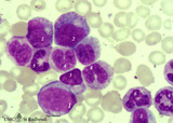 |
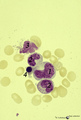
Neutrophilic myelopoiesis in bone marrow smear (human) | Stain: May-Grnwald-Giemsa (MGG). A group of neutrophilic myeloid cells are clustered together. (1) late myeloblast with nucleoli and hardly any azurophilic granules. (2) promyelocytes as the largest cells of the series, with ample cytoplasm filled with many azurophilic granules. (3) myelocytes with ... | Poja Histology Collection - Blood & Bone Marrow Subset | |
| 1796 |
 |
Basophilic erythroblast and polychromatic erythroblasts in bone marrow smear (human) | Stain: May-Grnwald-Giemsa (MGG). The basophilic erythroblast (1) shows slightly condensed chromatin and a strong basophilic (blue) cytoplasm. In the two polychromatic erythroblasts the chromatin condensation has progressed considerably, and the cytoplasm is much less basophilic. The cells are also s... | Poja Histology Collection - Blood & Bone Marrow Subset | |
| 1797 |
 |
Myeloblast | Scheme electron microscopy. A myeloblast is a large cell (10-20 μm) with a large nucleus (fine disperse chromatin) and nucleolus. In the cytoplasm the Golgi area is well developed, few large mitochondria and rough endoplasmic reticulum profiles with numerous free ribosomes and polysomes are shown a... | Poja Histology Collection - Blood & Bone Marrow Subset | |
| 1798 |
 |
Neutrophilic myelocytes with strong toxic granulation in peripheral blood smear (human) | Stain: May-Grnwald-Giemsa (MGG). Neutrophilic myelocytes at various differentiation stages, but all cells show strong toxic granulation. | Poja Histology Collection - Blood & Bone Marrow Subset | |
| 1799 |
 |
Platelets (peripheral blood, human) | Electron microscopy. These disc-shaped cells (2-4 m) without nucleus are derived from cytoplasmic fragments of the megakaryocyte. Free floating in the peripheral blood they develop thin extensions. They contain among others few mitochondria, smooth tubular systems, marginal localized microtubules, g... | Poja Histology Collection - Blood & Bone Marrow Subset | |
| 1800 |
 |
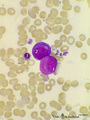
Mast cells in peripheral blood smear (human) | Stain: May-Grnwald-Giemsa (MGG). Mast cells (1) from two different smears. (2) Monocyte. Mast cells or tissue basophils are normally present in small numbers in bone marrow smears. They must not be confused with developing basophils. Mast cells are large cells (20-30 m in diameter) with an irregular... | Poja Histology Collection - Blood & Bone Marrow Subset |