The Health Education Assets Library (HEAL) is a collection of over 22,000 freely available digital materials for health sciences education. The collection is now housed at the University of Utah J. Willard Marriott Digital Library.
TO
Filters: Collection: "ehsl_heal"
| Title | Description | Subject | Collection | ||
|---|---|---|---|---|---|
| 901 |
 |
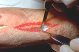
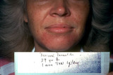
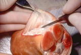
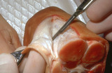
Necrosis of the skin | The necrotic skin must be removed. Normally the dermis is red and bleeds easily, and the underlying fat is yellow. If the tissues are a different color than that, then they need to be surgically removed. | Knowledge Weavers Dermatology | |
| 902 |
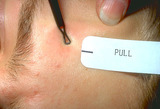 |
Opening comedos | The third step in opening comedos with a comedo extractor is to pull to the side while pushing and this generally forces out the comedo. | Comedo | Knowledge Weavers Dermatology |
| 903 |
 |
Lamellar ichthyosis | Lamellar ichthyosis. The abnormal stratum corneum has produced what appears as very thick scale on the skin, and with an abnormal barrier layer these patients commonly get secondary staphylococcal and yeast infections. Note that there are other associated ectodermal problems such as the teeth, hair,... | Knowledge Weavers Dermatology | |
| 904 |
 |
Excising with scalpel | When excising the scalpel should be held as if holding a pencil. A puncture should be made at one end of the ellipse. | Surgical Methods | Knowledge Weavers Dermatology |
| 905 |
 |
Necrotic tissue | If a transparent semipermeable dressing is to be used, the skin around the ulcer has to be cleansed with alcohol. | Knowledge Weavers Dermatology | |
| 906 |
 |
Nasal mask placement | This demonstrates placement of the nasal mask. | Knowledge Weavers Dermatology | |
| 907 |
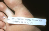 |
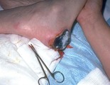
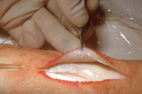
Puncture wounds of the foot | Puncture wounds of the foot need to be explored because they often contain fragments of shoe, stocking, or other foreign material. It is best to expore the wound by extending the opening sufficiently to be able to clearly see to the base of the wound to insure complete debridement/cleansing. The w... | Knowledge Weavers Dermatology | |
| 908 |
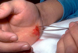 |
Puncture wounds | Visualization of the base of the wound and debridement is crucial. | Knowledge Weavers Dermatology | |
| 909 |
 |
Excision procedure | The wound that has been created in cross-section; note the beveling of the wound edges. | Knowledge Weavers Dermatology | |
| 910 |
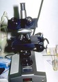 |
Scabies mite magnification | The scraping is viewed under the microscope at low magnification. The scabies mite measures about .1 to .3 mm in diameter, and is not visible with the naked eye, but very slight magnification will make it visible. | Knowledge Weavers Dermatology | |
| 911 |
 |
Milium extraction | Milium can be extracted by incision with a large needle and gently flipping out the contents. | Knowledge Weavers Dermatology | |
| 912 |
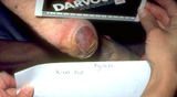 |
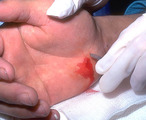
Necrosis of the skin | Pressure applied for too long to an area of skin compromises blood flow, and the skin necrosis. The purple/brown area on this heel is an area of necrosis of the skin. | Knowledge Weavers Dermatology | |
| 913 |
 |
Head lice | On close observation, you can often see the adult lice as, but it is much easier to see the pinhead size nits on the scalp hair. | Knowledge Weavers Dermatology | |
| 914 |
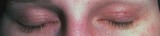 |
Atopic dermatitis | In adults who suffer atopic dermatitis, often the eyelids are red and scaly. | Knowledge Weavers Dermatology | |
| 915 |
 |
Nitric oxide pressure gauges | This demonstrates the pressure gauges attached to the tanks. | Knowledge Weavers Dermatology | |
| 916 |
 |
Nitric oxide | Copper tubing is used to allow for interrupted transfer of energy from the expanding gases. | Knowledge Weavers Dermatology | |
| 917 |
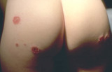 |
Nummular dermatitis | Lesions of acute (red and oozing) nummular dermatitis on the buttocks. | Knowledge Weavers Dermatology | |
| 918 |
 |
Nummular dermatitis | Lesions of nummular dermatitis. | Knowledge Weavers Dermatology | |
| 919 |
 |
Mild acne vulgaris | Mild acne vulgaris. | Knowledge Weavers Dermatology | |
| 920 |
 |
Recessive epidermolysis bullosa | An adult with recessive epidermolysis bullosa showing ulcers in the mouth and multiple dental caries secondary to inability to maintain proper dental hygiene. | Knowledge Weavers Dermatology | |
| 921 |
 |
Exaggerated insect bite reaction, possibly secondary to chigger bites | Exaggerated insect bite reaction. Other lesions were red plaques that measured 1.5 to 2.0 cm, and were very pruritic. | Knowledge Weavers Dermatology | |
| 922 |
 |
Chigger's mite | Chigger's mite. It is generally visible only with magnification. Its bite forms a red papule/wheal. | Chiggers | Knowledge Weavers Dermatology |
| 923 |
 |
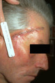
Basal cell carcinoma | This is a basal cell carcinoma on the edge of the nose. | Knowledge Weavers Dermatology | |
| 924 |
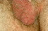 |
Halogenated steroids | Halogenated steroids, particularly fluorinated steroids, will often induce inflammation of the skin of the scrotum, vulvae, as well as the face. Halogenated steroids should never be used, with rare exception, on these areas, and then for the briefest period of time possible. | Knowledge Weavers Dermatology | |
| 925 |
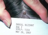 |
Ticks | The maximum incubation period for the tick-borne diseases seen in Utah would be seven days. If you were asked by someone who has had a tick how long they have to wait before they are out of the woods (so to speak), the answer is one week. | Knowledge Weavers Dermatology | |
| 926 |
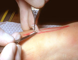 |
Scissors | The specimen can also be removed using scissors as one cuts beneath the dermis. | Knowledge Weavers Dermatology | |
| 927 |
 |
Suturing | This demonstrates the same maneuver using suture. | Knowledge Weavers Dermatology | |
| 928 |
 |
Basal cell carcinoma: excision removal | This demonstrates the wound one week after the surgery. The eversion will disappear within several weeks because of the downward and lateral pulling forces on the wound. | Surgical Methods | Knowledge Weavers Dermatology |
| 929 |
 |
Treating mild acne | This demonstrates two approaches to treating mild acne (comedos and less than about 10 red papules and pustules). The first treatment is shown on the left,and she applies Cleocin-T lotion to her face each morning, and Cleocin-T lotion and Retin-A 0.025% cream to her face each evening. A second alter... | Drug Effects | Knowledge Weavers Dermatology |
| 930 |
 |
Treatment of Scabies | Treatment of choice for scabies is to apply permethrin (Elimite) overnight from the neck down, and to ensure that everyone in the patient's family be treated at the same time. The bedding and clothing from the past week should be washed or dry cleaned, or it can be placed in a plastic bag and set as... | Knowledge Weavers Dermatology | |
| 931 |
 |
Stratum corneum | The thick stratum corneum of the palms and soles prevents chemicals from readily entering those areas. This patient worked around a chemical to which he became allergic, and you can see the line of demarcation along the sides of his hands indicating the thinner stratum corneum on the dorsum of the h... | Knowledge Weavers Dermatology | |
| 932 |
 |
Accutane | At the end of five months, virtually everyone clears, and only occasionally are six or seven months required. | Accutane | Knowledge Weavers Dermatology |
| 933 |
 |
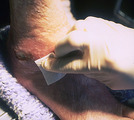
Administration of epinephrine into the subcutaneous fat | This demonstrates administration of epinephrine into the subcutaneous fat. | Dosage | Knowledge Weavers Dermatology |
| 934 |
 |
Accutane | This is the patient shown at the end of Accutane therapy (usually five months), and most patients who have completed five months of Accutane, 1 mg/kg/day obtain long-term remissions of their acne. | Accutane | Knowledge Weavers Dermatology |
| 935 |
 |
Atopic dermatitis involving the axilla | Atopic dermatitis involving the axilla. Note that the axillary fold is spared, and there is a ring of redness and scaling around the axillary fold. | Knowledge Weavers Dermatology | |
| 936 |
 |
Angiosarcoma | This patient had angiosarcoma. | Knowledge Weavers Dermatology | |
| 937 |
 |
Excision: suturing | The suture is tightened by pulling the suture with the hands ending in natural position. The knot tying procedures previously shown are done twice more until the four throws have been placed. Suture is then cut just above the knot. Deep dermals are placed roughly every cm along the under-surface (ba... | Knowledge Weavers Dermatology | |
| 938 |
 |
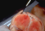
Undermining | The edges of the wound should pull together easily; if not, undermining needs to be done. Undermining means cutting the fibrous septae that connect the skin to the underlying fascia, and generally this is accomplished by using the scalpel to cut the septi just beneath the dermis as shown here. Under... | Knowledge Weavers Dermatology | |
| 939 |
 |
Scalpel | A puncture should be made at both ends of the wound to ensure that the points of the ellipse are very sharp and pointed. The specimen can then be removed by cutting beneath the dermis with a scalpel as shown here. | Knowledge Weavers Dermatology | |
| 940 |
 |
Seborrheic keratoses | The appearance of seborrheic keratoses immediately following freezing. The lesions should be frozen (kept white, or have a thaw time of) 30 seconds. | Knowledge Weavers Dermatology | |
| 941 |
 |
Suturing | This demonstrates the same maneuver using suture as the second throw is tightened with the left hand crossing over the right hand. This must be done to ensure that a square knot is formed. | Knowledge Weavers Dermatology | |
| 942 |
 |
Suturing | This third throw is initiated by placing the needle holder on the inside of the long arm (needle bearing end) of the V that is created. | Knowledge Weavers Dermatology | |
| 943 |
 |
Dermatitis biopsy | This is a biopsy of dermatitis. The epidermis (upper purple strip) is being infiltrated by lymphocytes (small purple dots), and they are also infiltrating into the dermis (lower pink area). The consequence of this lymphocytic attack is disruption of the maturation of the skin (scaling), vasodilation... | Knowledge Weavers Dermatology | |
| 944 |
 |
Suturing | The suture must be rotated in a right-hand direction to ensure that it is tightened along the long axis of the wound until the edges of the wound are pulled together and evert. | Knowledge Weavers Dermatology | |
| 945 |
 |
Nitric oxide | I generally use 50% nitric-oxide and 50% oxygen when I administer it to a patient through a nasal mask. For most adults and children I use a total flow of about 4 to 6 liters per minute. A general rule of thumb is that a person needs 100 cc's of gas per kg per minute. | Knowledge Weavers Dermatology | |
| 946 |
 |
Papular acne | When the closed comedos rupture, they can induce inflammation seen as red papules and pustules. When these inflammatory lesions form, an antibacterial needs to be used. | Anti-bacterial Agents | Knowledge Weavers Dermatology |
| 947 |
 |
Suturing | The needle holder is placed in the center of this V, and one can then begin to tie the suture. | Knowledge Weavers Dermatology | |
| 948 |
 |
Purpura | Purpura. This patient had vomited very vigorously, and the increased pressure within the capillaries caused some of them to rupture and red blood cells extravasated into the surrounding skin. Because there are red blood cells outside the vessels, compression does not cause this to blanch. | Knowledge Weavers Dermatology | |
| 949 |
 |
Androgenetic alopecia | Androgenetic alopecia often manifests as thinning of scalp hair on the vertex. | Knowledge Weavers Dermatology | |
| 950 |
 |
Wound dressings | This demonstrates the bulk dressing on the wound. The dressing should be changed twice daily, and the wound irrigated with sterile saline in an attempt to further remove any debris, including necrotic tissue. | Knowledge Weavers Dermatology | |
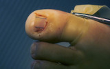
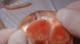
| 951 |
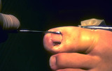 |
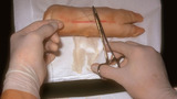
Ingrown nail | I use a curette to scrape back in the cul-de-sac beneath the proximal nail fold to physically remove all of the nail matrix I can. | Surgical Methods | Knowledge Weavers Dermatology |
| 952 |
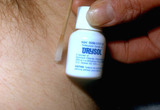 |
Skin tags | This shows Drysol being applied to the site of the skin tag removal to staunch bleeding. The Drysol hurts more than the snipping in my estimation. | Skin Tags | Knowledge Weavers Dermatology |
| 953 |
 |
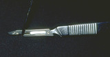
Scalpel | The #15 blade and other small blades should be used by holding the scalpel handle as one holds a pencil. | Knowledge Weavers Dermatology | |
| 954 |
 |
Impetigo | Infection just beneath the stratum corneum of streptococcus or staphylococcus is called impetigo. The stratum corneum is readily torn off, and generally the serous ooze from the dermal blood vessels dries on the skin and produces a honey-colored crust. | Knowledge Weavers Dermatology | |
| 955 |
 |
Ingrown nail | The portion of the nail plate that is to be removed is marked. | Knowledge Weavers Dermatology | |
| 956 |
 |
Excision procedure | This demonstrates the deep dermal suture ends that are exiting from the wound, and the order is crucial: loop-short-long. The long end is the needle bearing end. | Knowledge Weavers Dermatology | |
| 957 |
 |
Dermatofibroma | A benign growth called dermatofibroma. It can look like a nevus, but it feels rock hard to the touch. | Knowledge Weavers Dermatology | |
| 958 |
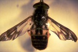 |
Bees | The sting of bees or wasps is a cause of anaphylaxis. | Knowledge Weavers Dermatology | |
| 959 |
 |
Scalpel | This cross-section shows the angles that are left on the edges that will allow for excellent eversion or puckering of the wound when the wound is closed. | Knowledge Weavers Dermatology | |
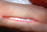
| 960 |
 |
Suturing | This demonstrates the vertical mattress suture as it first entered the skin. I enter and exit the skin approximately 2 mm away from the wound edge, and I penetrate about halfway down into the underlying dermis. The important point is that the needle should be the same distance from the wound edge on... | Knowledge Weavers Dermatology | |
| 961 |
 |
Scarring | Scarring occurs at sites where the epidermis tears off, and the digits normally scar together. | Knowledge Weavers Dermatology | |
| 962 |
 |
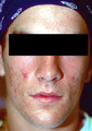
Rosacea | Patient with moderate Stage 2 rosacea. | Knowledge Weavers Dermatology | |
| 963 |
 |
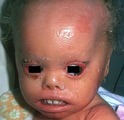
Ectodermal dysplasia | This patient has ectodermal dysplasia. The patient lacks normal body hair on the scalp. | Knowledge Weavers Dermatology | |
| 964 |
 |
Suturing | A double loop is placed around the needle holder. | Knowledge Weavers Dermatology | |
| 965 |
 |
Excision procedure | The suture is wrapped twice around the needle holder and the short arm of the suture is then grasped, and | Knowledge Weavers Dermatology | |
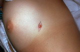
| 966 |
 |
Keloid | Once the keloid is anesthetized, the corticosteroid should be injected directly into the middle of the keloid until the keloid blanches. I find it easiest to advancethe needle to the tip of the keloid, and then as I withdraw the needle I inject into the canal that I have created with the needle. I d... | Knowledge Weavers Dermatology | |
| 967 |
 |
Mosquito bites | Mosquito bites. These are the legs of my sister and her daughter, and both develop an exaggerated insect bite reaction, which consist of striking swelling and itching, and it is much more pronounced than the average mosquito bite. | Knowledge Weavers Dermatology | |
| 968 |
 |
Suturing | A single loop is placed around the needle holder as shown. | Knowledge Weavers Dermatology | |
| 969 |
 |
Vasculitis | More severe damage to capillary sized vessels allows a significant amount of red blood cells and fluid to leak into the surrounding dermis, and this condition is called vasculitis. This is small vessel (capillary) vasculitis. | Knowledge Weavers Dermatology | |
| 970 |
 |
Androgenetic alopecia | Androgenetic alopecia often induces fine curly hair along the lateral margins of the scalp. | Knowledge Weavers Dermatology | |
| 971 |
 |
Bedbug | These are about 5 mm or more in length, are easily visible, but can be hard to flush out. | Knowledge Weavers Dermatology | |
| 972 |
 |
Ticks | The most common tick-borne disease in this area is Colorado Tick Fever, and second would be Rocky Mountain Spotted Fever, and rarely Tick Paralysis. The organism that causes Lyme disease (Borrelia burgdorferi) has not been shown to be carried by ticks in Utah. | Knowledge Weavers Dermatology | |
| 973 |
 |
Excision procedure | This demonstrates the 45 o angle in cross-section as one cuts through the dermis. | Surgical Methods | Knowledge Weavers Dermatology |
| 974 |
 |
Paraorificial dermatitis | periorificial dermatitis induced by the application of Vaseline to facial skin; it has been treated for one month with 1 gram of tetracycline per day. | Knowledge Weavers Dermatology | |
| 975 |
 |
Scalpel | The blade should be slid on the handle until it locks into place. | Knowledge Weavers Dermatology | |
| 976 |
 |
Rosacea | Patients with rosacea often have eye involvement, which can consist of styes as shown here. Dr. Randy Olson at the University of Utah recommends treating these with intralesional injection of Kenalog 40 mg/cc. He points out it requires only 1 to 2 drops of this intralesionally, and the inflammatory ... | Knowledge Weavers Dermatology | |
| 977 |
 |
Keloid | Keloids can be injected with a Depo form of injectable steroid and/or silicone gel sheet. The corticosteroid reduces the production of all components of the keloid, which include ground substance, collagen, and elastin. The silicone gel is thought to act by increasing the temperature of the skin and... | Knowledge Weavers Dermatology | |
| 978 |
 |
Excision procedure | This demonstrates placement of the deep dermal suture that is secured to the underside of the dermis as far away from the wound edge as possible. | Knowledge Weavers Dermatology | |
| 979 |
 |
Scalpel | This shows the outward angle of the scalpel in a cross-sectional view. | Knowledge Weavers Dermatology | |
| 980 |
 |
Wound dressing | This shows a bulky dressing applied on top of the non-adherent dressing to absorb the bleeding that will occur over the next hour or two after debridement. The wound should be gently cleansed once or twice daily with dilute Hibiclens (one part Hibiclens and three parts water), and then the Silvaden... | Knowledge Weavers Dermatology | |
| 981 |
 |
Suturing | The short arm of the suture is grasped. The throw is then tightened by pulling the hands back into their natural positions. | Knowledge Weavers Dermatology | |
| 982 |
 |
Suturing | This demonstrates the placement of the suture in a vertical mattress prior to tying. | Knowledge Weavers Dermatology | |
| 983 |
 |
Retin-A | During the first several weeks of use of Retin-A, some people experience a diffuse redness and scaling where it is applied, and acne lesions can become inflamed or more inflamed during that period of time. Simply reassure the patient that he/she should continue with the therapy and the inflammation ... | Retin-A; Drug Effects | Knowledge Weavers Dermatology |
| 984 |
 |
Kenalog | The solution should be injected directly into the lesion until there is slight blanching, which indicates saturation with the solution. You must warn the patient beforehand that there is some risk of atrophy of the skin and sometimes the underlying fat, and it can take 6 to 12 months for that to fil... | Knowledge Weavers Dermatology | |
| 985 |
 |
Excision procedure | Undermining on the opposite side of the wound. | Surgical Methods | Knowledge Weavers Dermatology |
| 986 |
 |
Suturing | The wound edges should pull together easily if wound has been properly undermined. The needle should be grasped two-thirds of the way from the point of the needle with the needle holders. This allows one to use a majority of the needle when suturing, but the needle is not grasped so far away from th... | Knowledge Weavers Dermatology | |
| 987 |
 |
Excision: suturing | The loops must be tightened by pulling and tightening along the long axis of the wound. | Knowledge Weavers Dermatology | |
| 988 |
 |
Puncture wounds | Puncture wounds often have to be enlarged in order to be explored. | Knowledge Weavers Dermatology | |
| 989 |
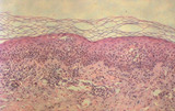 |
Dermatitis biopsy | This biopsy demonstrates basically what is happening within dermatitis. Lymphocytes (small purple dots) are attacking the epidermis (thick upper purple area), as well as the underlying dermis (fainter pink area at the base) and induce abnormal maturation of the epidermis (scaling), vasodilation (red... | Knowledge Weavers Dermatology | |
| 990 |
 |
Stasis dermatitis | Stasis dermatitis (shown here) that is not oozing, is best treated with a support hose. The optimum ankle pressure is around 30 to 40 mm Hg, and generally a hose that comes to just below the knee is adequate. The hose should be worn at all times when the extremity is in the dependent position, such ... | Knowledge Weavers Dermatology | |
| 991 |
 |
Excision procedure | Undermining is achieved by using scalpel or scissors to free the dermis from the fibrous septae that connect it to the underlying fascia. One undermines the wound until the wound edges pull together readily. | Surgical Methods | Knowledge Weavers Dermatology |
| 992 |
 |
Body louse | This is a body louse on the same patient as shown in slide 78. | Knowledge Weavers Dermatology | |
| 993 |
 |
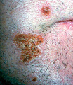
Keloid | Keloid. Keloids tend to form on areas where there is a significant amount of tension on the wound, and also in certain families, and in certain races, particularly those who are more darkly pigmented. A keloid is defined as scar tissue that extends beyond the boundaries of the original injury.(Summa... | Knowledge Weavers Dermatology | |
| 994 |
 |
Keloids | Keloids. These are composed of excessive amounts of collagen, elastin, and ground substance. They occur generally on the upper chest and back, and also over areas of stretch such as the elbows and knees. | Knowledge Weavers Dermatology | |
| 995 |
 |
Excision: suturing | The non-dominant (left) hand crosses over the dominant (right) hand on the second throw. | Knowledge Weavers Dermatology | |
| 996 |
 |
Periorificial dermatitis | Periorificial dermatitis. This eruption consists of erythema or sometimes discreet red papules with or without scale located on the eyelids, in a paranasal and paraoral distribution. In my experience, it most commonly occurs around the nose and mouth. The cause of this remains unknown, though it can... | Knowledge Weavers Dermatology | |
| 997 |
 |
Vitiligo | This patient has vitiligo. The melanocytes within the epidermis are destroyed presumably by a lymphocytic cell-mediated process. | Knowledge Weavers Dermatology | |
| 998 |
 |
Suturing | The second throw is made by placing the needle holder in the inside of the long arm (needle bearing) of the V, and | Knowledge Weavers Dermatology | |
| 999 |
 |
Scalpel | The other side of the ellipse is cut and the blade is angled outward. | Knowledge Weavers Dermatology | |
| 1000 |
 |
Suturing | The needle holder is then placed on the inside of the long arm (needle bearing end) of the V, and a single loop is thrown around the needle holder. The short end of the suture is then grasped, and | Knowledge Weavers Dermatology |