The Health Education Assets Library (HEAL) is a collection of over 22,000 freely available digital materials for health sciences education. The collection is now housed at the University of Utah J. Willard Marriott Digital Library.
TO
Filters: Collection: "ehsl_heal"
| Title | Description | Subject | Collection | ||
|---|---|---|---|---|---|
| 176 |
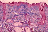 |
Lip (human), skin surface. | Stain: Azan. Keratinized squamous epithelium with hair follicles; submucosa with sebaceous glands, and extensions of skeletal muscle cells (orbicularis oris). | oral cavity | Poja Histology Collection - Oral Cavity Subset |
| 177 |
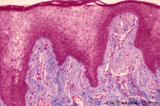 |
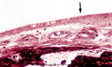
Lip (human), transitional zone (red zone or vermilion border) | Stain: Azan. Slightly cornified epithelium with high irregular dermal papillae and many capillaries. Note the epithelium is thicker, but less cornified than the epidermis. The red color of the lips is due to the rich vascularity of the lamina propria and the lucidity of the epithelium. | oral cavity; lining mucosa; red zone; vermilion border | Poja Histology Collection - Oral Cavity Subset |
| 178 |
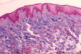 |
Lip (human), transitional zone (red zone or vermilion border) | Stain: Azan. Slightly cornified epithelium with high irregular dermal papillae and many capillaries. Fat cells and skeletal muscle cells are located in the submucosa. | oral cavity; lining mucosa; red zone; vermilion border | Poja Histology Collection - Oral Cavity Subset |
| 179 |
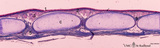 |
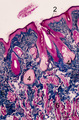
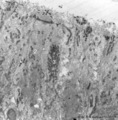
Longitudinal section of trachea (human, adult) | Stain: Azan. On top: the epithelium (1) is pseudostratified with cilia and goblet cells. The dense lamina propria (2, ↓, deep blue) is demarcated from the submucosa with seromucous tracheal glands (3). C-shaped cartilage rings (C, light blue) maintain patency. The cartilage edges are connected by ... | Pseudostratified epithelium ; Seromucous glands; Tracheal glands; Fibroelastic membrane ; Adventitia | Poja Histology Collection - Respiratory System Subset |
| 180 |
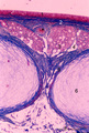 |
Longitudinal section of trachea (human, adult) | Stain: Azan. On top: the epithelium (1) is pseudostratified with cilia and goblet cells on a distinct basement membrane (↓). There is almost no demarcation (at this magnification) between lamina propria and submucosa due to the dense blue (2) staining of elastic fibers reinforced by collagen fiber... | Pseudostratified epithelium ; Tracheal glands; Fibroelastic membrane | Poja Histology Collection - Respiratory System Subset |
| 181 |
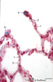 |
Lung alveoli (human) | Stain: Azan. Alveoli (diameter about 200 m) are formed by thin walls of epithelial cells (1) such as type I alveolar cells and (blue stained) fine collagen (3) and elastin intermingled with dilated capillaries (2). | Poja Histology Collection - Respiratory System Subset | |
| 182 |
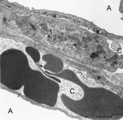 |
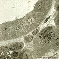
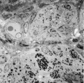
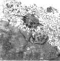
Lung capillaries and air-blood barrier (rat) | Electron microscopy. Two neighboring capillaries (C) and the alveolar spaces (A). The endothelial cell (1) of the upper capillary contains an organelle-rich cytoplasm, a centriole (2), contractile filaments (↓) and electron-dense membrane-bound granules, the so-called Weibel-Palade bodies. These b... | Poja Histology Collection - Respiratory System Subset | |
| 183 |
 |
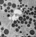
Multilamellar bodies of type II alveolar cell in the lung (mouse) | Electron microscopy. The cytoplasm of the type II alveolar cell (pneumocyte II) contains characteristic electron-dense multilamellar bodies (*) in different maturing stages. The lamellar bodies are responsible for the vacuolated appearance of these cells, and they give rise to surfactant (phospholip... | Pneumocyte II; Multilamellar bodies; Type II alveolar cell | Poja Histology Collection - Respiratory System Subset |
| 184 |
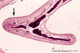 |
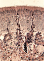
Nasal concha (dog, isolated turbinate bone) | Stain: Hematoxylin and eosin. This concha (turbinate with red-black-stained bone (1), is covered by a thin respiratory mucosa (3) and by a thick olfactory mucosa (2, arrow). Within the respiratory epithelium light stained goblet cells are visible between the low columnar/cuboidal epithelial cells (c... | Conchae nasales; Olfactory epithelium; Respiratory epithelium | Poja Histology Collection - Respiratory System Subset |
| 185 |
 |
Nasal concha with respiratory mucosa (dog, high magnification) | Stain: Hematoxylin and eosin. (↑) Indicates the transition of the olfactory epithelium (1) into the respiratory epithelium (2) with pseudostratified ciliated epithelium and goblet cells. The submucosa is richly vascularized (3). Lamellar bone of the turbinate (4) is stained reddish-black. | Conchae nasales; Olfactory epithelium; Respiratory epithelium | Poja Histology Collection - Respiratory System Subset |
| 186 |
 |
Nasal glands of the nasal vestibulum (rat) | Electron microscopy. At the top part of a striated draining duct with a wide lumen (*); these cuboidal ductal cells (1) contain many basolaterally located mitochondria. The ductal cells are enforced by interstitial fibroblast (2) and a capillary (4). Neighboring serous gland cells (5) contain dark s... | Nasal vestibulum; Seromucous glands; Nasal glands; Serous cells; Secretion granules; Intercalated duct; Striated duct | Poja Histology Collection - Respiratory System Subset |
| 187 |
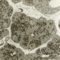 |
Nasal glands of the nasal vestibulum (rat) | Electron microscopy. A low-power magnification reveals serous acini of a seromucous gland; in the center and lower left corner lumina (*) of the acini are present and dense secretion granules of varying sizes represent the active cells. Between the acini the distended interstitium reveals a fibrobla... | Nasal vestibulum; Seromucous glands; Nasal glands; Serous cells; Secretion granules | Poja Histology Collection - Respiratory System Subset |
| 188 |
 |
Nasal glands of the nasal vestibulum (rat, higher magnification) | Electron microscopy. Part of a serous acinus of a seromucous nasal gland demonstrates the central lumen (*) formed by four gland cells. Note junctional complexes (↑) and the close proximity of the dense secretion granules (2) ready to be released into the central lumen. These active cells contain ... | Nasal vestibulum; Seromucous glands; Nasal glands; Serous cells; Secretion granules | Poja Histology Collection - Respiratory System Subset |
| 189 |
 |
Neuroepithelial body in terminal bronchiolus (golden hamster) | Electron microscopy. Three epithelial cells, as part of the N(euro) E(pithelial) B(ody), contribute to a cluster of neuroendocrine cells. They belong to the Amine Precursor Uptake and Decarboxylation cell system so-called APUD cells. The dense-core granules (↓) contain among others dopamine or ser... | Terminal bronchiolus ; Neuro-endocrine cells | Poja Histology Collection - Respiratory System Subset |
| 190 |
 |
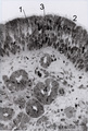
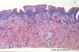
Nostril area of nasal vestibulum - cutaneous region - human | Stain: Azan. Slightly keratinized squamous epithelium (1) with thin red cornified layer; (2) hair follicles; (3) vibrissae and (4) associated sebaceous glands. At the bottom skeletal muscle fibers; (5) transverse part of the musculous nasalis; (6) connective tissue. | Poja Histology Collection - Respiratory System Subset | |
| 191 |
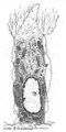 |
Odontoblast in tooth development - mammalian embryo | Scheme electronmicroscopy. A columnar and irregular shaped cell (mesenchymal origin) with a taperwise odontoblastic process within the predentin. The bottom side is adjacent to the future pulp cells. The cell body contains many organelles and especially a well-developed Golgi area with prosecretoy g... | oral cavity | Poja Histology Collection - Oral Cavity Subset |
| 192 |
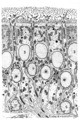 |
Olfactory epithelium in the nasal cavity (mammals) | Scheme electron microscopy. Four olfactory bulbs (1, vesicles) with radially sprouting cilia are present at the surface of the epithelium. Their slender cell bodies (2, bipolar neurons) are flanked by sustentacular (supporting) broad cells (3) whose apices are filled with well developed organelles, ... | Pseudostratified epithelium; Olfactory epithelium; Basal cells; Olfactory vesicle; Bipolar neuron | Poja Histology Collection - Respiratory System Subset |
| 193 |
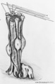 |
Olfactory epithelium in the nasal cavity (mammals) | Three-dimensional scheme electron microscopy. Six olfactory cells (1) surround one massive central supporting cell (2). A third, basal cell (3) is located at the basal plate aposing to the supporting cell. These olfactory cells are tall and slender bipolar nerve cells with chemoreceptive functions. ... | Pseudostratified epithelium; Olfactory epithelium; Basal cells; Olfactory vesicle; Bipolar neuron | Poja Histology Collection - Respiratory System Subset |
| 194 |
 |
Olfactory epithelium in the nose (human) | Electron microscopy. At the top (lumen) cross-sections of olfactory bulbs (1) (vesicles) with cilia/basal bodies. Sustentacular (supporting) cells (2) exhibit numerous microvilli in the lumen, their cytoplasms contain many dispersed mitochondria and in the lower half aggregations of endoplasmic reti... | Olfactory epithelium; Olfactory vesicle | Poja Histology Collection - Respiratory System Subset |
| 195 |
 |
Olfactory gland (Bowman) in olfactory region of concha (human) | Electron microscopy. Beneath the olfactory epithelium tubuloalveolar glands of Bowman (1) are found containing cells that produce dense secretion granules arranged around a large lumen (*) with greyish secretion product. These cells produce a serous fluid in which odoriferous substances are dissolve... | Olfactory epithelium; Bowman glands; Olfactory glands; Secretion granules | Poja Histology Collection - Respiratory System Subset |
| 196 |
 |
Olfactory mucosa in the nose (dog) | Stain: Iron Hematoxylin and eosin (Heidenhain). The pseudostratified epithelium of the olfactory mucosa contains sensory cells, support cells and basal cells. The upper darker stained nuclei (1) belong to the supporting (sustentacular) cells showing a dark apical line (↑) at the top. Below lighte... | Bowman glands; Olfactory glands; Olfactory mucosa; Pseudostratified epithelium | Poja Histology Collection - Respiratory System Subset |
| 197 |
 |
Olfactory mucosa in the nose (human) | Stain: Toluidine blue, one-micron Epon plastic section. Pseudostratified epithelium with light stained nuclei (1) (note distinct nucleoli) of olfactory cells. At the surface faintly stained cilia/microvilli (2). Nuclei of sustentacular (supporting) cells (3) are stained darker and predominantly in t... | Bowman glands; Olfactory glands; Olfactory mucosa; Pseudostratified epithelium | Poja Histology Collection - Respiratory System Subset |
| 198 |
 |
Olfactory vesicle (bulb) in the nose (gerbil) | DUPLICATE RECORD - FOR DELETION Electron microscopy. A distinct olfactory bulb (1, vesicle) with horizontally extended cross-sectioned cilia (*) and basal bodies (↑) is surrounded by numerous microvilli and free olfactory cilia. Close to the first bulb a second one is about to protrude above th... | Olfactory epithelium; Olfactory vesicle | Poja Histology Collection - Respiratory System Subset |
| 199 |
|
Olfactory vesicle (bulb) in the nose (gerbil) | Electron microscopy. A distinct olfactory bulb (1, vesicle) with horizontally extended cross-sectioned cilia (*) and basal bodies (↑) is surrounded by numerous microvilli and free olfactory cilia. Close to the first bulb a second one is about to protrude above the surface of the supporting cells b... | Olfactory epithelium; Olfactory vesicle | Poja Histology Collection - Respiratory System Subset |
| 200 |
 |
Papillae circumvallatae of the tongue (dorsal side, human) | Stain: Azan. Three broad papillae with taste buds facing the grooves in which the serous von Ebner glands drain. Striated skeletal muscles (musculus verticalis linguae and musculus longitudinalis superior) and lightly stained mucous glands (posterior lingual glands). | oral cavity; von Ebner; lingual muscles; lingual glands | Poja Histology Collection - Oral Cavity Subset |