The Health Education Assets Library (HEAL) is a collection of over 22,000 freely available digital materials for health sciences education. The collection is now housed at the University of Utah J. Willard Marriott Digital Library.
TO
| Title | Description | Subject | Collection | ||
|---|---|---|---|---|---|
| 1 |
 |
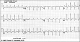
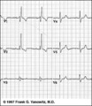
Nonconducted and aberrantly conducted PAC's | In A the slow sinus rhythm is actually caused by nonconducted PACs hidden in the ST segment. This is confirmed in B where some of the PACs are aberrantly conducted with LBBB, and some PACs are nonconducted. | Knowledge Weavers ECG | |
| 2 |
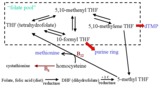 |
Folate Pool - The Role of B Vitamins in One-Carbon Metabolism | This figure depicts the pathway for folate utilization and the role of vitamins B6 and B12 in the metabolism of methyl-tetrahydrofolate and homocysteine. | Folate; Folicin | HEAL Open Review Collection |
| 3 |
 |
ECG components diagram - marquette | ECG components diagram - marquette | Knowledge Weavers ECG | |
| 4 |
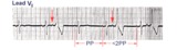 |
PAC's with RBBB aberrant conduction | PAC's are identified by the arrows. Note that the PP interval surrounding the PAC is less than 2x the basic sinus cycle indicating that the sinus node has been reset by the ectopic P wave. The pause after the PAC, therefore, is incomplete. | Knowledge Weavers ECG | |
| 5 |
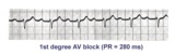 |
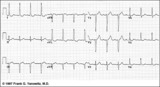
1st degree AV block | The normal PR interval is 0.12 - 0.20 sec, or 120 -to- 200 ms. 1st degree AV block is defined by PR intervals greater than 200 ms. This may be caused by drugs, such as digoxin; excessive vagal tone; ischemia; or intrinsic disease in the AV junction or bundle branch system. | Knowledge Weavers ECG | |
| 6 |
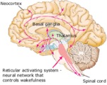 |
Neural Network Controlling Wakefulness (Labeled) | Brainstem reticular activating system. | Royal College of Surgeons in Ireland Illustrations | |
| 7 |
 |
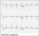
LVH - best seen in the frontal plane leads! | LVH - best seen in the frontal plane leads! | Knowledge Weavers ECG | |
| 8 |
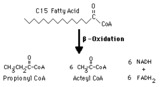 |
Propionyl CoA production from odd-chain fatty acids | Beta-oxidation of fatty acids with an odd number of carbons inthe chain yields the three-carbon propionyl CoA as the final fragment. | Biosynthesis | Knowledge Weavers Fatty Acids |
| 9 |
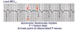 |
Isochronic ventricular rhythm | An isochronic ventricular rhythm is also called an accelerated ventricular rhythm because it represents an active ventricular focus. This arrhythmia is a common reperfusion arrhythmia in acute MI patients. It often begins and ends with fusion beats and there is AV dissociation. Treatment is usuall... | Knowledge Weavers ECG | |
| 10 |
 |
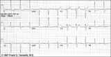
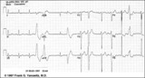
LVH: limb lead criteria | In this example of LVH, the precordial leads don't meet the usual voltage criteria or exhibit significant ST segment abnormalities. The frontal plane leads, however, show voltage criteria for LVH and significant ST segment depression in leads with tall R waves. The voltage criteria include 1) R in... | Knowledge Weavers ECG | |
| 11 |
 |
ST segment depression | ST segment depression is a nonspecific abnormality that must be evaluated in the clinical context in which it occurs. In a patient with angina pectoris ST depression usually means subendocardial ischemia and, unlike ST elevation, is not localizing to a particular coronary artery lesion. | Knowledge Weavers ECG | |
| 12 |
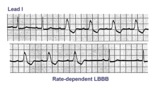 |
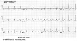
Rate-dependent LBBB | In this rhythm strip of sinus arrhythmia, the faster rates have a LBBB morphology. In some patients with a diseased left bundle branch, the onset of LBBB usually occurs initially as a rate-dependent block; i.e., the left bundle fails to conduct at the faster rate because of prolonged refractoriness... | Knowledge Weavers ECG | |
| 13 |
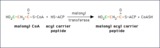 |
Transfer of a malonyl group to the acyl carrier peptide | During fatty acid synthesis the incoming two carbon fragment is introduced as the three-carbon malonyl group. It is added to the -SH group of the acyl carier peptide domain of fatty acid synthase. In a subsequent reaction the carbon shown in green will be lost as bicarbonate ion. | Knowledge Weavers Fatty Acids | |
| 14 |
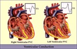 |
RV vs LV PVC's - marquette | RV vs LV PVC's - marquette | Knowledge Weavers ECG | |
| 15 |
 |
RBBB: Precordial leads | RBBB: Precordial leads | Knowledge Weavers ECG | |
| 16 |
 |
Inferior MI and RBBB | Inferior MI and RBBB | Knowledge Weavers ECG | |
| 17 |
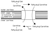 |
Transport of fatty acyl CoA into mitochondria by the carnitine shuttle | Fatty acyl CoA cannot cross the inner mitochondrial membrane, so it is carried in the form of fatty acyl carnitine. | Knowledge Weavers Fatty Acids | |
| 18 |
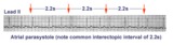 |
Atrial parasystole | Parasystolic rhythms involve an independent ectopic pacemaker resulting in nonfixed coupled premature beats. Parasystole may occur in the atria, as seen in this example, in the AV junction, and in the ventricles. Note the common inter-ectopic interval separating the parasystolic PAC's. | Knowledge Weavers ECG | |
| 19 |
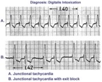 |
Digitalis intoxication: junctional tachycardia with and without exit block | In A the rhythm is junctional tachycardia with RBBB. In B there is 2nd degree exit block with a 3:2 conduction ratio; i.e., every 3rd junctional impulse fails to reach the ventricles... at least for the first two groupings on 1.4sec. | Knowledge Weavers ECG | |
| 20 |
 |
LVH and many PVCs | The combination of voltage criteria (SV2 + RV6>35mm) and ST-T abnormalities in V5-6 are definitive for LVH. There may also be LAE as evidenced by the prominent negative P terminal force in lead V1. Isolated PVCs and a PVC couplet are also present. | Knowledge Weavers ECG | |
| 21 |
 |
Frontal plane QRS axis = +150 degrees (RAD) | This is an unusual right axis deviation (RAD). Lead I is negative, which usually means RAD. Lead II is the isoelectric lead, which almost always means -30 degrees; but in this example the axis is 180 degrees away from -30, or +150 degrees. | Knowledge Weavers ECG | |
| 22 |
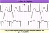 |
Pacemaker failure to capture - marquette | Pacemaker failure to capture - marquette | Knowledge Weavers ECG | |
| 23 |
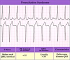 |
WPW type preexcitation - marquette | WPW type preexcitation - marquette | Knowledge Weavers ECG | |
| 24 |
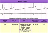 |
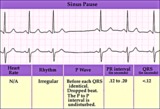
Sinus pause or arrest - marquette | Sinus pause or arrest - marquette | Knowledge Weavers ECG | |
| 25 |
 |
SA exit block - marquette | SA exit block - marquette | Knowledge Weavers ECG |