The Health Education Assets Library (HEAL) is a collection of over 22,000 freely available digital materials for health sciences education. The collection is now housed at the University of Utah J. Willard Marriott Digital Library.
TO
| Title | Description | Subject | Collection | ||
|---|---|---|---|---|---|
| 1 |
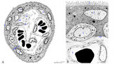 |
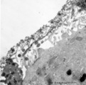
Scheme of electron microscopy of tertiary villus (human placenta, midpregnancy) | (See also POJA-L1227). (A) Survey and (B) detail of a tertiary placental villus lined by the multinucleated syncytiotrophoblast cell (1) (STC). The apex shows protrusions and extensive microvilli of varying sizes (brushborder, 2). Nuclei are sectioned at different levels and the cytoplasm contains a... | placenta; tertiary villi; placental barrier; electron microscopy | Poja Histology Collection - Placenta |
| 2 |
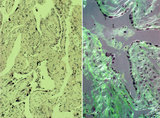 |
Spiral artery in endometrium (human, midpregnancy) | Stain: Hematoxylin-eosin. (A) cross-sections of spiral arteries in endometrium. (B) semi-polarized section of part of a spiral artery. Dark-stained large trophoblast cells (3), partly replacing the cuboidal hypertrophic (B2) endothelial cells. (4) smooth muscle cells. (L) lumen of vessel with cell... | placenta; spiral artery; endometrium; trophoblast | Poja Histology Collection - Placenta |
| 3 |
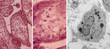 |
Tertiary villi (human placenta, midpregnancy) | (A) Lower and (B) higher magnification. Stain: Hematoxylin - azophloxine. (C) electron microscopy of Hofbauer cell. (A, B) show tertiary villi and intervillous spaces. Squeezed between villi fibrinoid clots (1). The large arrow (2) points to either a detached, free circulating large STC (syncyt... | placenta; chorionic villi; Hofbauer cell; fibrinoid; syncytiotrophoblast | Poja Histology Collection - Placenta |
| 4 |
 |
Tertiary villi (human placenta, full-term, cross-section) | Stain: Hematoxylin ? azophloxine. A few tertiary villi (1) within the intervillous space (2) contains capillary (3) and embryonic connective tissue (*). It remains covered by the postmitotic multinucleated syncytiotrophoblast cell (STC, 4). Distinct nuclear accumulation in the STC indicates the so-c... | placenta; tertiary villi; syncytiotrophoblast | Poja Histology Collection - Placenta |
| 5 |
 |
Villi of complete hydatidiform mole (human) | (A) Macroscopy aggregation (1) of abnormal villi of hydatidiform mole intermingled with blood clots (2). (B) After dissection of a complete mole grape-like aggregations of dilated chorionic villi are presented as numerous liquid-filled vesicles (1) varying in diameters (from few mm up to 1 cm) surr... | placenta; trophoblast; hydatiform mole | Poja Histology Collection - Placenta |
| 6 |
 |
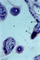
Trophoblast cell in lung alveolar interstitium (human, early midpregnancy) | Stain: Hematoxylin-eosin. It is well known that free circulating trophoblast cells can be found migrated into lung parenchyma during normal pregnancy. In this sample a large trophoblast cell (→) is localised within the normal alveolar interstitium. Alveolar phagocytes (*) are present in the alve... | placenta; trophoblast; lung | Poja Histology Collection - Placenta |
| 7 |
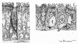 |
Epithelial lining of trachea and bronchiolus (mammals) | Left side trachea: Columnar ciliated cells (1) with up to 200 motile cilia with an organelle-rich apex and in the middle a goblet cell (2) with accumulation of mucus secretion granules in the apex. Note that all epithelial cells (i.e., junctions) contact the basal lamina. Four basal cells (3) do not... | Bronchiolus; Pseudostratified epithelium; Ciliated epithelium; Clara cells; Basal cells; Neuro-endocrine cells | Poja Histology Collection - Respiratory System Subset |
| 8 |
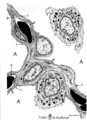 |
Air-blood barrier in the lung (mammals) | Scheme electron microscopy. (1, ↓) Represents type I pneumocytes lining alveolar spaces (A). Cell (2) represents a free alveolar macrophage. The type II pneumocyte (3) is adherent to type I pneumocyte extensions (note junctional connection), and contains multilamellar bodies (surfactant). A myofib... | Pneumocyte type I ; Pneumocyte type II | Poja Histology Collection - Respiratory System Subset |
| 9 |
 |
Neuroepithelial body in terminal bronchiolus (golden hamster) | Electron microscopy. Three epithelial cells, as part of the N(euro) E(pithelial) B(ody), contribute to a cluster of neuroendocrine cells. They belong to the Amine Precursor Uptake and Decarboxylation cell system so-called APUD cells. The dense-core granules (↓) contain among others dopamine or ser... | Terminal bronchiolus ; Neuro-endocrine cells | Poja Histology Collection - Respiratory System Subset |
| 10 |
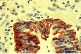 |
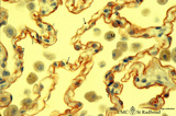
Keratin 7 in the bronchial epithelium of the lung (human, adult) | Stain: anti-keratin 7 antibody (Pan-Ck 7) immunoperoxidase staining (with aminoethylcarbazole (AEC) substrate). The epithelium of the bronchus (1) stains dark brown-red after the reaction with AEC indicating a positive reaction for cytokeratin 7. This antibody can be used in the study of development... | Bronchiolus; Immunoperoxidase; Immuno-reaction | Poja Histology Collection - Respiratory System Subset |
| 11 |
 |
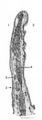
Surface of olfactory epithelium (rat) | Electron microscopy. Bottom left shows an olfactory bulb (1, vesicle) with cross-sectioned basal bodies. Right side part of the apex of a supporting cell (2) with microvilli (3). Parallel to the surface of the epithelium one long olfactory cilium (4) and several other cross-sectioned ones are detect... | Olfactory epithelium; Olfactory vesicle | Poja Histology Collection - Respiratory System Subset |
| 12 |
 |
Alveolar cells in the lung (mammals) | Scheme electron microscopy. (5) alveolar space; (6) type I Pneumocyte; (7) basal lamina; (8) myofibroblast; (9) collagen and elastin fibers; (10) mesothelial cell of the visceral pleura; (11) capillary with erythrocyte; (12) endothelial cell lining the capillary; (13) type II pneum... | Pneumocyte type I ; Pneumocyte type I I | Poja Histology Collection - Respiratory System Subset |
| 13 |
 |
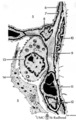
Scheme of the epiglottis (human, adult) | The laryngeal side of the epiglottis (1) is covered with respiratory epithelium, while the lingual side (2) is similar to the oral cavity epithelium (squamous type). The transitional zone to respiratory epithelium is marked by (3). The scaffold consists of elastic cartilage (4). Seromucous laryngea... | Laryngeal glands; Oral cavity | Poja Histology Collection - Respiratory System Subset |
| 14 |
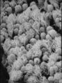 |
Epithelial lining of bronchiolus in the lung (rat) | Scanning electron microscopy. Bushes of cilia indicate the presence of ciliated cells (1). Clustered non-ciliated cells (2, Clara cells) are dome-shaped with stubby microvilli at the surface, they protrude into the lumen to the tips of the cilia. | Bronchiolus ; Ciliated epithelium ; Clara cells | Poja Histology Collection - Respiratory System Subset |
| 15 |
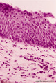 |
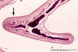
Transitional region of epiglottis (human, high magnification) | Stain: Hematoxylin and eosin. Transition of squamous epithelium into pseudostratified epithelium. Note diapedesis of lymphocytes (darker stained rounded cells) through the epithelial barrier (→). The proper lamina presents some lymphocyte infiltration. | Diapedesis | Poja Histology Collection - Respiratory System Subset |
| 16 |
 |
Keratin 7 in the lining alveolar epithelium of the lung (human, adult) | Stain: anti-keratin 7 antibody (Pan-Ck 7) immunoperoxidase staining (with aminoethylcarbazole (AEC) substrate). Both alveolar epithelial cell types I (1) and II (2) stain dark brown-red after the reaction with AEC indicating a positive reaction for cytokeratin 7. Note the white (non-stained) capilla... | Pneumocyte I; Pneumocyte II; Imunoperoxidase; Immuno-reaction | Poja Histology Collection - Respiratory System Subset |
| 17 |
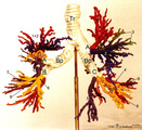 |
Arbor bronchialis, the bronchial tree (human, adult) | Resin corrosion cast of the lower trachea and bronchial tree (posterior aspect). The lobar and segmental bronchi and their main branches are coloured, different colours indicate areas supplied by different segmental bronchi. White-coloured lower trachea (Tr) divides into two principal bronchi (Bp)... | Segmental bronchi ; Macroscopy | Poja Histology Collection - Respiratory System Subset |
| 18 |
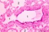 |
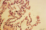
Respiratory bronchiolus (human) | Stain: Hematoxylin and eosin. A longitudinal section shows the lumen of a respiratory bronchiolus (1) with an irregular lining. It is covered by low columnar-cuboidal cells, a few pouches within the same lumen are lined by thin alveolar epithelium as found in the surrounding alveoli (2). Thin arrows... | Respiratory bronchiolus; Columnar epithelium; Cuboidal epithelium; Clara cells; Alveolar epithelium | Poja Histology Collection - Respiratory System Subset |
| 19 |
 |
Nasal concha (dog, isolated turbinate bone) | Stain: Hematoxylin and eosin. This concha (turbinate with red-black-stained bone (1), is covered by a thin respiratory mucosa (3) and by a thick olfactory mucosa (2, arrow). Within the respiratory epithelium light stained goblet cells are visible between the low columnar/cuboidal epithelial cells (c... | Conchae nasales; Olfactory epithelium; Respiratory epithelium | Poja Histology Collection - Respiratory System Subset |
| 20 |
 |
Small bronchus (golden hamster) | Electron microscopy. Two ciliated cells with nuclei (1). Supranuclearly electron-dense lysosomes (2, lipofucsin) are present, the apical parts contain mitochondria (3) and cross-sections of cilia (4) and microvilli (5) are visible in the lumen (*). Two mucoid goblet cells with packed secretion gran... | Ciliated epithelium | Poja Histology Collection - Respiratory System Subset |
| 21 |
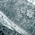 |
Striated duct of nasal gland (rat) | Electron microscopy. At (1) lumen of duct, (2) indicates nucleus of lining epithelial cell. Note the abundancy and palisade-like arrangement of mitochondria (3) in the basolateral regions, in the light microscope visible as a fine striation, hence the name striated duct or intralobular duct. (4) ind... | Nasal vestibulum; Nasal glands; Striated duct; Intralobular duct; Seromucous glands | Poja Histology Collection - Respiratory System Subset |
| 22 |
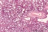 |
Late terminal sac period of developing lung (human, fetus, low magnification) | Stain: Hematoxylin and eosin. Cross-sections of a bronchus (1) with cartilage rings (2) closely associated with pulmonary arteries (3). The alveolar appearance is evident, and the open spaces represent future alveolar duct systems as well as alveolar sacs. The cellular septa are still thick due to n... | Lung development; Terminal sac period | Poja Histology Collection - Respiratory System Subset |
| 23 |
 |
Elastin in alveolus of lung tissue (human, adult) | Immuno-electron microscopy (embedded in Lowicryl HM 20). Elastin (E) is labeled with electron-dense gold particles of 10 nm. Note that the amorph elastin appears pale after embedding in this type of resin. (A) indicates alveolar space, (F) unlabelled myofibrolast, (C) collagen fibers, and (1) the th... | Alveolar cells type I; Myofibroblasts; Immuno-electron microscopy; Immuno-staining | Poja Histology Collection - Respiratory System Subset |
| 24 |
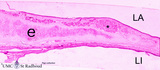 |
Survey of epiglottis (human) | Stain: Azan. Centrally light-stained elastic cartilage (e). Lingual side (LI) is covered with squamous epithelium. At the laryngeal side (LA) the squamous epithelium usually turns into respiratory epithelium with seromucous laryngeal glands (*). | Laryngeal gland; Oral cavity | Poja Histology Collection - Respiratory System Subset |
| 25 |
 |
Lamellar body of a type II alveolar cell in the lung (dog) | Electron microscopy. Extrusion of surfactant material out of a type II alveolar cell (pneumocyte II, great alveolar cell). After fusion of a multilamellar body (1) (cytosome) with the apical cell membrane (note microvilli 2) the electron-dense lamellar content or surfactant (consisting of phosphatid... | Type II alveolar cell; Pneumocyte II; Multilamellar bodies; Cytosomes; Tubular myelin | Poja Histology Collection - Respiratory System Subset |