TO
1 - 25 of 22
| Title | Curriculum | Description | Subject | Collection | ||
|---|---|---|---|---|---|---|
| 1 |
 |
Optic Disc Drusen | LEEpathologies; LEEopticdiscdrusen | Summary: ● Basics of optic disc drusen: ○ Name take from the German word, druse, meaning rock or crystal ○ Rocks of calcium and debris inside optic nerve > can cause optic neuropathy ○ Benign, but slowly progressive ○ Different from retinal disc drusen (seen in age-related macular de... | Optic Disc Drusen; Optic Neuropathy; Field Defects | Neuro-Ophthalmology Virtual Education Library: Andrew G. Lee Collection: https://novel.utah.edu/Lee/ |
| 2 |
 |
Patient Portal: Optic Disc Drusen | Optic disc drusen (ODD) are abnormal deposits of benign, usually calcified material within the optic disc, which is the front part of the optic nerve that connects each eye to the brain. We do not know the exact cause of optic disc drusen. They are present in 0.3-2% of people as an isolated case or ... | Optic disc drusen; Papilledema; Pseudopapilledema | Neuro-Ophthalmology Virtual Education Library: NOVEL http://NOVEL.utah.edu | |
| 3 |
 |
Novel Cellular and Animal Models of Optic Disc Drusen | Optic disc drusen (ODD) are calcified deposits of the anterior optic nerve and result in visual field loss in majority of patients.1 Most common cause of vision loss is anterior ischemic optic neuropathy (AION). Ultrastructurally, the hallmark of ODD is mitochondrial calcification,2 and energy dispe... | Optic Neuropathy; Genetic Disease | Neuro-Ophthalmology Virtual Education Library: NOVEL http://NOVEL.utah.edu | |
| 4 |
 |
Optic Disc Drusen in Young Adults with Anterior Ischemic Optic Neuropathy: A Multicenter Study (Video) | IC-D1hvi-optic-nerve-drusen | Anterior ischemic optic neuropathy (AION) usually occurs in patients over the age of 50 with systemic vascular disease. Lesscommonly, AION occurs in patients younger than 50 years with few or no vascular risk factors. Optic disc drusen (ODD), present in 2% of the general population, have occasio... | Optic Neuropathy, Vascular Disorders | Neuro-ophthalmology Virtual Education Library: NOVEL http://NOVEL.utah.edu |
| 5 |
 |
Optic Disc Drusen: Insights on Diagnosis | Guidelines for enhanced-depth imaging have improved the diagnosis of optic disc drusen and attempt to address confusion within the literature. In particular the presence of peripapillary mass-like structures have caused this confusion and potentially lead to misclassification of some disc oedema. Th... | Optic Disc Drusen; Peripapillary Hyperreflective Ovoid Mass-like Structures; Optical Coherence Tomography; Ocular Ultrasound; Visual Field | Neuro-ophthalmology Virtual Education Library: NOVEL http://NOVEL.utah.edu | |
| 6 |
 |
Pseudo Papilledema | LEEpathologies; LEEpseudopapilledema | Dr. Lee lectures medical students on pseudo-papilledema. | Papilledema Intracranial Pressure; Drusen; Diagnosis | Neuro-Ophthalmology Virtual Education Library: Andrew G. Lee Collection: https://novel.utah.edu/Lee/ |
| 7 |
 |
Mining for ODD Using Multimodal OCT | IC-D1ai-anterior | Optic disc drusen (ODD) are acellular, calcified deposits localized between the axons in the prelaminar optic nerve head of about 2% of the population. | Optic Disc Drusen; Optical Coherence Tomography; Enhanced Depth Imaging; Swept Source; Detection | Neuro-ophthalmology Virtual Education Library: NOVEL http://NOVEL.utah.edu |
| 8 |
 |
Ultrasound in Neuro-Ophthalmology | LEEultrasound; LEEimaging | Dr. Lee lectures medical students on ultrasound in neuro-ophthalmology. | Ultrasound; Neuro-Ophthalmology; Imaging | Neuro-Ophthalmology Virtual Education Library: Andrew G. Lee Collection: https://novel.utah.edu/Lee/ |
| 9 |
 |
The Elevated Optic Disc: When OCT Helps and When It Does Not. An Interactive Case Based Approach (video) | VBnflaopticalcoherencetomography; KBDburieddrusen; Medical Knowledge; Patient Care; Practice-Based Learning and Improvement | The differentiation of acquired optic disc edema representing a pathological process such as papilledema caused by increased intracranial pressure versus a usually benign, pre-existing condition such as optic disc drusen is one of the most classic, and most important, clinical challenges in neuro-op... | Spectral Domain Optical Coherence Tomography; Multi-Color Optical Coherence Tomography; Papilledema; Optic Disc Drusen; Epiretinal Membranes | Neuro-ophthalmology Virtual Education Library: NOVEL http://NOVEL.utah.edu |
| 10 |
 |
Long-term OCT Follow-up in Children with Optic Disc Drusen | A better understanding of optic disc drusen (ODD) etiology and pathophysiology might be possible by visualizing progression of ODD and potential precurser lesions. The purpose of this study was to examine the progression of ODD and scleral canal diameter in children previously diagnosed with ODD and... | Optic Neuropathy, Pediatric Neuro-Ophthalmology | Neuro-ophthalmology Virtual Education Library: NOVEL http://NOVEL.utah.edu | |
| 11 |
 |
Cases | OCT is capable of the highest resolution images of the retina and optic nerve clinically available of the eye, yet when clinicians examine its output, they frequently rely upon a few quantitative measures to determine whether the nerve fiber layer or ganglion cell complex is thinning or thickening. ... | OCT; NAION; Disc Swelling; Drusen | ||
| 12 |
 |
Deep Learning Can Accurately Distinguish Between True Papilledema and Optic Disc Drusen on Fundus Photographs | Identification of true papilledema due to raised intracranial pressure requires high neuro-ophthalmic expertise and expensive ancillary testing. Its ophthalmoscopic diagnosis can be easily mistaken with false papilledema due to optic disc drusen (ODD), especially if the latter are buried within the ... | High Intracranial Pressure/Headache; Optic Neuropathy; Pseudotumor Cerebri; Vascular Disorders | Neuro-ophthalmology Virtual Education Library: NOVEL http://NOVEL.utah.edu | |
| 13 |
 |
Reading an OCT Like We Read an MRI | OCT is capable of the highest resolution images of the retina and optic nerve clinically available of the eye, yet when clinicians examine its output, they frequently rely upon a few quantitative measures to determine whether the nerve fiber layer or ganglion cell complex is thinning or thickening. ... | OCT; NAION; Disc Swelling; Drusen | ||
| 14 |
 |
Transverse Axial / en Face OCT in the Assessment of Optic Disc Edema and Pseudopapilledma | OCT is capable of the highest resolution images of the retina and optic nerve clinically available of the eye, yet when clinicians examine its output, they frequently rely upon a few quantitative measures to determine whether the nerve fiber layer or ganglion cell complex is thinning or thickening. ... | OCT; NAION; Disc Swelling; Drusen | ||
| 15 |
 |
Frizzle Frazzled | Papilledema; Idiopathic Intracranial Hypertension (IIH); Ophthalmologic Disorders; Retinal Disorders; Optic Nerve Drusen | Neuro-ophthalmology Virtual Education Library: NOVEL http://NOVEL.utah.edu | ||
| 16 |
 |
Age Related Macular Degeneration | IC-E11aiv-age-related-macular-degeneration | Age-related macular degeneration (AMD) is a degenerative disease of the retina that causes central vision loss, and it is the leading cause of blindness in the developed world. Age is a strong nonmodifiable risk factor for AMD. Patients may have genetic susceptibility to AMD from mutations in genes ... | AMD; AREDS2; Exudative; Geographic Atrophy; Macular Degeneration; Nonexudative | Neuro-Ophthalmology Virtual Education Library: NOVEL http://NOVEL.utah.edu |
| 17 |
 |
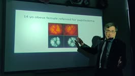
How Do I Evaulate Papilledema in a Child? (Video) | KBDpapilledema | LEARNING OBJECTIVES: 1. Identify etiologies of papilledema unique to children (craniosynostosis, IVH/premie; mastoiditis). 2. Explain the influence of age, gender and obesity on the incidence of papilledema/IIH. 3. Describe the appropriate diagnostic imaging methods for children with papilledema. | Pediatric Neuro-Ophthalmology; Papilledema; Drusen; Elevated ICP; Headache | Neuro-ophthalmology Virtual Education Library: NOVEL http://NOVEL.utah.edu |
| 18 |
 |
Peripapillary Hyperreflective Ovoid Mass-like Structures (PHOMS) in a Child Cohort | A peripapillary hyperreflective ovoid mass-like structure (PHOMS) is a common finding on optical coherence tomography (OCT) of patients with optic disc edema (ODE), optic disc drusen (ODD) or anomalous optic discs. The purpose of the study was to determine the prevalence of PHOMS in a population-bas... | Diagnostic Tests (ERG, VER, OCT, HRT, mfERG, etc); Retina | Neuro-ophthalmology Virtual Education Library: NOVEL http://NOVEL.utah.edu | |
| 19 |
 |
OCTA: What Can it Tell Me and How to Use It | Optical coherence tomography angiography (OCTA) compares the decorrelation signal (differences in the backscattered OCT light signal amplitude) between sequential OCT B-scans taken at a single cross section(motion contrast). Since only blood flow would be expected to create movement and differences ... | Optical Coherence Tomography Angiography (OCTA); Swept Source OCTA; Non-Arteritic Anterior Ischemic Optic Neuropathy (NAION); Non-Ischemic Optic Disc Edema; Optic Disc Drusen | Neuro-ophthalmology Virtual Education Library: NOVEL http://NOVEL.utah.edu | |
| 20 |
 |
Ocular Imaging, EDI, OCTA and Beyond: Where Are We, What Good Is It? | Enhanced depth imaging (EDI) is useful for discerning structural details in deeper layers of the retina, choroid and optic nerve head for identifying optic nerve drusen, deformation of Bruch's membrane in the peripapillary region and for assessing choroidal thickness changes. OCT angiography is limi... | OCT Enhanced Depth Imaging (EDI); OCT Angiography (OCT-A); Functional Retinal Imaging; Retinal Oximetry; Detection of Apoptosis of Retinal Ganglion Cells (DARC) | Neuro-Ophthalmology Virtual Education Library: NOVEL http://NOVEL.utah.edu | |
| 21 |
 |
Magnetic Resonance Imaging (MRI) Neuroop Pearls | LEEmripearls; LEEimaging; LEEmrinopearls | Dr. Lee lectures medical students on neuroop pearls. | MRI; Papilledema; Optic Neuritis | Neuro-Ophthalmology Virtual Education Library: Andrew G. Lee Collection: https://novel.utah.edu/Lee/ |
| 22 |
 |
Upbeat Nystagmus | IC-D7civ-upbeat-nystagmus | Upbeat Nystagmus; Saccadic Hypermetria; Saccadic Pursuit; Square Wave Jerks; Oscillopsia; Cerebellar Astrocytoma; Primary Position Upbeat Nystagmus; Horizontal Saccadic Dysmetria | Neuro-Ophthalmology Virtual Education Library - Shirley H. Wray Neuro-Ophthalmology Collection: https://novel.utah.edu/Wray/ |
1 - 25 of 22