John A. Moran Eye Center Neuro-Ophthalmology Collection: A variety of lectures, videos and images relating to topics in Neuro-Ophthalmology created by faculty at the Moran Eye Center, University of Utah, in Salt Lake City.
NOVEL: https://novel.utah.edu/
TO
1 - 200 of 127
| Title | Description | Type | ||
|---|---|---|---|---|
| 1 |
 |
3 Step Test | Demonstration of patient examination. | Image/MovingImage |
| 2 |
 |
A-scan Technique | This video describes and demonstrates the A-scan examination technique for examination of the eye using ultrasonography. | Image/MovingImage |
| 3 |
 |
Abducting (Dissociated) Nystagmus | Example of a patient with abducting (dissociated) nystagmus. Patient has a subtle internuclear ophthalmoplegia. Right eye has right-beating jerk nystagmus, with smaller oscillations in the left eye. Disease/Diagnosis: Abducting Nystagmus | Image/MovingImage |
| 4 |
 |
Aberrant Regeneration of the Lid | Patient with left third nerve palsy demonstrates anisocoria and mild vertical gaze limitation and aberrant movement of the left upper lid. Patient is instructed through all gaze positions. Left upper lid does not descend during downgaze but retracts instead. | Image/MovingImage |
| 5 |
 |
Aberrant Regeneration of the Seventh Nerve | Examples of patients with aberrant regeneration of the seventh nerve. First example is a patient with contractions around the mouth and dimpling, demonstrated with slow and rapid eye blinking. Second example shows contraction around nose with eye blink. | Image/MovingImage |
| 6 |
 |
Aberrant Regeneration of the Third | Patient with a right third nerve palsy demonstrates ptosis, anisocoria and ophthalmoplegia. During attempted downgaze, the right upper lid flutters back up (aberrant movement) and remains retracted. | Image/MovingImage |
| 7 |
 |
Aberrant Regeneration of the Third and Sixth Nerves | Image/MovingImage | |
| 8 |
|
Aberrant Regeneration of Third Nerve, Bilaterally (1 degree OD, 2 Digrees OS) | Example of patient with bilateral aberrancy of the third nerve. Shows lids popping up (synkinetic) with adduction. Patient had bilateral internal carotid artery aneurisms with third nerve compression. | Image/MovingImage |
| 9 |
 |
Amsler Grid Testing | Demonstration of Amsler Grid examination. | Text |
| 10 |
 |
B-scan Technique | This video describes and demonstrates the B-scan examination technique for examination of the eye using ultrasonography. | Image/MovingImage |
| 11 |
 |
Basic Eye Alignment Exam | Demonstration of basic eye alignment examination. Includes: a. Tools b. Cover-Uncover and SPCT c. Alternate Cover and APCT d. Maddox Rod Testing | Image/MovingImage |
| 12 |
 |
Basic Neurologic Exam | Demonstration of a basic neurologic examination. | Image/MovingImage |
| 13 |
 |
Basic Neurologic Exam: Coordination | Demonstration of a coordination examination. | Image/MovingImage |
| 14 |
 |
Basic Neurologic Exam: Cranial Nerves | Demonstration of a cranial nerve examination. | Image/MovingImage |
| 15 |
 |
Basic Neurologic Exam: Mental Status | Demonstration of a mental status examination. | |
| 16 |
 |
Basic Neurologic Exam: Motor Examination | Demonstration of a motor examination. | Image/MovingImage |
| 17 |
 |
Basic Neurologic Exam: Sensory | Demonstration of a sensory examination. | Image/MovingImage |
| 18 |
 |
Basic Neurologic Exam: Station and Gait | Demonstration of a station and gait examination. | Image/MovingImage |
| 19 |
 |
Before Tensilon | Example of patient with myasthenia gravis. Demonstration of baseline examination, followed by administration of 2mg of tensilon, which is a test dose. Procedure for administration of tensilon test is described, including variations. Patient is then shown after being given 4mg of tensilon, with very ... | Image/MovingImage |
| 20 |
 |
Bilateral Asynchronous Blepharospasm with Facial and Cervical Dystonia | Bilateral Asynchronous Blepharospasm with Facial and Cervical Dystonia. | Image/MovingImage |
| 21 |
 |
Bilateral Facial Myokymia | Example of a patient with a brain stem glioma. Shows bilateral facial myokymia. | Image/MovingImage |
| 22 |
 |
Bilateral Internuclear Ophthalmoplegia | Example of patient with bilateral internuclear ophthalmoplegia. Patient is led through instructions for direction and distance of gaze. | Image/MovingImage |
| 23 |
 |
Bilateral Ptosis | Video of patient with bilateral ptosis. | Image/MovingImage |
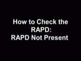
| 24 |
 |
Binocular Pendular Nystagmus | Example of a patient with binocular pendular nystagmus. Patient has somewhat dissociated nystagmus, with nystagmus seen more prominently in the left eye. Patient shows an occasional jerk nystagmus to the right in the right eye. Left eye oscillations are mostly pendular. | Image/MovingImage |
| 25 |
 |
Blepharospasm | Example of patient with blepharospasm. Patient is led through instructions for direction of gaze and opening and closing of eyes. Patient is led through same exercises again after receiving indomethacin treatment. | Image/MovingImage |
| 26 |
 |
Blepharospasm with Apraxia of the Eye | Image/MovingImage | |
| 27 |
 |
Brainstem Trauma | Image/MovingImage | |
| 28 |
 |
Brun's Nystagmus | Observation of patient with Brun's Nystagmus. Shows patient gazing to the right and the nystagmus beating in the direction of the gaze. | Image/MovingImage |
| 29 |
 |
Central Retinal Artery Occlusion | Video of central retinal artery occlusion. | Image/MovingImage |
| 30 |
 |
Cochet-Bonnet Esthesiometer (similar to Von Frey Hair) Exam | The Cochet-Bonnet esthesiometer , similar to Von Frey Hair testing, has been used in studies to quantify sensory changes in the trigeminal system, especially the cornea. The device uses a thin fiber to test sensation. The shorter the fiber, the more sensation is felt. The monofilament is applied per... | |
| 31 |
 |
Cogan's Lid Twitch | Example of a patient with Cogan's lid twitch, with discussion of how to detect it in an exam. | Image/MovingImage |
| 32 |
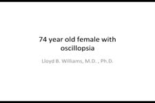
 |
Cogan's Lid Twitch | Image/MovingImage | |
| 33 |
 |
Color Vision Testing | Demonstration of color vision examination. | Text |
| 34 |
 |
Congenital Nystagmus | Example of patients with congenital nystagmus. First patient's nystagmus are mostly jerk and not pendular. Second patient's nystagmus are mostly pendular. Both patients show a uniform horizontal oscillation. Second patient also shows differences in frequency of oscillations depending on gaze, includ... | Image/MovingImage |
| 35 |
 |
Congenital Nystagmus | Patient with congenital nystagmus (no audio) | Image/MovingImage |
| 36 |
 |
Congenital Ocular Motor Apraxia | Two examples of congenital ocular motor apraxia. Patients have trouble initiating saccades, and compensate with head movement. Discussion of how to distinguish this condition from simply not seeing well. | Image/MovingImage |
| 37 |
 |
Convergence Retraction Nystagmus (Parinaud's Syndrome) | Examples of patients with convergence retraction nystagmus. Shows saccadic oscillations in patients looking upwards and following downwards moving targets. Also shows a side-view of the retracting movements of the globes. | Image/MovingImage |
| 38 |
 |
CPEO | Patient with Chronic Progressive External Ophthalmoplegia (CPEO) | Image/MovingImage |
| 39 |
 |
Cyclic Oculomotor Palsy | Example of patient with cyclic oculomotor palsy. | Image/MovingImage |
| 40 |
 |
Dilation Lag | Two examples of dilation lag (Horner's syndrome). In the first example, the right pupil dilates much faster than the left pupil when the light is turned out. In the second example, the left pupil dilates much faster than the right pupil when the light is turned out. Discussion of methods of document... | Image/MovingImage |
| 41 |
 |
Dissociated Nystagmus | Example of a patient with dissociated nystagmus. Demonstrates difference in movements between each eye. | Image/MovingImage |
| 42 |
 |
Downbeat Nystagmus | Example of patients with downbeating jerk nystagmus. Demonstrates how oscillations grow more prominent when the patient gazes down or laterally. Discusses some causes, including Arnold-Chiari malformation, infarction, and demyelination. | Image/MovingImage |
| 43 |
 |
Downbeat Nystagmus | Example of patient with downbeat nystagmus. Patient is led through instructions of where to gaze. (no audio) | Image/MovingImage |
| 44 |
 |
Downbeat Nystagmus | Example of patient with downbeat nystagmus. Patient is led through instructions of where to gaze. | Image/MovingImage |
| 45 |
 |
Duane's Retraction Syndrome Type 1: Lid Retraction | Example of patients with Duane's Retraction Syndrome, Type 1. Description of components of Duane's Syndrome: limitation of abduction, variable limitation of adduction, and palpebral fissure narrowing and globe retraction with attempted adduction. Type 1 includes limited or absent abduction with norm... | Image/MovingImage |
| 46 |
 |
Duane's Retraction Syndrome Type 3 | Example of a patient with Type 3 Duane's Retraction Syndrome, as well as bilateral Duane's Syndrome. Shows limitation of abduction in both eyes and adduction in the left eye. Also shows side-view of globe retraction in abduction. | Image/MovingImage |
| 47 |
 |
Duane's Syndrome | Example of patient with Duane's Syndrome. Patient is led through instructions for pursuit. | Image/MovingImage |
| 48 |
 |
Duane's Syndrome Type 1 | Clip of patient with Duane's Syndrome Type I. Presented at the Neurology Grand Rounds in Fall 2011 at the University of Utah. Presentation can be found in this collection at: Why Don't You See Double? http://content.lib.utah.edu/u?/EHSL-Moran-Neuro-opth,132 Disease/Diagnosis: Duane's Syndrome Type ... | Image/MovingImage |
| 49 |
 |
Duane's Syndrome Type 2: Aberrant Regeneration of the Third and Sixth Nerves | Example of a patient with Type 2 Duane's Syndrome. Demonstrates limitation of adduction in left eye with normal abduction. Discussion of limited pathological cases. | Image/MovingImage |
| 50 |
 |
Duane's Syndrome Type 3 | Clip of patient with Duane's Syndrome Type III. Presented at the Neurology Grand Rounds in Fall 2011 at the University of Utah. Presentation can be found in this collection at: Why Don't You See Double? http://content.lib.utah.edu/u?/EHSL-Moran-Neuro-opth,132 Disease/Diagnosis: Duane's Syndrome Ty... | Image/MovingImage |
| 51 |
 |
Exophthalmometry | Demonstration of exophthalmometry examination. | Text |
| 52 |
 |
Facial Myokymia Unilateral | Example of patient with facial myokymia, a disorder of the seventh nerve, probably due to brain stem involvement. Patient has multiple sclerosis. Discussion of characteristics, such as continuous, undulating, contractions in the distribution of the seventh nerve, and a spreading of these movements t... | Image/MovingImage |
| 53 |
 |
Facial Nerve Exam | Explanation of a facial nerve exam. | |
| 54 |
 |
Flutter in Downgaze | Examination of patient with flutter in downgaze (no audio) | Image/MovingImage |
| 55 |
 |
Fourth Nerve Palsy | Demonstration of examination of patient who experienced blurry vision and pain in the left eye. Demonstrates checking of eye movements, focusing on object while each eye is covered and uncovered, turning head both ways and repeating. Shows limitation of depression in adduction of left eye, left hype... | Image/MovingImage |
| 56 |
 |
Fusional Vergence Amplitudes | Demonstration of fusional vergence amplitudes examination. Incluudes: a. Convergence Amplitudes b. Divergence Amplitudes c. Vertical Ampitudes | Text |
| 57 |
 |
Gaze Palsy with Facial Weakness from Pontine AVM | Example of a patient with torsional nystagmus in both eyes and pendular nystagmus in the left eye. Patient is led through instructions for direction of gaze. | Image/MovingImage |
| 58 |
 |
Hemifacial Spasm | Example of patients with hemifacial spasm. First patient has a sequela of Bell's palsy, and is seen to have mainly clonic movements around the eye, with occasional tonic movements around the mouth. Second patient has a cerebellopontine angle epidurmoid tumor, and is seen to have movements around the... | Image/MovingImage |
| 59 |
 |
How to Measure the RAPD | This clip demonstrates the examination technique for measuring the Relative Afferent Pupillary Defect (RAPD). Demonstration of balancing an afferent papillary defect using filters in a patient with a resolving optic neuritis and an afferent papillary defect on the left. | Image/MovingImage |
| 60 |
 |
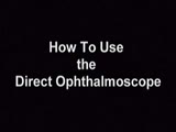
How to Use the Direct Ophthalmoscope in an Exam | Demonstration of using the direct ophthalmoscope to examine the optic disc. Covers hand placement , which eye to use, and distance from patient. | Image/MovingImage |
| 61 |
 |
Intermittent Square Wave Jerks | Patient with intermittent square wave jerks (no audio) | Image/MovingImage |
| 62 |
 |
Internuclear Ophthalmoplegia (2 Examples) | Two examples of patients with internuclear ophthalmoplegia. First patient has a right internuclear ophthalmoplegia. Patient had subacute bacterial endocarditis with a bacterial abscess in the brain stem. Ductions and gaze to the right look good, but when gazing to the left, the right eye does not ad... | Image/MovingImage |
| 63 |
 |
Introduction to Fogging Refraction | An introduction to fogging refraction. | |
| 64 |
 |
Introduction to Headache, Migraine and Secondary Headaches | Video lecture covering an introduction to headache, migraine, and secondary headaches by Kathleen Digre, MD. | |
| 65 |
 |
Introduction to the Basic Neurologic Exam | Introduction to the neurological examinations section of NExT. | |
| 66 |
|
Latent Nystagmus | Example of a patient with latent nystagmus. Demonstrates a lack of oscillations in forward gaze, followed by the occlusion of each eye, showing how this generates a jerking oscillation in the non-occluded eye away from the occluded eye. | Image/MovingImage |
| 67 |
 |
Levator Disinsertion | Example of patient with levator disinsertion, a lid disorder. Patient is pregnant and wears poorly fitting contacts. Discussion of characteristics, such as lid ptosis (shown in the left eye of patient), but with full levator function. | Image/MovingImage |
| 68 |
|
Light-near Dissociation | Example of patient with Argyll Robertson pupil with neurosyphilis. Shows a lack of pupillary response to light and some pupillary response to nearness of finger. | Image/MovingImage |
| 69 |
 |
Marcus Gunn Jaw Winking | Example of patient with Marcus Jaw Winking. Patient is led through instructions for movement of jaw (open, close, back and forth), with eyelid seen to be affected. Patient is then led through instructions for direction of gaze and pursuit. | Image/MovingImage |
| 70 |
 |
Measuring Visual Acuity | Demonstration on self of visual acuity exam, using a standard card. | Image/MovingImage |
| 71 |
 |
Migraine and Cluster Pathophysiology and Treatment | Video lecture covering pathophysiology and treatment of migraine and cluster headaches by Kathleen Digre, MD. | |
| 72 |
 |
Monocular Pendular Nystagmus | Example of a patient with monocular pendular nystagmus, with discussion of situations in which this condition is seen: acquired disorder of the visual-sensory pathway, and acquired disorder of the brain stem (e.g. multiple sclerosis). | Image/MovingImage |
| 73 |
 |
Multifocal Electroretinograms | The most important development in ERGs is the multifocal ERG (mfERG). Erich Sutter adapted the mathematical sequences called binary m-sequences creating a program that can extract hundreds of focal ERGs from a single electrical signal. This system allows assessment of ERG activity in small areas of ... | |
| 74 |
 |
Normal Eye Movements | This is an examination of a person with normal eye movements. Notice the patient has normal excursions. He has normal pursuit and saccades (horizontally and vertically). | Text |
| 75 |
 |
Normal Light Reflex without RAPD | This clip demonstrates the examination of the Relative Afferent Pupillary Defect (RAPD.) Demonstration of gauging the size of the pupil in light, testing light reflexes, swinging flashlight test for optic nerve abnormality. | Image/MovingImage |
| 76 |
 |
Ocular Flutter | Two examples of patients, the first with rotary, flutter-like movements, but not ocular flutter, and the second with genuine ocular flutter. Discussion of difference between ocular flutter and nystagmus, and how to elicit ocular flutter. | Image/MovingImage |
| 77 |
|
Ocular Lateropulsion (Wallenberg's Syndrome) | Example of patient with ocular lateropulsion. Patient also has central Horner syndrome and nystagmus in right gaze. When shifting gaze back to forward, eyes overshoot their mark. Eyes laterally deviate to the right upon opening. | Image/MovingImage |
| 78 |
 |
Ocular Myasthenia | Example of patient with myasthenia gravis. Demonstration of tensilon test. Patient shown to have bilateral ptosis, bilateral duction deficits, and left hypertropia. Discussion of techniques to observe subtle changes, such as bringing in a neutral observer or taking still photographs. Shows split-scr... | Image/MovingImage |
| 79 |
 |
Ocular Myotonia | Example of patient with ocular myotonia. Patient is led through instructions for direction of gaze and opening and closing of eyes. Right eye is shown to be stuck in position after held gaze to the left and right, with very slow relaxation back into forward gaze. | Image/MovingImage |
| 80 |
 |
Oculopalatal Myoclonus | Oculopalatal myoclonus (OPM) Rhythmic oscillations of eyes and palate. Occurred after specific brainstem injury from stroke, following stenting. Related PowerPoint Presentation: http://content.lib.utah.edu/u?/EHSL-Moran-Neuro-opth,129 Disease/Diagnosis: Oculopalatal myoclonus. | Image/MovingImage |
| 81 |
 |
Oculopalatal Myoclonus (PPT) | Oculopalatal myoclonus (OPM) Rhythmic oscillations of eyes and palate. Occurred after specific brainstem injury from stroke, following stenting. Related Video: http://content.lib.utah.edu/u?/EHSL-Moran-Neuro-opth,128 Disease/Diagnosis: Oculopalatal myoclonus | Image/MovingImage |
| 82 |
 |
Opsoclonus | Example of patients with opsoclonus, a saccadic abnormality. Discussion of characteristics of opsoclonus, such as involuntary, rapid, brief, random, conjugate saccades. Discussion of possible causes, including brain stem encephalitis (as in first patient), a paraneoplastic effect, tumors, and drug t... | Image/MovingImage |
| 83 |
 |
Opsoclonus | Example of patients with opsoclonus, a saccadic abnormality. | Image/MovingImage |
| 84 |
 |
The Orbital Exam | Comprehensive demonstration of the entire orbital examination. | |
| 85 |
|
Paradoxical Constriction of Pupils to Darkness (Flynn Phenomenon) | Example of patients both with and without paradoxical constriction of pupils. Observed in many congenital retinal disorders, such as achromatopsia, congenital stationary night-blindness, and Leber's congenital amaurosis. Sometimes seen in optic nerve disorders, such as dominant optic atrophy. | Image/MovingImage |
| 86 |
 |
Parinaud's Syndrome | Two examples of patients with Parinaud's syndrome, a dorsal midbrain syndrome. Discussion of hallmarks of this syndrome, including convergence retraction nystagmus, vertical gaze palsies, light-near dissociation, and Collier's Sign. Discussion of age-dependent disorders associated with this syndrome... | Image/MovingImage |
| 87 |
|
Periodic Alternating Nystagmus | Example of a patient with periodic alternating nystagmus, showing an alternation between left-beats and right-beats as the patient maintains forward gaze. Nystagmus maintain horizontal direction regardless of position of gaze. | Image/MovingImage |
| 88 |
 |
Physiologic (End-Gaze) Nystagmus | Demonstration of physiological nystagmus, where oscillations do not represent pathology, but occur when the patient's gaze is drawn too far laterally. | Image/MovingImage |
| 89 |
 |
Progressive Supranuclear Palsy | Progressive Supranuclear Palsy | Image/MovingImage |
| 90 |
|
Progressive Supranuclear Palsy | Example of patient with progressive supranuclear palsy. Discussion of difference between saccadic movement in supranuclear palsy and nystagmus. Shows saccadic intrusions in forward gaze, pursuit, saccades, and doll's head maneuver. | Image/MovingImage |
| 91 |
 |
Pulsating Exophthalmos | Example of a patient with neurofibromatosis with an absent sphenoid wing. Shows left eye pulsating back and forth with the pulse from front and side views. | Image/MovingImage |
| 92 |
 |
Pupil Exam | Demonstration of pupil examination. | Text |
| 93 |
 |
RAPD Present | This clip demonstrates the technique used to determine that Relative Afferent Pupillary Defect (RAPD) is present in a patient. | Image/MovingImage |
| 94 |
|
Rebound Nystagmus | Example of a patient with rebound nystagmus, where the oscillations alternate direction as the patient shifts gaze in different directions. Discussion of relationship to disease and disorders of the cerebellum, including degenerations of the cerebellum, infarction, and demyelination. | Image/MovingImage |
| 95 |
 |
Retraction Nystagmus | Patient with retraction nystagmus (no audio) | Image/MovingImage |
| 96 |
 |
Rotary Downbeat | Patient with rotary downbeat nystagmus (no audio) | Image/MovingImage |
| 97 |
|
Rotary Nystagmus | Example of a patient with rotary nystagmus, showing occasional counterclockwise rotary movements of both eyes. Seen more in intrinsic disorders of the brainstem. | Image/MovingImage |
| 98 |
 |
Sector Palsies and Light-Near Dissociation | Example of patient with bilateral Adie's pupils. Exam is performed with a slit-lamp. Shows iris stroma and focal segments of iris sphincter that retain their contractilty. Suggests post-ganglionic parasympathetic denervation. | Image/MovingImage |
| 99 |
 |
See-saw Nystagmus | Example of a patient with see-saw nystagmus, showing how one eye elevates as the other depresses, with the elevating eye intorting as the depressing eye extorts. Shows vertical oscillations with pendular waveforms. Suggests a large structural lesion in the pericellar region (associated with bi-tempo... | Image/MovingImage |
| 100 |
 |
See-saw Nystagmus | 7-year-old female whose mother noticed her eyes "bouncing" for 2 months. Visual acuity 20/70 OD and 20/40 OS, reduced color vision OU, and no afferent pupillary defect. See-saw nystagmus documented with videography. Manual perimetry revealed a complete right homonymous hemianopia. MRI revealed a lar... | Image/MovingImage |
| 101 |
 |
Spasm of the Near Reflex | Example of patient with spasm of the near reflex and voluntary nystagmus. Discussion of similar-looking conditions (e.g. six nerve palsy, limitation of abduction, lateral rectus muscle problems) and how to tell them apart from spasm of the near reflex by observing the myosis evoked by the near respo... | Image/MovingImage |
| 102 |
 |
Spasmus Nutans | Example of patient with spasmus nutans. Discussion of characteristics of this disorder, such as dissociated or monocular nystagmus, abnormal head position, and to-and-fro head oscillation. Sometimes an eccentric gaze is seen as well (as in patient). Patient has a monocular horizontal nystagmus in th... | Image/MovingImage |
| 103 |
 |
Spasmus Nutans | Example of patient with spasmus nutans. | Image/MovingImage |
| 104 |
 |
Spontaneous Venous Pulsations | This clips shows a spontaneous venous pulsation viewed during an ocular examination. | Image/MovingImage |
| 105 |
|
Square Wave Jerks | Example of patient with square wave jerks. Discussion of difference between square wave jerks (saccadic oscillations) and horizontal nystagmus. | Image/MovingImage |
| 106 |
 |
Stereoacuity Testing | Demonstration of examination for stereoacuity. | Text |
| 107 |
 |
Superior Oblique Myokymia | Close-up video of a patient with superior oblique myokymia (no audio.) | Image/MovingImage |
| 108 |
 |
Superior Oblique Myokymia | Example of patients with superior oblique myokymia, a saccadic intrusion. First patient is seen to have intermittent, intorting movements with superimposed slight vertical deviations in right eye. Discussion of disorder as benign, but frequently disabling, as patients experience episodes of diplopia... | Image/MovingImage |
| 109 |
 |
Test Duane | ||
| 110 |
 |
Testing the Visual Fields | Demonstration of various methods of testing visual fields, including counting fingers, motion, and color of several objects. | Image/MovingImage |
| 111 |
 |
Third Nerve Palsy | Patient with third nerve palsy (no audio) | Image/MovingImage |
| 112 |
 |
Third Nerve Palsy, Pupil Involving | Example of patient with third nerve palsy. Left eye shows pupilary involvement. Left eye doesn't immediately duct, but abducts well, with impaired superduction. Secondary and primary deviations are demonstrated. Anisocoria is more prominent when light is on, showing a parasympathetic defect to the p... | Image/MovingImage |
| 113 |
 |
Tour of the Direct Ophthalmoscope | This clip describes the parts and operation of the ophthalmoscope as an ocular examination tool. Includes adjustment of aperture size and adjustment of lenses. | Image/MovingImage |
| 114 |
 |
Tour of the Fundus | This clip demonstrates the funduscopic examination technique. | Image/MovingImage |
| 115 |
|
Transillumination - Ciliary Body Neurofibromas | Example of transillumination on a patient with neurofibromatosis, but without Lisch nodules. Shows suspected neurofibromas in the ciliary body. | Image/MovingImage |
| 116 |
|
Transillumination - Lisch Nodules | Demonstration of transillumination of the Lisch nodules on a patient with neurofibromatosis. Shows how Lisch nodules that were not very visible in slit-lamp examination are better seen with transillumination, which may therefore be useful in detecting Lisch nodules earlier in children where they are... | Image/MovingImage |
| 117 |
|
Transillumination Ocular Melanoma | Video describing condition. | Image/MovingImage |
| 118 |
 |
Trigeminal Nerve Exam | Explanation of a trigeminal nerve exam. | |
| 119 |
 |
Ultrasonography Techniques | This video describes and demonstrates the various techniques for examination of the eye using ultrasonography, including A-scan, B-scan and immersion. | Image/MovingImage |
| 120 |
 |
Ultrasonography: Immersion Technique | This video describes and demonstrates the immersion technique for examination of the eye using ultrasonography. | Image/MovingImage |
| 121 |
 |
Unilateral Blepharospasm | Example of patient with unilateral blepharospasm. | Image/MovingImage |
| 122 |
 |
Upbeat Nystagmus | Example of a patient with upbeat nystagmus. Shows vertical jerk nystagmus with fast phases in the up direction. Localizes to brain stem, and occurs with strokes, demyelination, and tumors. | Image/MovingImage |
| 123 |
 |
Vestibular Nystagmus | Example of patient with vestibular nystagmus. Patient is led through instructions for direction of gaze. Shown also with Frenzel goggles. | Image/MovingImage |
| 124 |
|
Vestibular Nystagmus | Discussion of vestibular nystagmus. Seen with peripheral disorders and central disorders, and in two varieties: spontaneous and positional. Horizontal jerk with small amplitude. | Image/MovingImage |
| 125 |
 |
Voluntary Nystagmus | Example of patient with voluntary nystagmus. Discussion of how a lack of uniform, patterned movement of the eyes along with associated lid movements suggests that activity is voluntary. | Image/MovingImage |
| 126 |
 |
Wall-Eyed Bilateral Internuclear Ophthalmoplegia (WEBINO) | Example of patient with horizontal binocular diplopia. Demonstration of exam, which shows alternating exotropia in cover test. As patient follows object, right eye does not pass the midline as the object moves to the left, while left eye go slightly past the midline, but does not abduct completely. ... | Image/MovingImage |
| 127 |
 |
Wall-Eyed Bilateral Internuclear Ophthalmoplegia (WEBINO) | Example of patient with Wall-Eyed Bilateral Internuclear Ophthalmoplegia. Patient is led through instructions for direction and distance of gaze. | Image/MovingImage |
1 - 200 of 127