The Health Education Assets Library (HEAL) is a collection of over 22,000 freely available digital materials for health sciences education. The collection is now housed at the University of Utah J. Willard Marriott Digital Library.
TO
| Title | Description | Subject | Collection | ||
|---|---|---|---|---|---|
| 51 |
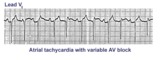 |
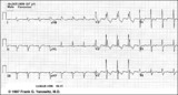
Atrial tachycardia with 3:2 AV block | In this rhythm the atrial rate from an ectopic focus is 160 bpm. Atrial activity can be seen on top of T waves, and before QRS's. Careful observation reveals a 3:2 Wenckebach relationship between P waves and QRS's. Atrial tachycardia with block is often a sign of digitalis intoxication. | Knowledge Weavers ECG | |
| 52 |
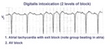 |
Atrial tachycardia with exit block and AV block | The ectopic P waves, easily seen in this example,occur in groups, separated by short pauses. This is likely due to an exit block just distal to the atrial pacemaker. Because not all of the P waves make it to the ventricles, there is also 2nd degree AV block. Therefore, two levels of block are pre... | Knowledge Weavers ECG | |
| 53 |
 |
Atypical LBBB with primary T wave abnormalities | Primary T wave abnormalities in LBBB refer to T waves in the same direction as the major deflection of the QRS. These are seen in leads I, III, aVL, V2-4. Most likely diagnosis is myocardial infarction. | Knowledge Weavers ECG | |
| 54 |
 |
Atypical LBBB with Q waves in leads I and aVL | In typical LBBB, there are no initial Q waves in leads I, aVL, and V6. If Q waves are present in 2 or more of these leads, myocardial infarction is present. | Knowledge Weavers ECG | |
| 55 |
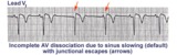 |
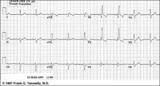
AV dissociation by default | If the sinus node slows too much a junctional escape pacemaker may take over as indicated by arrows. AV dissociation is incomplete, since the sinus node speeds up and recaptures the entricles. | Knowledge Weavers ECG | |
| 56 |
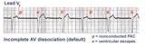 |
AV dissociation by default | The nonconducted PAC's set up a long pause which is terminated by ventricular escapes; note the wider QRS morphology of the escape beats indicating their ventricular origin. Incomplete AV dissociation occurs during the escape beats, since the atria are still under the control of the sinus node. | Knowledge Weavers ECG | |
| 57 |
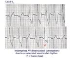 |
AV dissociation by usurpation | Normal sinus rhythm is interrupted by an accelerated ventricular rhythm whose rate is slightly faster than the sinus rhythm. Fusion QRS complexes occur whenever the sinus impulse enters the ventricles at the same time the ectopic ventricular focus initiates its depolarization. | Knowledge Weavers ECG | |
| 58 |
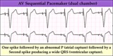 |
AV sequential pacemaker - marquette | (Summary) | Knowledge Weavers ECG | |
| 59 |
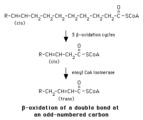 |
Beta-oxidation of a delta-9 fatty acyl CoA | Enoyl CoA isomerase is required to move the double bond in a Delta-9 fatty acyl CoA to a position where it can continue in beta-oxidation. | Knowledge Weavers Fatty Acids | |
| 60 |
 |
Bifascicular block: RBBB + LAFB | Bifascicular block: RBBB + LAFB | Knowledge Weavers ECG | |
| 61 |
 |
Bifascicular block: RBBB + LAFB | This is the most common of the bifascicular blocks. RBBB is most easily recognized in the precordial leads by the rSR' in V1 and the wide S wave in V6 (i.e., terminal QRS forces oriented rightwards and anterior). LAFB is best seen in the frontal plane leads as evidenced by left axis deviation (-50... | Knowledge Weavers ECG | |
| 62 |
 |
Bradycardia-dependent LBBB with carotid sinus massage | When carotid sinus massage slows the heart rate in this example, the QRS widens into a LBBB. This form of rate-dependent bundle branch block is thought to be due to latent pacemakers in the bundle undergoing phase 4 depolarization; when the sinus impulse enters the partially depolarized bundle, slow... | Knowledge Weavers ECG | |
| 63 |
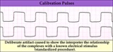 |
Calibration signal - marquette | Calibration signal - marquette | Knowledge Weavers ECG | |
| 64 |
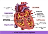 |
Cardiac conduction system diagram - marquette | Cardiac conduction system diagram - marquette | Knowledge Weavers ECG | |
| 65 |
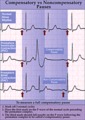 |
Compensatory vs. non-compensatory pauses - marquette | Compensatory vs. non-compensatory pauses - marquette | Knowledge Weavers ECG | |
| 66 |
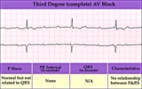 |
Complete AV block (3rd degree) with junctional rhythm | Complete AV block (3rd degree) with junctional rhythm | Knowledge Weavers ECG | |
| 67 |
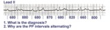 |
Complete AV block, junctional escape rhythm, and ventriculophasic sinus arrhythmia | Complete AV block is seen as evidenced by the AV dissociation. A junctional escape rhythm sets the ventricular rate at 45 bpm. The PP intervals vary because of ventriculophasic sinus arrhythmia; this is defined when the PP interval that includes a QRS is shorter than a PP interval that excludes a ... | Knowledge Weavers ECG | |
| 68 |
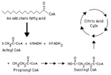 |
Complete oxidation of an odd-chain fatty acid -- overview | This diagram indicates production of propionyl CoA from an odd-chain fatty acid and the subsequent conversion of propionyl CoA to succinyl CoA, which can be metabolized through the citric (tricarboxylic) acid cycle. | Biosynthesis | Knowledge Weavers Fatty Acids |
| 69 |
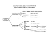 |
Conceptual framework: aArrhythmias and conduction abnormalities | Conceptual framework: aArrhythmias and conduction abnormalities | Knowledge Weavers ECG | |
| 70 |
 |
Condensation of an acyl group with a malonyl group | The acetyl group displaces the carboxyl of the malonyl group, forming a beta-ketoacyl group. This reaction is catalyzed by beta-ketoacyl Acyl Carrier Peptide synthase. The carboxyl released in the form of bicarbonate regenerates the bicarbonate used earlier in the acetyl CoA carboxylase reaction. | Knowledge Weavers Fatty Acids | |
| 71 |
 |
Dehydrogenation of fatty acyl CoA | Fatty acyl CoA is dehydrogenated by FAD in a reaction catalyzed by one of the acyl CoA dehydrogenases. Note that the dehydrogenation occurs between the alpha- and beta-carbons. | FAD | Knowledge Weavers Fatty Acids |
| 72 |
 |
Diagram: frontal plane leads | Diagram: frontal plane leads | Knowledge Weavers ECG | |
| 73 |
 |
Diagram: stages of acute Q-wave MI | Diagram: stages of acute Q-wave MI | Knowledge Weavers ECG | |
| 74 |
 |
Diagram: type I vs. type II 2nd degree AV block | In type I 2nd degree AV block the PR progressively lengthens until a nonconducted P wave occurs. The PR gets longer by smaller and smaller increments; this results in gradual shortening of the RR intervals. The RR interval of the pause is usually less than the two preceding RR intervals. The RR i... | Knowledge Weavers ECG | |
| 75 |
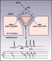 |
Diagram: AV nodal reentrant tachycardia | The AV node often has dual pathways; in this diagram the alpha pathway is fast, but has a long refractory period; the beta pathway is conducts more slowly, but recovers faster.In sinus rhythm the faster alpha pathway is used and accounts for the normal PR interval. When a PAC occurs, however, the i... | Knowledge Weavers ECG |