|
|
Title | Date | Type |
| 1 |
 |
30 | 2021 | Image |
| 2 |
 |
6-16 | 2021 | Image |
| 3 |
 |
6-16 Non-Arteritic | 2021 | Image |
| 4 |
 |
6-4 Prime | 2021 | Image |
| 5 |
 |
6th Nerve Orbital Negative | 2021 | Image |
| 6 |
 |
80 Infiltrative Orbital Negative | 2021 | Image |
| 7 |
 |
81-b | 2021 | Image |
| 8 |
|
81-b Glaucoma | 2021 | Image |
| 9 |
|
81-b Glaucoma (again) | 2021 | Image |
| 10 |
 |
81-b Glaucoma with Arrow | 2021 | Image |
| 11 |
 |
81-b Glaucoma with Arrow (cropped) | 2021 | Image |
| 12 |
|
81-b Glaucoma with Arrow (cropped, again) | 2021 | Image |
| 13 |
 |
9-27B | 2021 | Image |
| 14 |
 |
9-27C | 2021 | Image |
| 15 |
 |
Absent Physiologic Cup (cropped) | 2021 | Image |
| 16 |
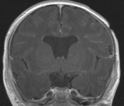 |
Absent Septum Pellucidum | 2021 | Image |
| 17 |
 |
Absent Septum Pellucidum with Arrow | 2021 | Image |
| 18 |
|
Absent Septum Pellucidum with Arrow (again) | 2021 | Image |
| 19 |
 |
Aicardi | 2021 | Image |
| 20 |
 |
Altitudinal Disc Pallor (cropped) | 2021 | Image |
| 21 |
 |
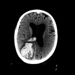
Aneurysm (CT) | 2021 | Image |
| 22 |
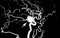 |
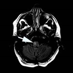
Aneurysm, Angio with Arrow | 2021 | Image |
| 23 |
 |
Anisocoria | 2021 | Image |
| 24 |
 |
Anisocoria with logo | 2021 | Image |
| 25 |
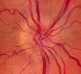 |
Anomalous Vasculature Optic Disc (cropped) | 2021 | Image |
| 26 |
|
Anomalous Vasculature Optic Disc (cropped, again) | 2021 | Image |
| 27 |
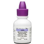 |
Apraclonidine Bottle | 2021 | Image |
| 28 |
 |
Apraxia of Lid Opening (cropped) | 2021 | Image |
| 29 |
 |
Aqueductal Stenosis | 2021 | Image |
| 30 |
 |
Arteritic Ischemic Optic Neuropathy (OU) | 2021 | Image |
| 31 |
 |
Asymmetric Papilledema (OU) | 2021 | Image |
| 32 |
 |
Atrophic Disc | 2021 | Image |
| 33 |
 |
Atrophic Disc with Cataract (cropped) | 2021 | Image |
| 34 |
 |
Atrophic EOMs | 2021 | Image |
| 35 |
|
Atrophic EOMs with Arrows | 2021 | Image |
| 36 |
 |
Atrophic EOMs, CPEO, CT | 2021 | Image |
| 37 |
 |
Atrophic Papilledema | 2021 | Image |
| 38 |
 |
Autopsy Alzheimer | 2021 | Image |
| 39 |
 |
AV with Fistula Arrow | 2021 | Image |
| 40 |
 |
Axial Enhancing Optic Nerve (MRI) | 2021 | Image |
| 41 |
 |
Axial Enhancing Optic Nerve MRI with Arrow | 2021 | Image |
| 42 |
 |
Balint Biparietal Infarcts | 2021 | Image |
| 43 |
 |
Bihemispheric Innervation | 2021 | Image |
| 44 |
 |
Bilateral End Organ Lesion | 2021 | Image |
| 45 |
 |
Bilateral Occipital Infarctions (CT) | 2021 | Image |
| 46 |
 |
Bilateral Orbital Inflamation | 2021 | Image |
| 47 |
 |
Bilateral Orbital Inflammation in GCA with Arrows | 2021 | Image |
| 48 |
 |
Bilateral Pontine Infarction | 2021 | Image |
| 49 |
 |
Bilateral Restricted Diffusion Optic Nerves | 2021 | Image |
| 50 |
 |
Bilateral Restricted Diffusion Optic Nerves with Arrows | 2021 | Image |
| 51 |
|
Bilateral Restricted Diffusion Optic Nerves with Arrows (new image) | 2021 | Image |
| 52 |
 |
Bilateral Temporal Legion | 2021 | Image |
| 53 |
 |
Biparietal Abscesses Balint | 2021 | Image |
| 54 |
 |
Biparietal Infarcts (DWI) | 2021 | Image |
| 55 |
|
Biparietal Infarcts (DWI) 1 | 2021 | Image |
| 56 |
|
Biparietal Infarcts (DWI) 2 | 2021 | Image |
| 57 |
 |
Bitemporal - 1 | 2021 | Image |
| 58 |
 |
Bitemporal Hemorrhage | 2021 | Image |
| 59 |
 |
Bithalamic Infarcts with Arrows | 2021 | Image |
| 60 |
 |
Blepharospasm (cropped) | 2021 | Image |
| 61 |
 |
Blue | 2021 | Image |
| 62 |
 |
Brain Hemorrhage | 2021 | Image |
| 63 |
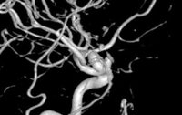 |
Carotid Aneurysm | 2021 | Image |
| 64 |
 |
Carotid Aneurysm 3D | 2021 | Image |
| 65 |
|
Carotid Aneurysm 3D (again) | 2021 | Image |
| 66 |
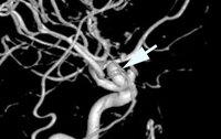 |
Carotid Aneurysm with Arrow | 2021 | Image |
| 67 |
 |
Carotid Fasicula | 2021 | Image |
| 68 |
 |
Carotoid | 2021 | Image |
| 69 |
 |
Cataract | 2021 | Image |
| 70 |
 |
Caudal Quadrigeminal Glioma with Arrow | 2021 | Image |
| 71 |
 |
Cavernoma Medulla with Arrow | 2021 | Image |
| 72 |
 |
Cavernous 6th Lesion | 2021 | Image |
| 73 |
 |
Cerebellar Atrophy | 2021 | Image |
| 74 |
 |
Cerebellar Atrophy in SCA with Arrow (MRI) | 2021 | Image |
| 75 |
 |
Cerebellar Hem with Arrow | 2021 | Image |
| 76 |
 |
Cerebral Metamorphopsia | 2021 | Image |
| 77 |
 |
Cerebral Polyopia | 2021 | Image |
| 78 |
 |
Choriorentinitis | 2021 | Image |
| 79 |
 |
Choriorentinitis (cropped) | 2021 | Image |
| 80 |
|
Choriorentinitis (cropped, again) | 2021 | Image |
| 81 |
 |
Classic Hemisphereic Signs of MS (MRI) | 2021 | Image |
| 82 |
|
Classic Hemisphereic Signs of MS (MRI, again) | 2021 | Image |
| 83 |
 |
Clear Margins Elevated Optic Disc (cropped) | 2021 | Image |
| 84 |
 |
Close Up Relieving Diplopia (figure b) | 2021 | Image |
| 85 |
 |
Coarse Visual Snow | 2021 | Image |
| 86 |
 |
Coloboma (cropped) | 2021 | Image |
| 87 |
|
Coloboma (cropped, again) | 2021 | Image |
| 88 |
|
Coloboma -1 (cropped) | 2021 | Image |
| 89 |
 |
Coloboma Optic Disc | 2021 | Image |
| 90 |
 |
Coloboma with Arrow (cropped, new image) | 2021 | Image |
| 91 |
 |
COMA Hamm (MRI) | 2021 | Image |
| 92 |
|
COMA Hamm (MRI, again) | 2021 | Image |
| 93 |
 |
Comet Tail | 2021 | Image |
| 94 |
 |
Completely Excavated Optic Disc Cropped | 2021 | Image |
| 95 |
 |
Compressive Optic Disc Pallor | 2021 | Image |
| 96 |
 |
Compressive Optic Neuropathy (MRI) | 2021 | Image |
| 97 |
 |
Coronal Atrophy (MRI, R, SOM) | 2021 | Image |
| 98 |
 |
Cranio Coronal with Arrow (MRI) | 2021 | Image |
| 99 |
 |
Cranio Cyst | 2021 | Image |
| 100 |
 |
CRVO | 2021 | Image |