The Health Education Assets Library (HEAL) is a collection of over 22,000 freely available digital materials for health sciences education. The collection is now housed at the University of Utah J. Willard Marriott Digital Library.
| Title | Description | Subject | Collection | ||
|---|---|---|---|---|---|
| 1 |  | Adrenal Adenoma in Clinical Conn's Syndrome | Adrenal adenoma, aldosterone producing associated with Conn's syndrome. Gross photograph showing 2 contiguous slices of adrenal gland with cortical adenoma. Patient had hyperaldosteronism (Conn's syndrome). | Adrenal Adenoma; Conn Syndrome | HEAL Reviewed Collection |
| 2 | 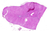 | Adrenal cortical hyperplasia | Patient with ectopic ACTH production and diffuse adrenal cortical hyperplasia. | ACTH | HEAL Reviewed Collection |
| 3 | | Adrenal gland with pheochromocytoma | Patient with clinical symptoms of hypertension. Image shows adjacent normal adrenal gland with paraganglioma (pheochromocytoma) involving adrenal medulla. | Adrenalin; Noradrenalin | HEAL Reviewed Collection |
| 4 | | Adrenal myelolipoma | Gross photograph with ruler showing residual normal adrenal and mixture of yellow adipose tissue and red myeloid areas of myelolipoma. | benign | HEAL Reviewed Collection |
| 5 | | Adrenal myelolipoma | Gross photograph showing residual normal adrenal and mixture of yellow adipose tissue and red myeloid areas of myelolipoma. | benign | HEAL Reviewed Collection |
| 6 | | Adrenal myelolipoma | Close up showing residual normal adrenal and mixture of yellow adipose tissue and red myeloid areas of myelolipoma. | Benign | HEAL Reviewed Collection |
| 7 |  | Bladder carcinoma with lymphovascular invasion | Lymphovascular invasion by a bladder carcinoma away from the main tumor mass. | Urothelial Carcinoma | HEAL Reviewed Collection |
| 8 | 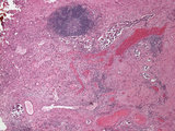 | Bladder carcinoma with lymphovascular invasion | Lymphovascular invasion by a bladder carcinoma away from the main tumor mass. | Urothelial Carcinoma | HEAL Reviewed Collection |
| 9 | 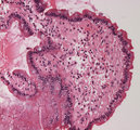 | Duodenum amyloid deposition | Duodenum with amyloid deposition in lamina propria. Amyloid etiology unknown. Amyloid shows up as homogenous pink material in lamina propria and around blood vessels 20X. | Small Bowel | HEAL Reviewed Collection |
| 10 | 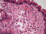 | Duodenum amyloid deposition | Duodenum with amyloid deposition in lamina propria. Amyloid etiology unknown. Amyloid shows up as homogenous pink material in lamina propria and around blood vessels 40X. | Small Bowel | HEAL Reviewed Collection |
| 11 |  | Duodenum amyloid deposition - Congo Red | Duodenum with amyloid deposition in lamina propria. Amyloid etiology unknown. Amyloid shows up as homogenous bright pink material in lamina propria and around blood vessels after staining with congo red 20X. | Small Bowel | HEAL Reviewed Collection |
| 12 | 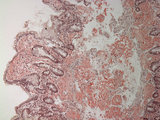 | Duodenum amyloid deposition - Congo Red | Duodenum with amyloid deposition in lamina propria. Amyloid etiology unknown. Amyloid shows up as homogenous bright pink material in lamina propria and around blood vessels after staining with congo red 10X. | Small Bowel | HEAL Reviewed Collection |
| 13 | 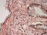 | Duodenum amyloid deposition - Congo Red | Duodenum with amyloid deposition in lamina propria. Amyloid etiology unknown. Amyloid shows up as homogenous bright pink material in lamina propria and around blood vessels after staining with congo red 40X. | Small Bowel | HEAL Reviewed Collection |
| 14 |  | Eosinophilic granuloma mastoid | High power view (40X) of a neoplastic proliferation of Langerhan's cells (Histiocytosis) in mastoid bone stained with CD1a an immunohistochemical stain for Langerhans cells | HEAL Reviewed Collection | |
| 15 | 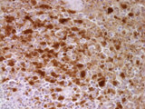 | Eosinophilic granuloma mastoid | High power view (40X) of a neoplastic proliferation of Langerhan's cells (Histiocytosis) in mastoid bone stained for S100 an immunohistochemical stain dendritic cells | HEAL Reviewed Collection | |
| 16 | 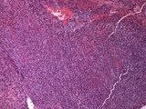 | Eosinophilic granuloma mastoid | Low power view (10X) of a neoplastic proliferation of Langerhan's cells (Histiocytosis) in mastoid bone | HEAL Reviewed Collection | |
| 17 |  | Eosinophilic granuloma mastoid | High power view (40X) of a neoplastic proliferation of Langerhan's cells (Histiocytosis) in mastoid bone. | HEAL Reviewed Collection | |
| 18 |  | Eosinophilic granuloma mastoid | High power view (40X) of a neoplastic proliferation of Langerhan's cells (Histiocytosis) in mastoid bone | HEAL Reviewed Collection | |
| 19 | 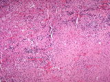 | Gummatous inflammation from tertiary syphilis in adrenal gland | Granulomatous (Gumma) inflammation from tertiary syphilis involving the adrenal gland. | spirochete; gumma; gummatous inflammation | HEAL Reviewed Collection |
| 20 | 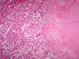 | Gummatous inflammation from tertiary syphilis in adrenal gland | Granulomatous (Gumma) inflammation from tertiary syphilis involving the adrenal gland. | spirochete; gumma; gummatous inflammation | HEAL Reviewed Collection |
| 21 | 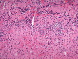 | Gummatous inflammation from tertiary syphilis in adrenal gland | Granulomatous (Gumma) inflammation from tertiary syphilis involving the adrenal gland. | spirochete; gumma; gummatous inflammation | HEAL Reviewed Collection |
| 22 | 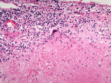 | Gummatous inflammation from tertiary syphilis in adrenal gland | Granulomatous (Gumma) inflammation from tertiary syphilis involving the adrenal gland. | spirochete; gumma; gummatous inflammation | HEAL Reviewed Collection |
| 23 | 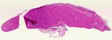 | Gummatous inflammation from tertiary syphilis in adrenal gland | Granulomatous (Gumma) from tertiary syphilis involving the adrenal gland. | spirochete; gumma; gummatous inflammation | HEAL Reviewed Collection |
| 24 | 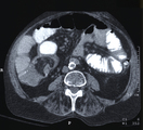 | Intussusception | This image depicts Intussusception. Intussusception can be found in an abdominal CT. The CT may show nothing if early in the disease or may show a perforation, an obstruction, or a soft tissue mass. | Intestinal Invagination; Intussuscipiens; Intussusceptum; Lead Point; Concentric Rings | HEAL Reviewed Collection |
| 25 |  | Molluscum contagiosum | Photomicrograph showing cytopathic changes of pox virus (Molluscum contagiosum) in HIV patient. | AIDS | HEAL Reviewed Collection |