The Health Education Assets Library (HEAL) is a collection of over 22,000 freely available digital materials for health sciences education. The collection is now housed at the University of Utah J. Willard Marriott Digital Library.
TO
| Title | Description | Subject | Collection | ||
|---|---|---|---|---|---|
| 1 |
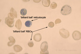 |
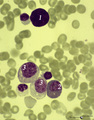
'Billiard Ball' Cells | 'billiard ball' cells in Hb SC disease with supravital stain | Fe01aT; Hb SC | Albert Einstein College of Medicine Gallery of Hematology Images |
| 2 |
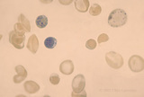 |
'Billiard Ball' Cells | 'billiard ball' cells in Hb SC disease with supravital stain | Fe01a0; Hb SC | Albert Einstein College of Medicine Gallery of Hematology Images |
| 3 |
 |
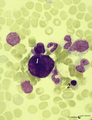
(Pro)myelocyte and neutrophilic granulocytes in bone marrow smear (human) | Stain: May-Grnwald-Giemsa (MGG). The promyelocyte (1) contains coarse primary, azurophilic granules in the basophilic cytoplasm. The absence of nucleoli indicates the late stage of the promyelocyte. (2) segmented neutrophilic granulocyte with toxic granulation (large irregular granules). (3) indicat... | Poja Histology Collection - Blood & Bone Marrow Subset | |
| 4 |
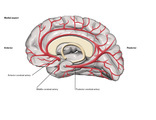 |
3 Major Cerebral Arteries - Midsagittal (Labeled) | Anterior cerebral, middle cerebral, and posterior cerebral arteries. | Royal College of Surgeons in Ireland Illustrations | |
| 5 |
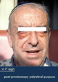 |
4 P' Sign of Amyloid | 4 P' sign of amyloid | Kg010T | Albert Einstein College of Medicine Gallery of Hematology Images |
| 6 |
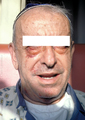 |
4 P' Sign of Amyloid | 4 P' sign of amyloid | Kg0100 | Albert Einstein College of Medicine Gallery of Hematology Images |
| 7 |
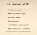 |
The 5q-syndrome in Myelodysplastic Syndrome | the 5q-syndrome in MDS | Mb1200 | Albert Einstein College of Medicine Gallery of Hematology Images |
| 8 |
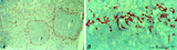 |
: Localization of ED3-positive subpopulation of macrophages in spleen (rat) | Stain: Immunohistochemistry of Vector red staining using the antibody ED3. The survey in (A) shows that the ED3-positive macrophages are found as a ring in the marginal zone border, as well as spread in the red pulp area (2). The cells are sometimes referred as marginal metallophilic macrophages. T... | metallophilic macrophages; ED3 antibody; marginal zone | Poja Histology Collection - Lymphatic Tissues and Organs Subset |
| 9 |
 |
: Lymph node (human) | Stain: Azan. Specialized venules (1) or so-called high endothelial venules (HEV) are here located in the paracortical area (4) close to the lymphatic follicle (2+3). The HEVs are lined by cuboidal or columnar endothelial cells that possess specific homing receptors for antigen-primed B- and T ly... | paracortex; high endothelial venule (HEV); germinal center | Poja Histology Collection - Lymphatic Tissues and Organs Subset |
| 10 |
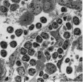 |
: Lymph node (rat) | Electron microscopy. A low magnification of a part of the medulla showing medullary cords surrounded by labyrinthine medullary sinus (*). In this picture the medullar cord runs from left bottom corner to right top corner, and is lined by flat reticular cell types. Within the cord one finds a star-sh... | medulla; electron microscopy; sinusoid | Poja Histology Collection - Lymphatic Tissues and Organs Subset |
| 11 |
 |
A bare megakaryocyte nucleus and myelocytes in bone marrow smear (human) | Stain: May-Grnwald-Giemsa (MGG). (1) Indicates a bare large-sized polyploid nucleus of a megakaryocyte which has totally shed the cytoplasm. These cells are often seen in normal marrow and are ultimately removed by macrophages. There is no cytoplasm left, nor platelets are left around the nucleus. T... | Poja Histology Collection - Blood & Bone Marrow Subset | |
| 12 |
 |
A bare megakaryocyte nucleus in bone marrow smear (human) | Stain: May-Grnwald-Giemsa (MGG). (1) indicates a naked, large-sized polyploid nucleus of a megakaryocyte with visible lobes. No cytoplasm nor platelets are left around the nucleus. Most surrounding cells are of myeloid origin, except one orthochromatic erythroblast (2). | Poja Histology Collection - Blood & Bone Marrow Subset | |
| 13 |
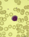 |
A plasmacytoid lymphocyte in peripheral blood smear (human) | Stain: May-Grnwald-Giemsa (MGG). The plasmacytoid lymphocyte is an activated B lymphocyte to be transformed into a plasma cell. The cytoplasm is more basophilic and the chromatin pattern is more clumped than in a virgin small lymphocyte. When the cell contains numerous immunoglobulin inclusions (glo... | Poja Histology Collection - Blood & Bone Marrow Subset | |
| 14 |
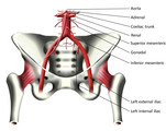 |
Abdominal Aorta and Arteries of the Pelvis (Labeled) | Abdominal aorta and pelvic arteries. | Adrenal Artery; Coeliac Trunk; Gonadal Artery; Left External Iliac Artery; Left Internal Iliac Artery | Royal College of Surgeons in Ireland Illustrations |
| 15 |
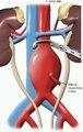 |
Abdominal Aortic Aneurysm (Labeled) | Left renal vein and inferior mesenteric artery adjacent to aortic aneurysm. | Royal College of Surgeons in Ireland Illustrations | |
| 16 |
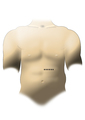 |
Abdominal Incision Line for Malecot Catheter Insertion | Abdominal incision for Malecot catheterization. | Malecot Catheter | Royal College of Surgeons in Ireland Illustrations |
| 17 |
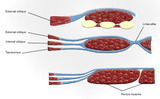 |
Abdominal Muscles (Labeled) | Abdominal muscles including external oblique, internal oblique, linea alba, transversus, and rectus muscles. | External Oblique Muscle; Internal Oblique Muscle; Transversus Abdominis; Linea Alba | Royal College of Surgeons in Ireland Illustrations |
| 18 |
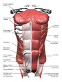 |
Abdominal Muscles (Labeled) | Abdominal muscles and adjacent structures. | Superior Epigastric Vessels; External Oblique Muscle; Rectus Sheath; Linea Alba; External Oblique Aponeurosis; Superficial Inguinal Ring; Deep Inguinal Ring; Transversalis Fascia; Medial Umbilical Ligament; Iliohypogastric Nerve | Royal College of Surgeons in Ireland Illustrations |
| 19 |
 |
Abducens nerve | Abducens nerve. Yellow nerve added to digitized image from graphic output. Photograph. Multimedia. | Abducens nerve; Central nervous system; Cranial nerves; Anatomy | Slice of Life |
| 20 |
 |
Abducens nerve, intracranial portion passing through tegmentum | Abducens nerve, intracranial portion passing through tegmentum. Transverse plane. Photograph. Multimedia. | Abducens nerve; Tegmentum mesencephali; Pons; Central nervous system; Anatomy | Slice of Life |
| 21 |
 |
Abducens nucleus | Abducens nucleus. Transverse plane. Photograph. Multimedia. | Slice of Life | |
| 22 |
 |
Abducens nucleus VI | Abducens nucleus VI. Pons. Transverse plane. Photograph. Multimedia. | Abducens nerve; Pons; Cranial nerves; Central nervous system; Anatomy | Slice of Life |
| 23 |
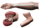 |
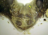
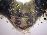
Above the Wrist Amputation of Hand - Forearm Cross Section | Above the wrist amputation of hand. | Amputation of Hand | Royal College of Surgeons in Ireland Illustrations |
| 24 |
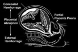 |
Abruptio placenta | Abruptio placenta | Knowledge Weavers Human Reproduction | |
| 25 |
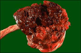 |
Abruptio placenta - Gross | This large retroplacental blood clot is known as abruptio placenta. Such abnormal hemorrhage prior to delivery can lead to sudden onset of pain in the mother. | Knowledge Weavers Human Reproduction |