The Health Education Assets Library (HEAL) is a collection of over 22,000 freely available digital materials for health sciences education. The collection is now housed at the University of Utah J. Willard Marriott Digital Library.
TO
| Title | Description | Subject | Collection | ||
|---|---|---|---|---|---|
| 1 |
 |
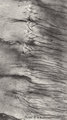
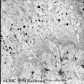
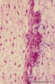
Cross-section of tooth in alveolar bone (cat, adult) | Stain: Picric acid and hematoxylin. From left to right: Periodontal ligament with blood vessels. Acellular cementum (dark purple rim). Dentin with radiair arrangement of dentinal tubules; fine incremental (imbrication) lines of von Ebner run at right angles to these tubules. These lines represent di... | oral cavity; incremental lines; von Ebner | Poja Histology Collection - Oral Cavity Subset |
| 2 |
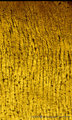 |
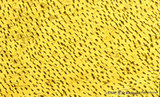
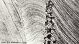
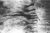
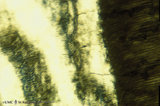
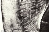
Dentinal tubules in longitudinal section of tooth - human, adult. Thin ground section, polarizing microscopy - optical axes of the polarizing plates are crossed at 60. | Slightly oblique section shows dentinal tubules depicted by brown-stained hollow fiber-like structures containing odontoblastic processes (Tomes' fibers) in a semi-three dimensional way. Numerous fine secondary branches of these processes are anastomosing with those of neighboring tubules. | oral cavity; Tomes' Fibers; dentinal tubules | Poja Histology Collection - Oral Cavity Subset |
| 3 |
 |
Cross section of tooth in alveolar bone - cat; low magnification | Stain: Picric acid and hematoxylin. From left to right: alveolar bone tissue with osteons; periodontal ligament with blood vessels; acellular cementum (dark purple rim); dentin with (purple) radiair arrangement of dentinal tubules; fine incremental (imbrication) lines of von Ebner run at right angl... | oral cavity; incremental lines; alveolar bone | Poja Histology Collection - Oral Cavity Subset |
| 4 |
 |
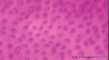
Dentinal tubules in longitudinal section of tooth - human, adult. Thin ground section, polarizing microscopy - optical axes of the polarizing plates are crossed at 60. | Slightly oblique section shows dentinal tubules depicted by brown-stained hollow fiber-like containing odontoblastic processes (Tomes' fibers) in a semi-three dimensional way ('stubble-field' aspect). Darker stained clusters represent anastomosing secondary branches. | oral cavity; Tomes' Fibers; dentinal tubules | Poja Histology Collection - Oral Cavity Subset |
| 5 |
 |
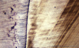
Dentinoenamel junction in longitudinal section of tooth (human, adult). Thin ground section. | From left to right: Enamel with fine striation (course of enamel rods or prisms). Few enamel tufts (left, dark) consisting of hypocalcified enamel rods and interprismatic substance arise from the junction. Scallop-like course of dentinoenamel junction. Dentin with dentinal tubules to the dentinoenam... | oral cavity; enamel tufts; dentinoenamel junction; dentinal tubules | Poja Histology Collection - Oral Cavity Subset |
| 6 |
 |
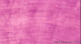
Dentinal tubules in longitudinal section of tooth - human, adult. Thin ground section, polarizing microscopy - optical axes of the polarizing plates are crossed at 60. | Slightly oblique section shows dentinal tubules depicted by brown-stained stubble structures containing odontoblastic processes (Tomes' fibers) in a semi-three dimensional way. Numerous fine branches of these processes are obvious in dentin. | oral cavity; Tomes' Fibers; dentinal tubules | Poja Histology Collection - Oral Cavity Subset |
| 7 |
 |
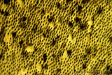
Dentin in cross section of tooth - human, adult | Stain: Hematoxylin and eosin. Cross-sectioned dentinal tubules demonstrate: a light stained center with the odontoblastic process (Tomes' fiber); darker stained peritubular dentin (highly mineralized), also called Neuman's sheath. Intertubular dentin (less mineralized) is present between the tubule... | oral cavity; dentinal tubules; secondary dentin | Poja Histology Collection - Oral Cavity Subset |
| 8 |
 |
Dentin in cross section of tooth (human, adult) | Stain: Hematoxylin and eosin. Longitudinally sectioned dentinal tubules are parallely arranged, and numerous side branches are visible. | oral cavity; dentinal tubules | Poja Histology Collection - Oral Cavity Subset |
| 9 |
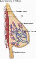 |
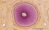
Breast - Cross Section - Ductal Carcinoma (Labeled) | Ductal carcinoma of breast. | Pectoralis Major; Breast Lobule; Ductule | Royal College of Surgeons in Ireland Illustrations |
| 10 |
 |
Dentinoenamel junction in the tooth (human, adult). Thin ground section of crown. | From left to right: Enamel with fine striation (composition of enamel rods or prisms); darker zones almost perpendicular to the striation are the incremental lines (Retzius) due to successive apposition of layers of enamel as the crown is formed. Dentinoenamel junction is shown as a narrow fissure f... | oral cavity; dentinal tubules; dentinoenamel junction; interglobular dentin; lines of Retzius | Poja Histology Collection - Oral Cavity Subset |
| 11 |
 |
Dentinoenamel junction in the tooth - human, adult. Thin ground section of crown. | From left to right: Superficial dentin (bluish) in the crown with S-shaped course of dentinal tubules; They pass uninterrupted through the irregular black structures (due to filling with air) representing hypocalcified areas (interglobular dentin); Mineralization of dentin starts in small globular ... | oral cavity; dentinal tubules; dentinoenamel junction; interglobular dentin; lines of Retzius | Poja Histology Collection - Oral Cavity Subset |
| 12 |
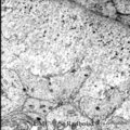 |
Dentinogenesis in tooth development - bell stage, gerbil, postnatal | Electronmicroscopy. At the top right corner side the distal cytoplasmic parts of presecretory ameloblasts resting on a thin grey basal lamina. In the central area predentin with collagen fibers (grey patches) and cross-sectioned small odontoblastic branches. In between them dispersed numerous dark-s... | oral cavity; predentin; matrix vesicles | Poja Histology Collection - Oral Cavity Subset |
| 13 |
 |
Dentinogenesis in tooth development - bell stage, gerbil, postnatal | Electronmicroscopy. At the bottom side of the predentin partly cross-sectioned odontoblasts with some organelles and many vesicular structures, the dark ones containing hydroxyapatite. Close to the odontoblasts a high concentration of secreted collagen fibers. Further away numerous matrix vesicles (... | oral cavity; predentin; matrix vesicles | Poja Histology Collection - Oral Cavity Subset |
| 14 |
 |
Dentinoenamel junction in longitudinal section of tooth - human, adult. Thin ground section. | From left to right: enamel with fine striation (composed of stalks of enamel rods or prisms); enamel tufts (dark) arise at the dentinoenamel junction, and these tufts consist of hypocalcified enamel prisms and interprismatic substance; dentin with dentinal tubules up to the dentinoenamel. | oral cavity; enamel tufts; dentinoenamel junction; dentinal tubules | Poja Histology Collection - Oral Cavity Subset |
| 15 |
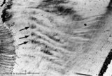 |
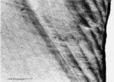
Enamel in longitudinal section of tooth - human, adult. Thin ground section. | From left to right: dentin with dentinal tubules; dentinoenamel junction; enamel with arrows pointing to bands (lines) of Hunter-Schreger; these alternating light and dark strips originate at the dentinoenamel junction and do not reach the enamel surface. This optical phenomenon is the result of the... | oral cavity; Hunter-Schreger bands; Retzius | Poja Histology Collection - Oral Cavity Subset |
| 16 |
 |
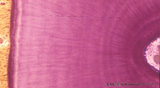
Enamel in longitudinal section of tooth - human, adult. Thin ground section, polarizing microscopy - optical axes of the polarizing plates are crossed at 60. | Using polarizing microscopy the birefringence of the crystalline structure of enamel is colorful demonstrated. Incremental lines (striae) of Retzius are well shown as straight oblique zones. Note the parallel lines of enamel stacks at the left corner of the picture. At the right side the lightly col... | oral cavity; Retzius; dentinoenamel junction | Poja Histology Collection - Oral Cavity Subset |
| 17 |
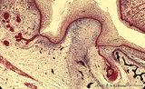 |
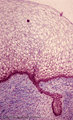
Early cap stage in tooth development - human, embryo; low magnification | Stain: Azan. From top to bottom: top left vestibular groove with gland formation; stratified ectoderm with dark red rim of basal cells from which a dental lamina sprouts downwards into the dental crypt (bony cavity) (bottom right corner); bone stains dark blue; connective tissue/mesenchym stains lig... | oral cavity; tooth development; dental lamina | Poja Histology Collection - Oral Cavity Subset |
| 18 |
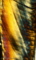 |
Enamel in longitudinal section of tooth - human, adult. Thin ground section, polarizing microscopy - optical axes of the polarizing plates are crossed at 60. | Using polarizing microscopy the birefringence of the crystalline structure of enamel is colorful demonstrated. Incremental lines (striae) of Retzius are well shown as straight oblique zones. Top right a long dark fissure-like structure representing a crack. | oral cavity; Retzius; dentinoenamel junction | Poja Histology Collection - Oral Cavity Subset |
| 19 |
 |
Enamel in longitudinal section of tooth - human, adult. Thin ground section, polarizing microscopy - optical axes of the polarizing plates are crossed at 60. | Left side enamel and at right side dark-stained striation of dentin. Wavy course of enamel prisms from dentinoenamel junction (right side, scalloped appearance) to the left. The transparent areas represent enamel lanes at a different polarizing angle. | oral cavity; dentinoenamel junction | Poja Histology Collection - Oral Cavity Subset |
| 20 |
 |
Enamel in longitudinal section of tooth - human, adult. Thin ground section. | From left to right: incremental lines (striae) of Retzius are distinctly shown as oblique broad zones (at the left) to the enamel surface; during formation of the crown successive apposition of layers of enamel is deposited and results in these so-called incremental grow lines; surface of enamel in ... | oral cavity; incremental lines; Retzius | Poja Histology Collection - Oral Cavity Subset |
| 21 |
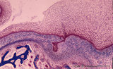 |
Early cap stage in tooth development - human, embryo; low magnification | Stain: Azan. From top to bottom: top left vestibular groove with gland formation; stratified ectoderm with ingrowth of the dental lamina (in the middle); bulbous growing end of dental lamina; bottom left alveolar bone formation (dark blue); and connective tissue/mesenchym stains light blue. | oral cavity; tooth development; dental lamina | Poja Histology Collection - Oral Cavity Subset |
| 22 |
 |
Enamel (odontogenic) organ in tooth development - bell stage, human, embryo | Stain: Azan. Outer surface of bell; from left to right: (avascular) Stellate reticulum; capillaries in this stage proliferate and invaginate between the outer dental epithelial cells; Part of fibrous tooth follicle. | oral cavity | Poja Histology Collection - Oral Cavity Subset |
| 23 |
 |
Early cap stage in tooth development - human, embryo | Stain: Azan. From top to bottom: stratified ectoderm with ingrowth of the dental lamina; knob-like end of the dental lamina; and collagen fibers of lamina propria are blue. | oral cavity; tooth development; dental lamina | Poja Histology Collection - Oral Cavity Subset |
| 24 |
 |
Enamel in longitudinal section of tooth - human, adult. Thin ground section. | At the left side surface of enamel in cuspal region; the parallel horizontal arrangement of stacks of rods (prisms) is evident. At the right side area of dentin (dark area). Incremental lines (striae) of Retzius run as curved lines (from bottom to middle top) and presented successive apposition of l... | oral cavity; incremental lines; Retzius; Hunter-Schreger bands | Poja Histology Collection - Oral Cavity Subset |
| 25 |
 |
Enamel in longitudinal section of tooth - human, adult. Thin ground section. | Enamel is compact and acellular, and consists of vertical stacks of rods (prisms) as well as interrod (interprismatic) regions with less calcifying substance parallel to each other. Each prism is surrounded by an enamel sheath (a non-mineralized organic substance). From left to right: surface of ... | oral cavity; enamel rods; enamel prisms | Poja Histology Collection - Oral Cavity Subset |