John A. Moran Eye Center Neuro-Ophthalmology Collection: A variety of lectures, videos and images relating to topics in Neuro-Ophthalmology created by faculty at the Moran Eye Center, University of Utah, in Salt Lake City.
NOVEL: https://novel.utah.edu/
TO
| Identifier | Title | Description | Subject | ||
|---|---|---|---|---|---|
| 1 |
 |
The_3_Step_Test_Digre.pdf | The 3 Step Test: Looking for a 4th Nerve Palsy | Description of the three step test (3 step test) used when looking for a 4th nerve palsy. | 3 Step Test |
| 2 |
 |
Digre_AION | Anterior Ischemic Optic Neuropathy | PPT describing Anterior Ischemic Optic Neuropathy (AION). Covers clinical signs, such as monocular vision loss, swollen nerve, and visual field defects, as well as risk factors. | Anterior Ischemic Optic Neuropathy |
| 3 |
 |
Basal encephalocele | Basal Encephaloceles | Basal Encephaloceles | |
| 4 |
 |
Basic Headache.pdf | Basic Headache | Presentation covering an overview of headache and migraine. | Migraine, Headache |
| 5 |
 |
benign_episodic_unilateral_mydriasis | Benign Episodic Unilateral Mydriasis | Presentation covering benign episodic mydriasis. | Benign Episodic Mydriasis |
| 6 |
 |
Warner_Clover-leaf_Visual_Field_Defects | Clover-leaf Visual Field Defects | Description of clover-leaf visual field defects. | Visual Field Defects |
| 7 |
 |
Cone Dystrophy.pdf | Cone Dystrophy | PPT covering Cone Dystrophy - An inherited degeneration that presents between 10 - 30 years of age. Symptoms are decreased visual acuity, poor color vision, and sometimes light sensitivity. | Cone Dystrophy; Central Cone Dystrophy |
| 8 |
 |
Dissection of the Carotid Artery | Dissection of the Carotid Artery | Vascular; Dissection | |
| 9 |
 |
External_photography_pupils_extra_ocular_muscles_Digre.pdf | Documenting the Neuro-ophthalmic Patient: External Photography | Description of documenting the neuro-ophthalmic patient using external photography. This covers pupils and extra ocular muscles. | External Photography |
| 10 |
 |
weinberg_1.pdf | Dysthyroid Optic Neuropathy: A Preventable Cause of Blindness | Dysthyroid Optic Neuropathy (DON) is a treatable cause of visual loss in ~5% of pts w/ ted. Monitor closely those pts with risk factors (proptosis, tight orbit, restricted motility, strabismus, smoker, diabetic). Oral prednisone is often effective, but frequent relapses after tapering. Orbital xrt ... | Dysthyroid Ophthalmopathy; Thyroid Orbitopathy; Thyroid Eye Disease; Thyroid Associated Ophthalmopathy (TAO); Graves' Disease; Restrictive Orbitopathy |
| 11 |
 |
Webvision-EOG-Creel | The Electro-oculogram: Clinical Applications | The electrooculogram measures the potential that exists between the cornea and Bruch's membrane at the back of the eye. The potential produces a dipole field with the cornea approximately 5 millivolts positive compared to the back of the eye, in a normally illuminated room. Although the origin of th... | Electro-oculogram |
| 12 |
 |
Webvision-ERG-Creel | The Electroretinogram and Electro-oculogram: Clinical Applications | The global or full-field electroretinogram (ERG) is a mass electrical response of the retina to photic stimulation. The ERG is a test used worldwide to assess the status of the retina in eye diseases in human patients and in laboratory animals used as models of retinal disease. | Electroretinogram; Electro-oculogram |
| 13 |
 |
FluoreseinAngiography | Fluoresein Angiography | Comprehensive description of using fluoresein angiography in examinations. | Fluoresein Angiography |
| 14 |
 |
Glaucoma the basics.pdf | Glaucoma: The Basics | Glaucoma is the most common optic neuropathy. Progressive cupping of the optic disc due to increased intraocular pressure together with visual field abnormalities and local disc susceptibility factors characterize this neuropathy. This PowerPoint lecture covers the basics of Glaucoma and includes ma... | Glaucoma; Optic Neuropathy |
| 15 |
 |
Herpes Zoster: Zoster Ophthalmicus with Third Nerve Palsy | Herpes Zoster Ophthalmicus with Third Nerve Palsy | Images showing presentation of Herpes Zoster (Zoster Ophthalmicus). | Herpes Zoster (Zoster Ophthalmicus) |
| 16 |
 |
Hydroxychloroquine Maculopathy (Plaquenil).pdf | Hydroxychloroquine Maculopathy (Plaquenil) | An overview of Chloroquine Maculopathy. | Maculopathy; Hydroxychloroquine; Plaquenil |
| 17 |
 |
Leber's Hereditary Optic Neuropathy | Leber's Hereditary Optic Neuropathy | Images and visual fields from a boy with acute visual loss. | Leber's Optic Neuropathy |
| 18 |
 |
Macula.pdf | Macula | Overview of the structure and viewing of the macula. | Macula; Retina |
| 19 |
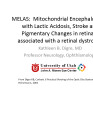 |
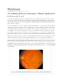
MELAS and RP.pdf | MELAS and RP | MELAS; Mitochondrial Encephalopathy with Lactic Acidosis, Stroke and Pigmentary Changes in retina-associated with a retinal dystrophy. This 53 year old man had seizures, encephalopathy and lactic acidosis typical of MELAS. His fundus examination showed granularity and some slight pigmentary changes ... | Mitochondrial Encephalopathy with Lactic Acidosis; MELAS Syndrome |
| 20 |
 |
Mimics of Atrophy | Mimics of Atrophy | Pseudo Atrophy | |
| 21 |
 |
Webvision-mfERG-Creel | The Multifocal Electroretinogram: Clinical Applications | The most important development in ERGs is the multifocal ERG (mfERG). Erich Sutter adapted the mathematical sequences called binary m-sequences creating a program that can extract hundreds of focal ERGs from a single electrical signal. This system allows assessment of ERG activity in small areas of ... | Multifocal Electroretinogram |
| 22 |
 |
Near_Reflex_and_Accomodation | Near Reflex and Accomodation | Description of testing the near reflex and accomodation. | Near Reflex; Accomodation |
| 23 |
 |
Normal optic disc.pdf | Normal Optic Disc | Overview of the structure and function of the normal optic disc. | Normal Optic Disc Anatomy |
| 24 |
 |
Nutritional amblyopia.pdf | Nutritional Amblyopia | Example of patient with amblyopia with nutritional causes. | Nutritional Optic Atrophy; Wernicke's Encephalopathy |
| 25 |
 |
Optic Disc pallor pseudo and real.pdf | Optic Disc Pallor Pseudo and Real | Discussion of the causes of optic disc pallor. | Optic Disc; Optic Atrophy; Pallor |